
Translate this page into:
Synovial Hemangioma in the Knee: MRI Findings
Address for correspondence: Dr. Harun Arslan, Department of Radiology, Van Education and Hospital, Van, Turkey. E-mail: harun.ars75@gmail.com
-
Received: ,
Accepted: ,
This is an open-access article distributed under the terms of the Creative Commons Attribution License, which permits unrestricted use, distribution, and reproduction in any medium, provided the original author and source are credited.
This article was originally published by Medknow Publications & Media Pvt Ltd and was migrated to Scientific Scholar after the change of Publisher.
Abstract
Synovial hemangiomas are rare benign tumors of vascular origin. A 23-year-old boy presented with knee pain and swelling. The boy had developed symptoms 18-months earlier. He was diagnosed with synovial hemangioma based on magnetic resonnance imaging examination and histopathologic findings of the arthroscopic biopsy tissue. We present the magnetic resonance imaging and histopathologic findings of synovial hemangioma of the knee.
Keywords
Hemangioma
magnetic resonance imaging
synovium

INTRODUCTION

Synovial hemangiomas are uncommon, localized, benign tumoral lesions of vascular origin, which can sometimes spread to other joints and muscular areas. They are seen in less than 1% of all hemangiomas.[1] They are most commonly seen during childhood and early adulthood.[2] They most commonly settle in the knee joint, and at times in the elbow, wrist, and ankle joint. Usually preoperative diagnosis in a group of patients presenting with arthritis of a single joint involvement is relatively difficult to make. But one-third of the cases can be diagnosed during this period.[3] We present the magnetic resonance imaging findings in a 23-year-old male patient who was diagnosed with synovial hemangiomas of the left knee.
CASE REPORT
A 23-year-old male patient presented to our department with pain and swelling in the left knee that had persisted for 5 days. On physical examination, mild swelling in the knee, palpation, and mild pain in the suprapatellar region were noted. Clinical history showed the patient had had similar discomfort over a period of 1.5 years, but he ignored it as the symptoms were not continuous. X-ray did not show any pathology. Ultrasound examination of the suprapatellar region and knee joint showed increased perfusion and a hypoechoic, solid, soft tissue mass. Computed tomography of the knee did not provide any further information. It confirmed increased nodular soft tissue in the joint. Magnetic resonance imaging of the knee showed intra-articular nodular soft tissue filling the medial suprapatellar bursa, also extending into the vastus medialis. Sagittal T1 and axial proton density fat suppression magnetic resonance images [Figure 1] and coronal proton density fat suppression sequences [Figure 2] showed the mass exhibiting a heterogeneous signal and containing fibrous septa and irregular margins. The lesion was hypointense on T1-weighted sequences and hyperintense on proton density fat suppression; after injection of contrast material, the mass showed diffuse contrast enhancement [Figure 3]. There was no pathological appearance in adjacent bone structures. Based on the patient's clinical history and the magnetic resonance imaging findings, preliminary diagnosis of hemangioma and synovial sarcoma was made. Diagnosis of hemangioma was confirmed on histopathologic examination of the arthroscopic biopsy sample [Figure 4].
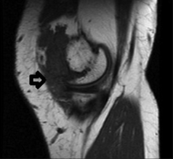
- 23-year old male with swelling and pain in his knee which was subsequently diagnosed as synovial hemangioma. Sagittal T1 weighted spin echo (SE) image shows hypointense intra-articular nodular soft tissue mass filling medial suprapatellar bursa, extending into the vastus medialis (arrow).
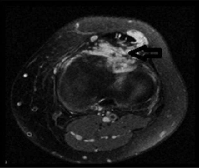
- 23-year old male with swelling and pain in his knee which was subsequently diagnosed as synovial hemangioma. Coronal proton density fat-suppressed image shows the mass lesion with heterogeneous signal containing fibrous septa and irregular margin (arrow).
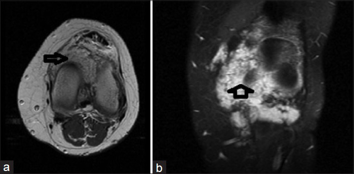
- 23-year old male with swelling and pain in his knee which was subsequently diagnosed as synovial hemangioma. (a) Axial T1 SE and (b) coronal T1 fat-suppressed images show diffuse contrast enhancement (arrow).
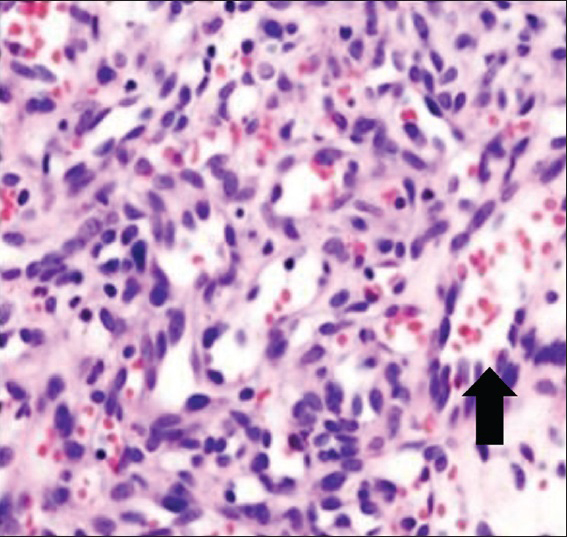
- 23-year old male with swelling and pain in his knee which was subsequently diagnosed as synovial hemangioma. Biopsy tissue stained with hematoxylin and eosin ×100 shows hemangioma in the arthroscopic biopsy (arrow).
DISCUSSION
Synovial hemangiomas first reported by Bouchut in 1856, is quite a rare benign tumor often seen in the knee joint. In rare cases, it can be seen in the ankle, elbow, and shoulder joint. The lesion can be diffuse or localized. Close to 200 cases have been reported in the literature.[45] Synovial hemangioma is a benign, difficult-to-diagnose tumor of vascular origin. In patients, recurrent intra-articular effusion may occur without trauma. When the diagnostic joint aspiration fluid is examined, generally blood cells are detected. However, in patients bleeding and coagulation rates are normal. Although this situation may be helpful in diagnosis, similar feature may also occur in pigmented villonodular synovitis. Synovial hemangiomas, usually seen in adolescence, progress with exacerbations and remissions, and occur with swelling of the knee and pain.[6] Synovial hemangiomas cases can be misdiagnosed clinically and histologically. They show similarity with especially pigmented villonodular synovitis, both macroscopically and microscopically. Sometimes, clinicians and pathologists experience difficulty in the differential diagnosis of both pathologies. However, often the only method for distinguishing these two pathologies is histopathologic examination.[27] Synovial tumors are not seen frequently in clinical practice, as they are localized in the joint. Generally clinical diagnosis is difficult. Synovium consists of mesenchymal tissue and is found in all diarthrodial joints. In the case of knee pain, swelling, and sometimes restricted movement, especially in the absence of specific trauma, and if the conventional radiological examinations are normal, synovial tumors should be considered in the differential diagnosis. Synovial hemangioma, as in this case, is one of the reasons for recurrent knee effusions and is a rare benign lesion. Often it occurs in the knee joint of children and young adults. Rather than being a true neoplasm or hamartoma, hemangioma is a form of congenital vascular malformation. The maximum size of lesions that are sometimes seen in phleboliths is between 0 and 8 cm. Histologically, they may be of capillary (25%), cavernous (50%), mixed (20%), or pure venous (5%) type. Many of those in the synovial membrane are of mixed capillary and cavernous types. In pathological differential diagnosis, nonspecific synovitis/bursitis, pigmented villonodular synovitis, nodular synovitis, and organized hemorrhage should be considered. Often for a long period of time before diagnosis, patients are symptomatic. Characteristic symptoms are painful swelling, muscular atrophy, limitations in movements of the knee, leg length increase, and a locked knee due to tumor. It occurs as recurrent swelling of the knee and intermittent pain usually depends on hemarthrosis. In our case, the symptoms were traced back to 1 year earlier. Other than pain and mild swelling in the knee, there were no specific symptoms and signs.
CONCLUSION
Synovial hemangiomas, besides having a low rate of correct diagnosis on preoperative clinical and radiological examinations, can also be misdiagnosed after histopathologic examination. Though not much emphasized in the literature, it may form massive soft tissue mass on invasion. Therefore, in patients presenting with complaints in joint that are suggestive of this diagnosis, the correct diagnosis is very important. In the diagnosis of synovial hemangioma of the knee, magnetic resonance imaging is non-invasive and is the diagnostic method often used. In addition, magnetic resonance imaging can demonstrate better the spread of lesion, so it is an effective and valuable method in the planning of surgical treatment.
Available FREE in open access from: http://www.clinicalimagingscience.org/text.asp?2015/5/1/23/156129
Source of Support: Nil
Conflict of Interest: None declared.
REFERENCES
- Diffuse synovial hemangioma of the knee: A case report. Acta Orthop Traumatol Turc. 2004;38:224-8.
- [Google Scholar]
- Synovial hemangioma: A report of 20 cases with differential diagnostic considerations. Hum Pathol. 1993;24:737-45.
- [Google Scholar]
- Benign synovial tumors of the knee: A diagnostic problem. J Bone Joint Surg Am. 1966;48:1350-8.
- [Google Scholar]
- Recurrent hemarthrosis of the knee mimicking pigmented villonodular synovitis. Isr Med Assoc J. 2005;7:50-1.
- [Google Scholar]


