
Translate this page into:
Anatomical bone structure differences in patients with hemophilic arthropathy of the knee

*Corresponding author: Serkan Bayram, MD, Department of Orthopaedics and Traumatology, Istanbul University Istanbul Faculty of Medicine, Istanbul, Turkey. dr.serkanbayram89@gmail.com
-
Received: ,
Accepted: ,
How to cite this article: Ekinci M, Akgül T, Arzu U, Bayram S, Yağcı TF, Kılıçoğlu Ö. Anatomical bone structure differences in patients with hemophilic arthropathy of the knee. J Clin Imaging Sci 2022;12:46.
Abstract
Objectives:
The anatomical differences of the bony structure of the knee joint in patients with hemophilia were evaluated, and the results were compared with the knees of patients with primary gonarthrosis and no arthrosis.
Material and Methods:
This study reviewed 41 knees in 21 patients (with an Arnold-Hilgartner classification of Stages 4 and 5 hemophilic arthropathy) who underwent total knee arthroplasty in single center. Two control groups including 21 asymptomatic patients (42 knees) and 21 primary knee osteoarthritis patients (42 knees) were formed to compare the measurements with hemophiliacs. Femoral mediolateral width, femoral anteroposterior width, femur and tibia diaphysis width, adductor tubercle-joint line distance, tibial plateau width, and medial and lateral tibia plateau width were measured separately.
Results:
Femoral mediolateral width was significantly narrow comparing with healthy individuals and primary knee osteoarthritis group. Tibial plateau was similar to asymptomatic group but significantly narrow compared with primary knee osteoarthritis group. With the correlation, the tibial plateau measurements and medial and lateral plateau were significantly narrow at hemophilic arthropathy group (P < 0.05). The slope was less in hemophilic patients as compared with asymptomatic individuals (P: 0.001). Hemophilic patients had larger femoral aspect ratios than asymptomatic group but there were no observable differences with the primary osteoarthritis group. For the tibial aspect ratios, hemophilic had a smaller ratio than the primary osteoarthritis group but there were no significant differences with the asymptomatic group.
Conclusion:
Hemophilic knee has a mismatch between femoral and tibial side while comparing with the other groups.
Level of Evidence:
Level IV, cross-sectional study.
Keywords
Hemophilic arthropathy
Primary knee osteoarthritis
Anatomic bone structure
Knee
Radiological feature

INTRODUCTION
Hemophilia is a coagulation disorder caused by deficiencies in specific coagulation factors.[1,2] The joints are the most common sites for bleeding in hemophilia patients, and the knee is the most affected joint. This is thought to be due both to the large size of the synovial membrane and to rotational forces.[3,4] Repetitive intra-articular bleeding and iron accumulation in the synovium can cause blood-induced synovitis and cartilage injury, which results in progressive joint damage and hemophilic arthropathy.[3,5,6]
Total knee arthroplasty (TKA) is an effective treatment for end-stage hemophilic arthropathy with severe joint deterioration.[3,7] However, during TKA surgery for hemophilic arthropathy, it was observed that the knees of hemophilic patients exhibited different anatomies for which standard implants were not always suitable; standard TKA implants matching the anterior-posterior (AP) plane were not appropriate for use in the mediolateral plane. Based on the authors’ experience and the difficulties encountered during surgery, a comparison was undertaken; the bone structures of the knee joints of TKA patients with a diagnosis of hemophilic arthropathy were compared with those of primary knee osteoarthritis patients using standard implants.
In this study, the anatomical differences of the bony structure of the knee joint in patients with hemophilia were evaluated, and the results were compared with the knees of patients with primary gonarthrosis and no arthrosis.
MATERIAL AND METHODS
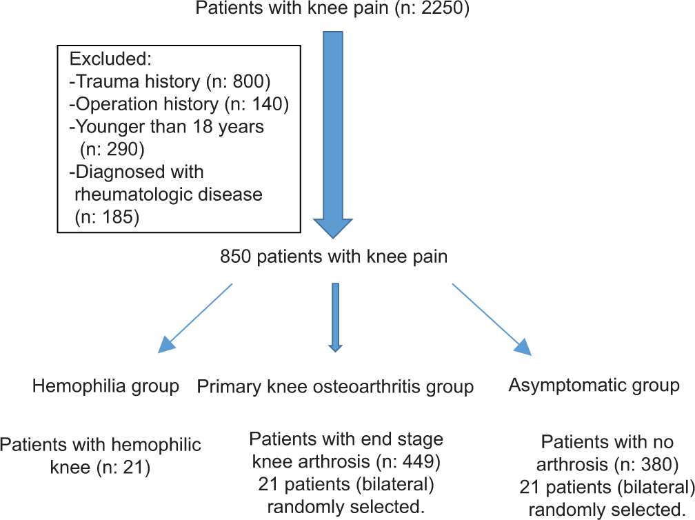
This study reviewed 41 knees in 21 patients (with an Arnold-Hilgartner classification[8] of Stages 4 and 5 hemophilic arthropathy) who underwent TKA in the authors’ institution. This research has been approved by the IRB (Institutional Review Board 2017/146) of the authors’ affiliated institutions. Informed consent was obtained from each patient. Two control groups including asymptomatic patients and primary knee osteoarthritis patients were formed to compare the measurements with the group of patients with hemophilia. All the hemophilic patients were male who were diagnosed with hemophilia A; the patients in both control groups were randomly selected (see flaw chart). All asymptomatic patients were recruited through their presentation at the authors’ outpatient clinic with knee pain; they had no arthrosis findings on X-rays. The patients with primary knee osteoarthritis were selected from those patients in the clinic who had primary TKA with the diagnosis of primary knee osteoarthritis [Table 1].
| Parameters | Asymptomatic | Gonarthrosis | Hemophilic |
|---|---|---|---|
| FAW | 83.9±28 mm | 67.4±5.7 mm | 62.5±5.1 mm |
| FMW | 90.8±8 mm | 86.36±4.9 mm | 87±6.6 mm |
| FD | 35.2±3.6 mm | 35.8±4.6 mm | 25±3.2 mm |
| TD | 28.2±2.7 mm | 30±5.2 mm | 22.7±3.2 mm |
| ATJLD | 47.2±6.1 mm | 50.2±5.6 mm | 49.3±6.5 mm |
| TPW | 85.6±5 mm | 88.8±6.4 mm | 83.56±5.4 mm |
| MTPW | 42.2±3.2 mm | 44.6±3.96 mm | 41.83±3.4 mm |
| LTPW | 42.8±4.1 mm | 44.2±3.3 mm | 41.87±3.5 mm |
| SLOPE | 15.3±19.9º | 9.2±2.3° | 5.6±6.4° |
| MPTA | 87.6±1.6° | 84.4±3.3° | 84.6±4.8° |
| TAW | 54.5±4.8 mm | 50.6±5.2 mm | 53.1±5.5 mm |
FAW: Femoral anteroposterior width, FMW: Femoral mediolateral width, FD: Femur diaphysis, TD: Tibia diaphysis, ATJLD: Adductor tubercle-joint line distance, TPW: Tibial plateau width, MTP: Tibial anteroposterior width, LTPW: Lateral tibia plateau width, MPTA: Medial proximal tibial angle, TAW: Tibial anteroposterior width
Radiographic measurements
Pre-operative full-leg radiographs were obtained, including AP and lateral images of both knees with patients in a bipedal stance with their feet in neutral rotation and the patellae pointing forward. A picture archiving and communication system (Extreme PACS, Ankara, Turkey) was used for all measurements. All measurements were recorded independently by the same author (M.E.K). The distance between the beam origin and the detector plate was 120 cm in all cases. This software made it possible to measure the distances on the AP and lateral knee radiographs with full accuracy and to eliminate changes related to the magnification. Patients were excluded from the study if any of the following applied: They had X-rays taken because of knee trauma or a fracture extending to the knee joint, they had undergone previous knee surgery, they were diagnosed with a metabolic or inflammatory rheumatological disease, they were prescribed steroid therapy for more than 6 months for any reason, or they exhibited an advanced degree of lower extremity malalignment (more than 20° of varus or 10° of valgus deformity).
The following items were measured:
Femoral mediolateral width (FMW): The line between the medial and lateral epicondyles at their most prominent points (|AB|) [Figure 1].
Femoral AP width (FAW): The distance between the anterior and posterior tangential and perpendicular line according to diaphysis on the distal femoral condyles (|IJ|) [Figure 2].
Femur diaphysis (FD) width and tibia diaphysis (TD) width: The width of the femur and tibia on their middiaphysis.
Adductor tubercle joint line distance (ATJLD): The perpendicular distance between the adductor tubercle and the joint line of the knee (|EF|) [Figure 3].
Tibial plateau width (TPW): The line between the most prominent points of the medial and lateral edge of the plateau (|CD|) [Figure 1].
Tibial AP width (TAW): The distance between two lines on the tibial plateau that is perpendicular to the diaphysis of the tibia and tangent to the anterior and posterior edges of the tibial plateau on the lateral radiographs (|KL|) [Figure 2].
Medial TPW and lateral TPW: The distances between the medial and lateral tibial eminences and the most prominent points of the medial and lateral edges of the plateau (|CD| - |GH| and |GH|) [Figure 3].
Medial proximal tibial angle (MPTA): The angle between the tibial plateau and the tibial mechanical axis [Figure 4].
Tibial slope: The angle between the line of the diaphyseal axis and the line drawn using the highest two points of the anterior and posterior edges of the medial plateau [Figure 2].

- Demonstration of radiographic measurement of femoral mediolateral width (|AB|) and tibial plateau width (|CD|).

- Demonstration of radiographic measurement of femoral anteroposterior width (|IJ|), tibial anteroposterior width (|KL|), and tibial slope (TS).

- Demonstration of radiographic measurement of adductor tubercle joint line distance (|EF|), medial tibial plateau width, and lateral tibial plateau width (|CD| - |GH| and |GH|).

- Demonstration of radiographic measurement of medial proximal tibial angle.
The femoral and tibial aspect ratios were calculated by dividing the measured ML widths by the measured AP widths on the X-rays (femoral aspect ratio: FMW/FAW and tibial aspect ratio: TPW/TAW). The aspect ratio allows for the estimation of prosthesis shape and the underhanging/ overhanging of the components.[8]
The ratio between the height of the femur metaphysis and the FMW (distal femoral ratio: ATJLD/FMW), the ratio of the FD and the FMW (FD/FMW), and the tibial plateau (TD/TPW) distances were evaluated to better assess the knee anatomy.
PCL-substituting Genesis II knee arthroplasty (Smith and Nephew, Memphis, TN, USA) was used in all patients through a medial parapatellar approach. Patella resurfacing prosthesis was not utilized, while patellar denervation was done in all patients [Figures 5-8].

- Pre-operative anteroposterior radiograph of patient with hemophilic arthropathy.

- Pre-operative lateral radiograph of patient with hemophilic arthropathy.

- Post-operative anteroposterior radiograph of patient with hemophilic arthropathy.

- Post-operative lateral radiograph of patient with hemophilic arthropathy. The mismatch was especially evident in the tibial component.
Statistical analysis
The data were analyzed using SPSS software (version 22.0 for Windows) (SPSS Inc., Chicago, USA). Mean, median, standard deviation, maximum, and minimum were used as descriptive statistical methods. Normality of distribution was tested using the Shapiro–Wilk test. Comparisons were made using a one-way ANOVA test, and the significance value was accepted as P < 0.05 at a 95% confidence interval. For comparisons between the groups, Tukey’s range test was used among the post hoc assessments.
RESULTS
The FMW was measured as 87 ± 6.6 mm, and the knee joints were significantly narrower compared with both healthy individuals and the primary knee osteoarthritis group (P < 0.05). However, the TPW was measured at 83.56 ± 5.4 mm, which was similar to the asymptomatic group (P = 0.9) but significantly narrower compared with the primary knee osteoarthritis group (P < 0.05). With the correlation in the TPW measurements, the medial and lateral plateaus were significantly narrower in the hemophilic arthropathy group (P < 0.05).
There was no difference in the primary knee osteoarthritis and hemophilic arthropathy groups for tibial slope measurements (P = 0.213). However, a smaller slope was evident in hemophilic patients compared with asymptomatic individuals (P < 0.05).
No significant difference in the MPTA measurements was detected between the hemophilic and primary knee osteoarthritis groups (P = 0.811), but the MPTA was less when compared with the asymptomatic group (P < 0.05). There were no significant differences in ATJLD between all the groups. For the hemophilic group, the TD and FD were significantly narrower – as in the FD and TD measurements (P < 0.05) [Table 1].
The hemophilic patients demonstrated larger femoral aspect ratios than the asymptomatic group (1.38 [95% CI, 1.13–1.79] vs. 1.32 [95% CI, 1.20–1.65], P < 0.05); however, there were no observable differences with the primary knee osteoarthritis group. For the tibial aspect ratios, the hemophilic group had a smaller ratio than the primary knee osteoarthritis group (1.58 [95% CI, 1.24–2.19] vs. 1.76 [95% CI, 1.50–2.04], P < 0.05), and there were no significant differences with the asymptomatic group. This evidence reveals that the knees of patients with hemophilia are mismatched between the femoral and tibial sides in comparison with the other groups [Figure 9].

- Graphic showing the comparison of the femoral and tibial aspect ratios between all groups.
DISCUSSION
The knee is the most commonly involved joint in end-stage hemophilic arthropathy, and TKA is a complex and challenging procedure.[9,10] The knee’s anatomy is distorted due to physeal overgrowth in childhood, widening of the femoral intercondylar notch, large osteophytes, and squaring of the patella.[10,11] The larger femoral and tibial surfaces cause a mismatch in implant sizing as they are wider in the mediolateral plane compared with the AP plane.[10,11] Restoring the mechanical axis and achieving accurate alignment are difficult in such a scenario.[11] Using conventional jigs, standard operation techniques and implants that rely on visual confirmation of alignment accuracy can lead to high possibilities of error, unsuitable implant size, poor implant fit, and an increased incidence of outliers.
One of the most important factors for successful TKA is that the components to be implanted must be in proper rotational alignment.[12-14] The previous studies recommend that the femoral component should be inserted parallel to the transepicondylar axis or the AP axis.[14-18] Trochlear wear or intercondylar osteophytes sometimes make it difficult to detect both the medial and lateral epicondyles or to accurately locate the AP axis.[16,19]
Almost all of the TKA implants are produced in accordance with the anthropometric characteristics of Western, male,[19] White patients,[20,21] although TKA is regarded as a highly successful surgery resulting in pain reduction, an improved quality of life, and enhanced knee joint function.[22]
The use of TKA implants that do not fit the anthropometrical features of the patient may cause more blood loss, overhang/ underhang, early loosening, irritation, unstable implant fixation, and ROM restriction. To prevent these complications and obtain better results, implants suitable for a patient’s own knee morphometry should be used.
The anthropometric characteristics of the bony structures of the knee (distal femur and proximal tibia) between different ethnicities and genders have been evaluated in numerous studies.[23-27] Kim et al. found that the AP dimensions and the mediolateral width of the distal femur and plateau tibia of Korean patients were smaller compared with Western patients.[28] They discovered that the knees of black patients had larger AP dimensions than the knees of White patients in their study.[27] The anatomical differences of the distal femur and proximal tibia were also compared between males and females. The distal femurs and proximal tibias of the males tended to be larger than in the females.[21,29] However, according to the best of the authors’ knowledge, this is the first study to evaluate the bony dimensions of the knee in hemophilic patients suffering with severe hemophilic arthropathy, arthritic patients who have undergone TKA, and asymptomatic healthy individuals. It was anticipated that this study would simplify research for the clinical implications of these anthropometric differences and ascertain whether TKA manufacturers would give price to address the potential for compromised implant fit.
The aspect ratios were described as the mediolateral width divided by the AP width of the distal femur or the proximal tibia. These calculations can be used as a guide for femoral component sizing. A higher ratio means a greater mediolateral width for the AP size, while a lower ratio means a smaller mediolateral dimension for the AP size.
It is important to note that aspect ratios can be useful for predicting component sizing, and femoral and tibial shapes can be better understood and maintain a measure of the knee dimension between patients. In this study, the knees of hemophilia patients had smaller AP dimensions compared with the asymptomatic patients’ knees in terms of femoral aspect ratio. This correlates with the relatively higher ML/ AP aspect ratio. This analysis of tibial aspect ratios revealed that the knees of hemophilic patients had larger AP dimensions than did the knees of patients suffering from primary arthrosis (which results in a smaller tibial aspect ratio). This could result in mismatches where standard tibial components would be relatively small and incompatible for patients with hemophilia in terms of AP dimensions. Ultimately, an unsuitable fit could also result in the underhang/overhang of the components, which could lead to soft-tissue impingements, ROM restrictions, pain, and early loosening.[30]
The limitations of this research are that it is a retrospective study and the measurements were performed on AP and lateral radiographs. In addition, the results could be more useful if they were supplemented with pre-operative anatomical measurements or 3D computed tomography (CT) analysis of the knees. Although the bony anatomy of the knee joint is better determined by CT analysis, the templating of the TKA designs is performed preoperatively on standard AP and lateral radiographs of the knee to obtain the proper size and position of the TKA implant. Therefore, analysis of the anatomical measurements on standard radiographs is not a misapplication in clinical practice.
The findings confirm that the knees of hemophilic patients have a mismatch between the femoral and tibial sides in comparison with the other groups. The use of unsuitable implants may lead to complications, such as early loosening and limitation of knee motion, and TKA implants produced for standard patient populations may not be suitable for patients with hemophilia. For satisfactory results, patient-specific implants should be considered for use in hemophilic patients.
CONCLUSION
The hemophilic knees have a mismatch between the femoral and tibial sides. The hemophilic patients demonstrated larger femoral aspect ratios than the asymptomatic group. For the tibial aspect ratios, the hemophilic group had a smaller ratio than the primary knee osteoarthritis group, and there were no significant differences with the asymptomatic group.
Acknowledgment
The authors would like to thank Enago (www.enago.com) for the English language review.
Declaration of patient consent
Institutional Review Board (IRB) permission obtained for the study.
Financial support and sponsorship
Nil.
Conflicts of interest
There are no conflicts of interest.
References
- Pathophysiology, diagnosis and prevention of arthropathy in patients with haemophilia. Haemophilia. 2011;17:571-8.
- [CrossRef] [PubMed] [Google Scholar]
- Survivorship analysis of cementless meniscal bearing total knee arthroplasty. Clin Orthop Relat Res. 1997;338:119-23.
- [CrossRef] [PubMed] [Google Scholar]
- Joint protection in haemophilia. Haemophilia. 2011;17(Suppl 2):1-23.
- [CrossRef] [PubMed] [Google Scholar]
- Musculoskeletal complications of hemophilia. HSS J. 2010;6:37-42.
- [CrossRef] [PubMed] [Google Scholar]
- Iron deposits and catabolic properties of synovial tissue from patients with haemophilia. J Bone Joint Surg Br. 1998;80:540-5.
- [CrossRef] [PubMed] [Google Scholar]
- Biochemical and physiological aspects of degenerative joint diseases with special reference to hemophilic arthropathy. Ann N Y Acad Sci. 1975;240:285-90.
- [CrossRef] [PubMed] [Google Scholar]
- Total joint arthroplasty: The final solution for knee and hip when synovitis could not be controlled. Haemophilia. 2007;13(Suppl 3):49-58.
- [CrossRef] [PubMed] [Google Scholar]
- Hemophilic arthropathy, Current concepts of pathogenesis and management. J Bone Joint Surg Am. 1977;59:287-305.
- [CrossRef] [Google Scholar]
- Morphometry of the proximal tibia to design the tibial component of total knee arthroplasty for the Korean population. Knee. 2007;14:295-300.
- [CrossRef] [PubMed] [Google Scholar]
- Correction of fixed contractures during total knee arthroplasty in haemophiliacs. Haemophilia. 1999;5(Suppl 1):33-8.
- [CrossRef] [PubMed] [Google Scholar]
- Effects of hemophilia on articulations of children and adults. Clin Orthop Relat Res. 1996;328:7-13.
- [CrossRef] [Google Scholar]
- Effect of femoral and tibial component position on patellar tracking following total knee arthroplasty: 10-year follow-up of Miller-Galante I knees. Am J Knee Surg. 2001;14:152-6.
- [Google Scholar]
- Malrotation causing patellofemoral complications after total knee arthroplasty. Clin Orthop Relat Res. 1998;356:144-53.
- [CrossRef] [PubMed] [Google Scholar]
- Effect of rotational alignment on patellar tracking in total knee arthroplasty. Clin Orthop Relat Res. 1999;366:155-63.
- [CrossRef] [PubMed] [Google Scholar]
- Femoral rotational alignment, based on the anteroposterior axis, in total knee arthroplasty in a valgus knee. A technical note. J Bone Joint Surg Am. 1995;77:1331-4.
- [CrossRef] [PubMed] [Google Scholar]
- The posterior condylar angle in osteoarthritic knees. J Arthroplasty. 1998;13:812-5.
- [CrossRef] [Google Scholar]
- Determining the rotational alignment of the femoral component in total knee arthroplasty using the epicondylar axis. Clin Orthop Relat Res. 1993;286:40-7.
- [CrossRef] [Google Scholar]
- Determining femoral rotational alignment in total knee arthroplasty: Reliability of techniques. J Arthroplasty. 2001;16:301-5.
- [CrossRef] [PubMed] [Google Scholar]
- Will gender-specific total knee arthroplasty be a better choice for women? A systematic review and meta-analysis. Eur J Orthop Surg Traumatol. 2014;24:1341-9.
- [CrossRef] [PubMed] [Google Scholar]
- Asian-specific total knee system: 5-14 year follow-up study. BMC Musculoskelet Disord. 2011;12:251.
- [CrossRef] [PubMed] [Google Scholar]
- Clinical implications of femoral anthropometrical features for total knee arthroplasty in Koreans. J Arthroplasty. 2015;30:1220-7.
- [CrossRef] [PubMed] [Google Scholar]
- Health-related quality of life in total hip and total knee arthroplasty. A qualitative and systematic review of the literature. J Bone Joint Surg Am. 2004;86:963-74.
- [CrossRef] [PubMed] [Google Scholar]
- The female knee: Anatomic variations. J Am Acad Orthop Surg. 2007;15(Suppl 1):S31-6.
- [CrossRef] [PubMed] [Google Scholar]
- Human knee joint anatomy revisited: Morphometry in the light of sex-specific total knee arthroplasty. J Arthroplasty. 2011;26:346-53.
- [CrossRef] [PubMed] [Google Scholar]
- Gender-specific design in total knee arthroplasty. J Arthroplasty. 2007;22:27-31.
- [CrossRef] [PubMed] [Google Scholar]
- Knee alignment differences between Chinese and Caucasian subjects without osteoarthritis. Ann Rheum Dis. 2008;67:1524-8.
- [CrossRef] [PubMed] [Google Scholar]
- What differences in morphologic features of the knee exist among patients of various races? A systematic review. Clin Orthop Relat Res. 2017;475:170-82.
- [CrossRef] [PubMed] [Google Scholar]
- Are Korean patients different from other ethnic groups in total knee arthroplasty? Knee Surg Relat Res. 2015;27:199-206.
- [CrossRef] [PubMed] [Google Scholar]
- Anthropometric measurements of the human knee: Correlation to the sizing of current knee arthroplasty systems. J Bone Joint Surg Am. 2003;85(Suppl 4):115-22.
- [CrossRef] [Google Scholar]


