Translate this page into:
Successful coil embolization of pseudo pulmonary sequestration: A report of two cases

*Corresponding author: Wafa Yahya Qatomah, College of Medicine, King Saud bin Abdulaziz University for Health and Sciences, Riyadh, Kingdom of Saudi Arabia. dr.wafa91@hotmail.com
-
Received: ,
Accepted: ,
How to cite this article: Qatomah WY, Ali RM, Qatomah AY, Arabi M. Successful coil embolization of pseudo pulmonary sequestration: A report of two cases. J Clin Imaging Sci 2022;12:36.
Abstract
Pseudo pulmonary sequestration is a rare congenital anomaly, which entails systemic arterial supply to the basal segment of the lung in the absence of pulmonary arterial supply. Diagnosis is often made by radiographic appearance without specific clinical symptoms. The mainstay treatment is surgical resection; however, embolization can be considered as an alternative approach.
Herein, we present a report of two females who presented with nonspecific chronic chest pain. Both patients were diagnosed with pseudo pulmonary sequestration on CT scan and completed uneventful pregnancies prior to successful management with coil embolization.
Keywords
Systemic arterial supply to the normal lung
Pseudo pulmonary sequestration
Embolization
Case report
INTRODUCTION
Pseudo pulmonary sequestration is a rare congenital anomaly, which entails systemic arterial supply to the basal segment of the lung in the absence of pulmonary arterial supply, without associated parenchymal abnormalities. Pryce has classified it as type 1 sequestration.[1,2] Most patients are diagnosed based on the radiographic appearance in the absence of clinical findings.[2] The mainstay treatment is surgical resection of the affected segment and aberrant systemic artery. Alternatively, transarterial coil embolization can be used as a noninvasive approach.
CASE REPORTS
Case 1
A 20-year-old pregnant female presented with chronic shortness of breath and nonspecific chest pain. The patient is known for a history of uncontrolled bronchial asthma since childhood.
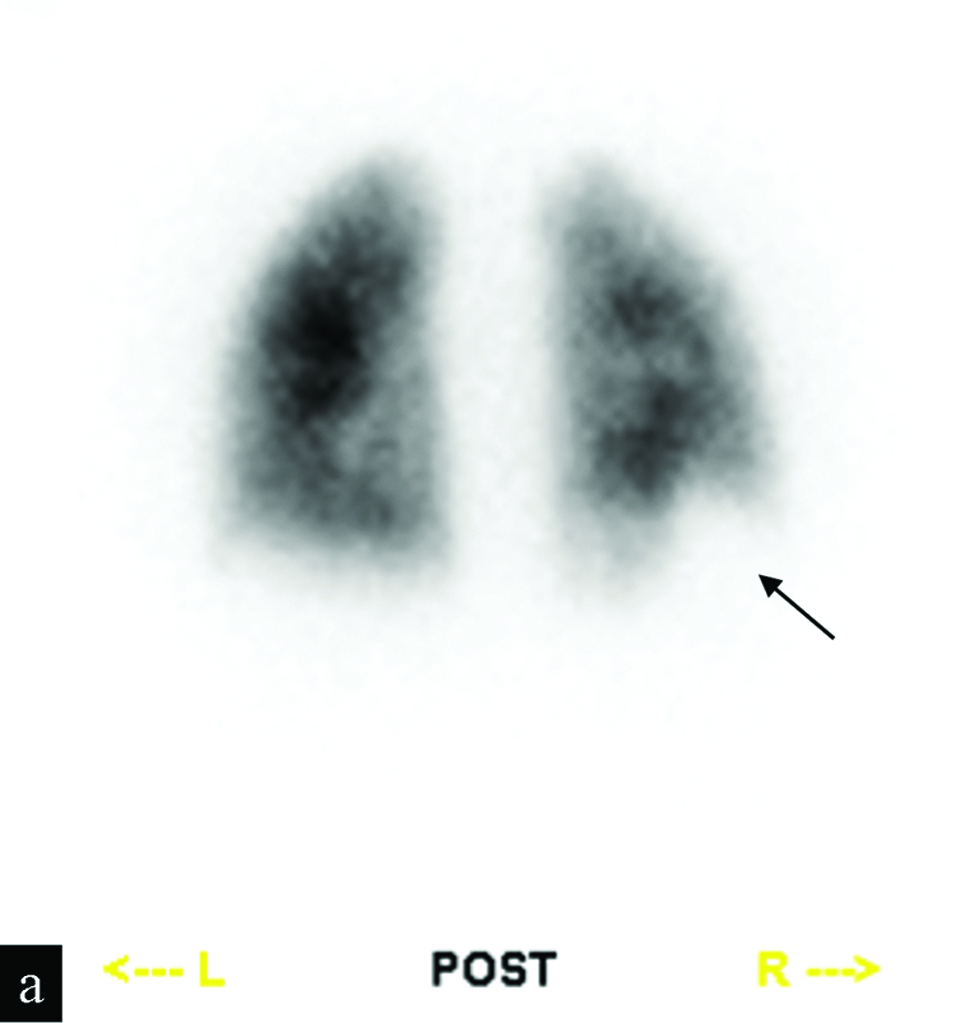
Workup showed slightly elevated D-dimer 0.72 mg/L (Normal 0-0.5 mg/L) and V/Q scan which showed a single wedge-shaped perfusion defect in the right basal segment [Figure 1a].

- A 20-year-old female presented with chronic shortness of breath and nonspecific chest pain. (A) Posterior view of Tc99m MAA lung perfusion scan shows right basal perfusion defect (arrow).
CT chest angiography showed no evidence of pulmonary embolism; however, there was an incidental finding of anomalous systemic arterial supply from the abdominal aorta to the right base with a large vein draining into the pulmonary veins with no associated parenchymal abnormalities [Figure 1b]. The plan was to continue with the pregnancy and proceed with transarterial embolization later. The patient completed her pregnancy with uneventful normal vaginal delivery.

- A 20-year-old female presented with chronic shortness of breath and nonspecific chest pain. (B) Axial CT scan of the lower chest shows prominent right phrenic artery (arrow) supplying the right lung base.
Three months later, the patient underwent angiography that demonstrated a prominent right phrenic artery supplying the lung base. Pulmonary angiography showed no arterial supply to the right base [Figure 1c]. The right phrenic artery was super selectively cannulated with a Lantern microcatheter (Penumbra Inc, CA, USA) [Figures 1d and e].

- A 20-year-old female presented with chronic shortness of breath and nonspecific chest pain. (C) Pulmonary angiography shows lack of perfusion to the right base (arrows) compared to the left.

- A 20-year-old female presented with chronic shortness of breath and nonspecific chest pain. (D) Selective right phrenic angiography shows the vascular supply to the right base.

- A 20-year-old female presented with chronic shortness of breath and nonspecific chest pain. (E) Venous drainage from the right base drains into the right lower pulmonary vein (arrows).
Multiple detachable Ruby coils (Penumbra Inc, CA, USA) were deployed (6 mm x 50 cm, 8 mm x 60 cm, 8 mm x 60 cm, 3 mm x 12 cm).
Post embolization, the patient-reported improved shortness of breath but complained of right shoulder pain that was managed with analgesics and discharged on the same day. Chest CT 1 month later revealed complete obliteration of the systemic arterial supply to the base of the right lung with minimal basal atelectatic changes. Clinical follow-up 18 months later showed no active respiratory complaints.
Case 2
A 29-year-old pregnant female presented with chronic shortness of breath and nonspecific chest pain. She had a history of chronic cough and recurrent chest infections.
Chest CT angiography showed anomalous phrenic artery coursing into the right lower lobe with draining vein into the pulmonary circulation in keeping with intralobar sequestration without associated parenchymal abnormalities [Figure 2a]. The patient was managed conservatively while pregnant and completed uneventful pregnancy with normal vaginal delivery. One year later, the patient presented for embolization due to persistent symptoms. Initial aortography showed aberrant arterial supply to the right lung base through the right phrenic and venous drainage through the right pulmonary vein [Figures 2b to d]. Embolization for the right phrenic artery and its two branches was done using RUBY coils (Penumbra Inc, CA, USA) of variable diameters, and the main phrenic trunk was embolized with 6 mm Amplatzer Plug 4 (Abbott, MN, USA) [Figure 2e].

- A 29-year-old female presented with chronic shortness of breath and nonspecific chest pain. (A) Color 3D Volume rendering image of the lower thorax and upper abdomen shows prominent right phrenic artery supplying (arrow) the right base with venous drainage via the right lower pulmonary (short arrows).

- A 29-year-old female presented with chronic shortness of breath and nonspecific chest pain. (B) Initial descending thoracic aortography.

- A 29-year-old female presented with chronic shortness of breath and nonspecific chest pain. (C, D) selective right phrenic angiography showed aberrant arterial supply to the right lung base and venous drainage through the right pulmonary vein (arrow).

- A 29-year-old female presented with chronic shortness of breath and nonspecific chest pain. (E) Celiac angiography showed complete embolization of the systemic supply to the right lung base.
Postprocedure, the patient complained of chest discomfort, dyspnea, and right shoulder pain. CXR was unremarkable, and she was managed with analgesics and discharged on the same day. She reported improved chest tightness 2 weeks later but no significant improvement in shortness of breath and exertional dyspnea. She also had a low-grade fever for a few days post embolization. During a 1-year phone call follow-up, the patient reported improvement in chest symptoms.
DISCUSSION
Pseudo pulmonary sequestration is a rare congenital anomaly in which the basal segment of the lower lobe of the lung is supplied by an anomalous systemic artery in the absence of pulmonary supply. Also known as Pryce’s type 1 sequestration in which the lung is connected with the tracheobronchial tree.[1,2]
The anomalous artery that supplies the sequestration most commonly arise from the thoracic aorta and less commonly from the abdominal aorta, or celiac trunk.[2,3]
Most patients with this condition are clinically asymptomatic and usually diagnosed based on radiographic findings only.[2] If symptomatic, patients may present with a murmur, dyspnea, recurrent infection, hemoptysis, volume overload cardiac failure, and pulmonary venous hypertension.[2,4]
Pseudo pulmonary sequestration may mimic PE on V/Q scan due to the large perfusion defect that is not supplied by the pulmonary artery.
CT angiography is helpful to provide details of the anomalous artery and its origin.[4]
Surgical excision is the treatment of choice. However, transarterial embolization is reported as an effective and noninvasive procedure. Proximal coil or plug embolization occludes the anomalous artery and maintains the lung parenchyma. Although particles were used in the management of sequestration, they may need to be avoided in pseudo sequestration[4,5] to minimize the risk of pulmonary infarction and subsequent parenchymal inflammation.
In conclusion, transarterial embolization may be offered as an alternative noninvasive procedure in the case of pseudo pulmonary sequestration. Further studies are needed to compare the effectiveness of such a noninvasive technique to the conventional surgical approach.
Declaration of patient consent
Institutional Review Board (IRB) permission obtained for the study.
Financial support and sponsorship
Nil.
Conflict of interest
There are no conflicts of interest.
References
- Lower accessory pulmonary artery with intralobar sequestration of lung: A Report of seven cases. J Pathol Bacteriol. 1946;58:457-67.
- [CrossRef] [PubMed] [Google Scholar]
- Pseudosequestration of the left lung. Tex Heart Inst J. 2007;34:195-8.
- [PubMed] [PubMed Central] [Google Scholar]
- Pulmonary sequestration: What the radiologist should know. Clin Imaging. 2021;73:61-72.
- [CrossRef] [PubMed] [Google Scholar]
- Successful endovascular embolization of an intralobar pulmonary sequestration. Radiol Case Rep. 2018;13:125-9.
- [CrossRef] [PubMed] [Google Scholar]
- Systemic arterial supply to the normal basal segments of the left lower lobe of the lung—Treatment by coil embolization—and a literature review. Cardiovasc Intervent Radiol. 2011;34:S117-21.
- [CrossRef] [PubMed] [Google Scholar]


