
Translate this page into:
Role of HRCT Temporal Bone in Pre-operative Assessment of Tegmen Height in Chronic Otitis Media Patients
 , Poonam Yadav1, Ahmad Umar Khan1, Anchal Singh1, Mazhar Khan1

*Correspondence author: Sachin Khanduri, Department of Radiodiagnosis, Era’s Lucknow Medical College and Hospital, Sarfarazganj, Lucknow - 226 016, Uttar Pradesh, India.drsachinrad@gmail.com
-
Received: ,
Accepted: ,
How to cite this article: Husain M, Khanduri S, Faiz SM, Abbas SZ, Yadav P, Khan AU, et al. Role of HRCT temporal bone in pre-operative assessment of tegmen height in chronic otitis media patients. J Clin Imaging Sci 2020;10:79.
Abstract
Objectives:
High-resolution CT (HRCT) temporal bone has emerged as a useful option in pre-operative assessment of tegmen height in chronic otitis media patients.
Material and Methods:
A total of 60 patients with clinical suspicion of chronic otitis media were enrolled in the study. HRCT evaluation was done using Siemens Somatom Force 384 slice multidetector computed tomography machine. We radiologically assess tegmen height using lateral semicircular canal as a reference point on HRCT. Final result has been based on correlation of radiological and intraoperative findings. Diagnostic efficacy of HRCT temporal bone was evaluated in terms of sensitivity, specificity, PPV, NPV, and accuracy for pre-operative assessment of tegmen height.
Results:
The correlation between actual tegmen height and estimated tegmen height (by equation) was 0.457 which is highly significant (P < 0.001). In the study, the mean tegmen height of exposed dura (ED) was 5.81 ± 1.71 (95% CI 4.91–6.70) while the mean tegmen height of non-exposed dura (NED) was 8.40 ± 1.31 (95% CI 8.02– 8.78). Highly significant difference was found in mean tegmen height between ED and NED cases (P < 0.001).
Conclusion:
Pre-operative CT assessment of tegmen height is an important parameter in assessing risk of dural injury during tympanomastoid surgeries.
Keywords
HRCT temporal bone
Chronic otitis media
Tegmen height

INTRODUCTION
Chronic otitis media (COM) is a condition in otorhinolaryngology and it is characterized by chronic, intermittent, or persistent ear discharge through a perforated tympanic membrane.[1] It is associated with chronic inflammation of the middle ear cleft characterized by persistent perforation of the tympanic membrane with recurrent or persistent mucopurulent otorrhea.
Ossicular erosion, a frequent complication of chronic otitis media (COM), may lead to total failure of middle ear mechanics and resulting in substantial hearing loss.[2] X-rays, CT scans, or MRI scans are often helpful in the diagnosis of chronic otitis media, to rule out a cholesteatoma or complications related to chronic otitis media. A lumbar puncture may be necessary in persons in whom meningitis is suspected.[3]
Advent of HRCT and improvements in radiological technique has definitely improved study of the temporal bone in patients with COM, which includes evaluation of the extent and sites of involvement and inter-relationship of the tympanomastoid compartment with adjacent neurovascular structures. Therefore, HRCT can be recommended not only in cases suspected with potential complications but also in all cases of COM to know the extent of disease, and the presence of anatomical variations, which should alert the surgeon and guide in surgical approach and treatment plan of COM.[4]
The tegmen tympani is the roof of the middle ear, and the tegmen mastoideum is the roof of the mastoid.[5] Determining the height of the tegmen tympani also has practical implications relating to surgical access and may influence the surgical approach used. This is particularly useful when deciding whether an intact canal versus canal wall down mastoidectomy should be performed.[6]
The tegmen separates the surgeon’s drill from the dura, cerebrospinal fluid and the brain, and injury can result in cerebrospinal fluid leak,[7] pneumocephalus,[8] brain herniation,[9] and cerebellar abscess.[10] Middle fossa tegmen plate is an important anatomic landmark in tympanomastoidectomy operations. The tegmen may be lower than expected. It is important that such situations with a risk of surgical complications be determined in pre-operative period.[11]
MATERIAL AND METHODS
We conducted this prospective study in the Department of Radio-Diagnosis in collaboration with Department of Otorhinolaryngology, Era’s Lucknow Medical College and Hospital, Lucknow, from January 2018 to November 2019. The study protocol was cleared for ethics by research Institutional Review Board.
This research project sets out to answer the role of high-resolution computed tomography (HRCT) temporal bone in pre-operative assessment of tegmen height in chronic otitis media patients. The goal of the study was pre-operative assessment of tegmen height by HRCT temporal bone in chronic otitis media patients and correlates the findings of HRCT temporal bone with observations made during chronic otitis media surgery. A total of 60 patients with clinical suspicion of chronic otitis media were enrolled in the study. HRCT evaluation was done using Siemens Somatom Force 384 slice multidetector computer tomography machine. We radiologically assess tegmen height using lateral semicircular canal as a reference point on HRCT. Only those who were willing to participate in the study and gave their written consent for the same were included in the study. Pregnant patients, patients with congenital ear disease, COM patients with intracranial complications, and those who had undergone previous temporal bone surgeries or with a history of trauma to temporal bone were excluded from the study.
The sample size was calculated as 60 at 90% confidence and 10% error allowance. All patients were then subjected to imaging evaluation using HRCT temporal bone as a modality and tympanomastoid surgery was planned. Data so obtained were subjected to statistical analysis. Correlation with intraoperative findings was done to achieve the final interpretation.
RESULTS
HRCT is widely used in diagnosis of inflammatory middle ear diseases. Out of 60 patients, 22 (36.7%) patients were age group of 15–24 years. Twenty-two (36.7%) patients were the age group of 25–34 years. Twelve (20.0%) patients were the age group of 35–44 years, and remaining 4 (6.7%) patients were the age group of 45 and above years. Most of the patients were below the age 35 years. Out of 60 patients, COM without cholesteatoma was found in 24 (40.0%) cases in the right ear and 13 (21.7%) cases in the left ear. Acute otitis media (AOM) was found in 6 (10.0%) cases in the right ear while COM with cholesteatoma was found in 7 (11.7%) cases of the right ear and 13 (21.7%) cases in the left ear. The right ear was normal in 38.3% of cases while the left ear was normal in 56.7% of cases.
Overall, the tegmen height of an individual aged above 15 years lies in the range of 7.43–8.16 with 95% probability [Table 1].
| Age (years) | Tegmen height (mm) | 95% CI | F-value | P-value | ||
|---|---|---|---|---|---|---|
| Mean | SD | Lower | Upper | |||
| 15–24 | 7.80 | 1.16 | 7.44 | 8.15 | 4.76 | 0.004 |
| 25–34 | 7.27 | 2.09 | 6.64 | 7.90 | ||
| 35–44 | 9.01 | 2.13 | 8.11 | 9.91 | ||
| ≥45 | 7.01 | 3.27 | 4.28 | 9.75 | ||
| Total | 7.79 | 2.00 | 7.43 | 8.16 | ||
During intraoperative, exposed dura (ED) was found in 14 (23.3%) cases while non-exposed dura (NED) was found in 46 (76.7%) cases [Table 2].
| Intra-OP findings | No | % |
|---|---|---|
| ED | 14 | 23.3 |
| NED | 46 | 76.7 |
| Total | 60 | 100.0 |
In the study, the mean tegmen height of normal ear was 8.38 ± 1.36 (95% CI 8.03–8.73). Among the problematic ears, the mean tegmen height was minimum in case of COM with cholesteatoma patients with value 6.65 ± 1.96 (95% CI 5.79– 7.51) and maximum in case of AOM patients with value 8.45 ± 0.61 (95% CI 7.96–8.94). The significant difference was found in mean tegmen height among cases of different ear pathologies (P = 0.003) [Table 3].
| Pathology | Tegmen height (MM) | 95% CI | F-value | P-value | ||
|---|---|---|---|---|---|---|
| Mean | SD | Lower | Upper | |||
| Normal | 8.38 | 1.36 | 8.03 | 8.73 | 18.06 | 0.003 |
| COM without choles | 7.40 | 2.61 | 6.56 | 8.24 | ||
| AOM | 8.45 | 0.61 | 7.96 | 8.94 | ||
| COM with choles | 6.65 | 1.96 | 5.79 | 7.51 | ||
| Overall | 7.79 | 2.00 | 7.44 | 8.15 | ||
Table 4 shows the descriptive summary of tegmen height together with the two significant parameters ear pathology and age. According to the summary, minimum tegmen height was observed in the group of ≥45 years with COM with cholesteatoma as ear pathology. The correlation between actual tegmen height and estimated tegmen height (by equation) was 0.457 which is highly significant (P < 0.001) [Table 5].
| Age | Patho | Mean | SD | CIL | CIU |
|---|---|---|---|---|---|
| 15–24 | Normal | 8.10 | 0.85 | 7.74 | 8.45 |
| COM without choles | 8.14 | 0.71 | 7.65 | 8.63 | |
| AOM | 8.45 | 0.65 | 7.82 | 9.08 | |
| COM with choles | 6.61 | 1.48 | 5.69 | 7.52 | |
| Total | 7.80 | 1.16 | 7.45 | 8.14 | |
| 25–34 | Normal | 7.90 | 1.56 | 7.20 | 8.60 |
| COM without choles | 5.98 | 2.55 | 4.73 | 7.23 | |
| AOM | 8.45 | 0.78 | 7.37 | 9.53 | |
| COM with choles | 8.18 | 0.65 | 7.70 | 8.66 | |
| Total | 7.27 | 2.09 | 6.65 | 7.89 | |
| 35–44 | Normal | 9.48 | 1.24 | 8.78 | 10.18 |
| COM without choles | 8.53 | 2.73 | 6.99 | 10.08 | |
| Total | 9.01 | 2.13 | 8.15 | 9.86 | |
| ≥45 | Normal | 8.96 | 1.30 | 7.69 | 10.23 |
| COM without choles | 10.50 | 10.50 | 10.50 | ||
| COM with choles | 3.25 | 0.05 | 3.19 | 3.31 | |
| Total | 7.01 | 3.27 | 4.75 | 9.28 | |
| Overall | Normal | 8.38 | 1.36 | 8.03 | 8.73 |
| COM without choles | 7.40 | 2.61 | 6.56 | 8.24 | |
| AOM | 8.45 | 0.61 | 7.96 | 8.94 | |
| COM with choles | 6.65 | 1.96 | 5.79 | 7.51 | |
| total | 7.79 | 2.00 | 7.44 | 8.15 |
| Tegmen height | Estimated |
|---|---|
| Observed by CT | |
| Pearson correlation (r) | 0.457 |
| P-value | <0.001 |
The correlation between actual tegmen height and estimated tegmen height (by equation) was 0.457 which is highly significant (P<0.001).
The linear regression equation to estimate tegmen height (TH) on the basis of age range and ear pathology will be, tegmen height = 7.68 + 0.61 (age 15–24) + 0.21 (age 25–34) + 1.89 (age 35–44) – 1.12 (COM without choles) + 0.29(AOM) – 1.41 (COM with choles).
Where in the right side of the above equation, the each of the bracketed terms is Boolean variables having value 1 for presence and 0 for absence.
In the study, the mean tegmen height of exposed dura (ED) was 5.81 ± 1.71 (95% CI 4.91–6.70) while the mean tegmen height of non-exposed dura (NED) was 8.40 ± 1.31 (95% CI 8.02–8.78). Highly significant difference was found in mean tegmen height between ED and NED cases (P < 0.001) [Table 6] [Figures 1-4].
| ED/NED | Tegmen height (MM) | 95% CI | t-value | P-value | ||
|---|---|---|---|---|---|---|
| Mean | SD | Lower | Upper | |||
| ED | 5.81 | 1.71 | 4.91 | 6.70 | 6.03 | <0.001 |
| NED | 8.40 | 1.31 | 8.02 | 8.78 | ||

- A 40-year-old woman who presented with fever and ear pain with diagnosis of chronic otitis media. CT scan coronal image showing right sided tegmen height measuring ~ (5.7) mm.
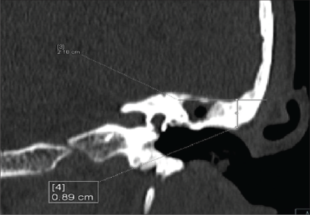
- A 55-year-old woman who presented with ear discharge and ear pain with diagnosis of chronic otitis media. CT scan coronal image showing left sided tegmen height measuring ~ (8.9) mm.
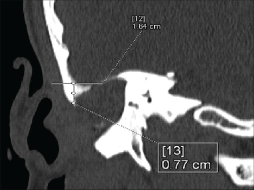
- A 50-year-old man who presented with fever and hearing loss with diagnosis of chronic otitis media. CT scan coronal image showing right sided tegmen height measuring ~ (7.7) mm.
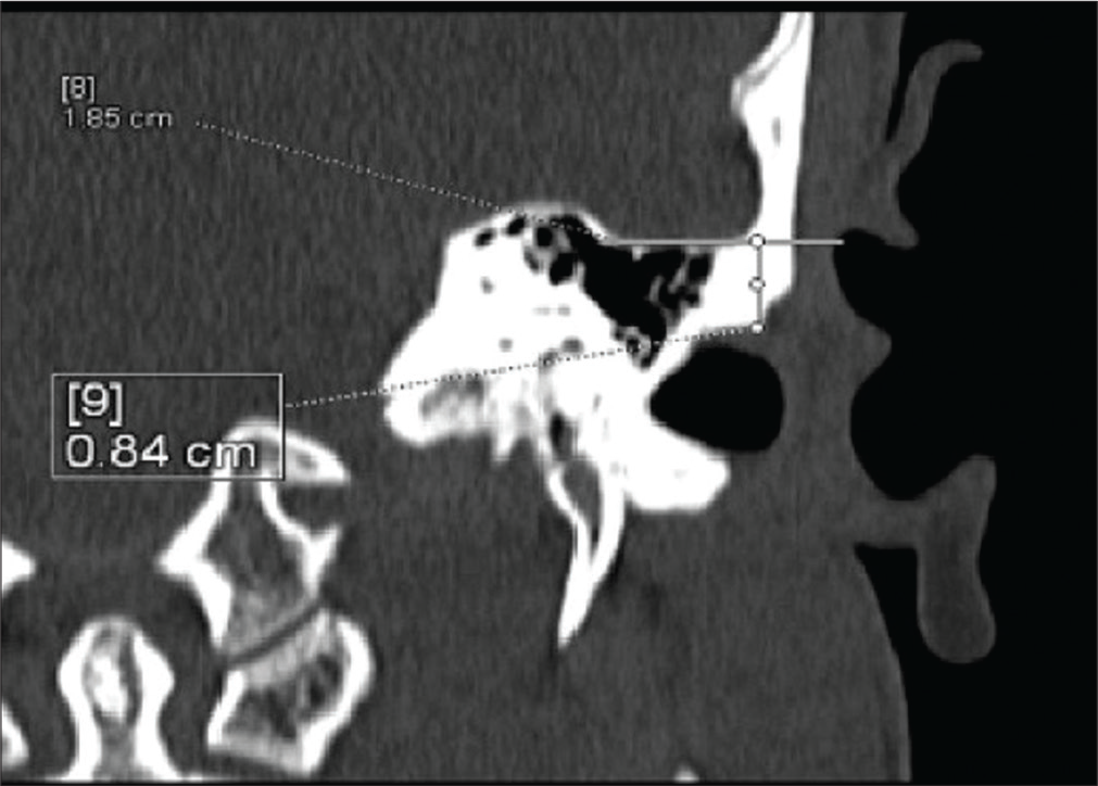
- A 40-year-old man who presented with fever and ear pain with diagnosis of chronic otitis media. CT scan coronal image showing left sided tegmen height measuring ~ (8.4) mm.
DISCUSSION
HRCT imaging acquires an important role in the radiographic assessment of temporal bone. HRCT is widely used in diagnosis of inflammatory middle ear diseases, in particular, chronic otitis media and in evaluation of middle ear following mastoidectomy or tympanoplasty.
According to the study by Parida et al. study, the infectious diseases are almost equally distributed in the young and middle age group in male patients. Fewer infections are seen in the elder age group. (13) The present study includes 60 cases, of which 33 (55%) are male and 27 (45%) are female. Idris et al. comprised approximately equal percent distribution of male and female patients.[6] Gomaa et al. did a prospective study that included 56 consecutive patients with chronic suppurative otitis media, unsafe type cholesteatomas.[12]
In the present study, out of total patients, approximately equal percent (36.7%) of patients belongs to the age group of 15–24 years and 25–34 years, respectively. The majority of the patients were below the age of 35 years. Our study comprised 60 patients. Future studies need to be done with large number of patients over a longer time period so that better analysis and correlation can be done.
A thorough understanding of the shape of the tegmen is important when performing surgery in the mastoid and middle ear.[13] A low-lying tegmen plate may increase the risk of breaching the middle cranial fossa when surgically exploring the epitympanum or mastoid. This may result in dural injury, with possible CSF leak and/or neural tissue damage. The present study demonstrated that tegmen height has significant deviation with age. Furthermore, some studies recommended that HRCT is the preferred method for evaluating most patients with cholesteatomas of the temporal bone.[11]
In the study, the mean tegmen height of exposed dura (ED) was 5.81 ± 1.71 while the mean tegmen height of non-exposed dura (NED) was 8.40 ± 1.31. Highly significant difference was found in mean tegmen height between ED and NED cases.
CONCLUSION
This study was undertaken to assess the role of HRCT in pre-operative assessment of tegmen height in chronic otitis media patients. The study concluded that the cases where exposed dura was an intraoperative finding showed lower tegmen height on HRCT temporal bone as compared to the ones where dura was found non-exposed intraoperatively. Therefore, pre-operative CT assessment of tegmen height is an important parameter in avoiding risk of dural injury during tympanomastoid surgeries. Even though it will not change the surgical procedure but will make the surgeon more careful while operating.
Declaration of patient consent
The authors certify that they have obtained all appropriate patient consent.
Financial support and sponsorship
Nil.
Conflicts of interest
There are no conflicts of interest.
References
- Day-case paediatric mastoid surgery. Int J Pediatr Otorhinolaryngol. 2003;67:771-5.
- [CrossRef] [Google Scholar]
- Chronic Suppurative Otitis Media-Burden of Illness and Management options Geneva: World Health Organization; 2004. p. :13-4.
- [Google Scholar]
- Chronic Otitis Media. 2018. Available from: https://www.dizziness-and-balance.com/disorders/unilat/chronic%20otitis%20media.htm [Last accessed on 2020 Aug 22]
- [Google Scholar]
- Radiological study of the temporal bone in chronic otitis media: Prospective study of 50 cases. Indian J Otol. 2014;20:48-55.
- [CrossRef] [Google Scholar]
- Imaging review of the temporal bone: Part I. Anatomy and inflammatory and neoplastic processes. Radiology. 2013;269:17-33.
- [CrossRef] [PubMed] [Google Scholar]
- A new simple radiological classification measuring the height of the tegmen tympani. Glob J Otolaryngol. 2018;16:555944.
- [CrossRef] [Google Scholar]
- Canal-down mastoidectomy: Experience in 81 cases. Otol Neurotol. 2001;22:451-6.
- [CrossRef] [PubMed] [Google Scholar]
- Lateralization of the tympanic membrane as a complication of canal wall down tympanoplasty: A report of four cases. Otol Neurotol. 2003;24:145-8.
- [CrossRef] [PubMed] [Google Scholar]
- Cerebellar abscess following mastoidectomy for chronic otitis media. Pediatr Neurosurg. 2007;43:327-9.
- [CrossRef] [PubMed] [Google Scholar]
- Tegmen height: Preoperative value of CT on preventing dural complications in chronic otitis media surgery. Clin Imaging. 2014;38:246-8.
- [CrossRef] [PubMed] [Google Scholar]
- High resolution computed tomography in the evaluation of temporal bone cholesteatoma. J Med Sci Clin Res. 2017;5:26614-20.
- [CrossRef] [Google Scholar]
- Evaluation of temporal bone cholesteatoma and the correlation between high resolution computed tomography and surgical finding. Clin Med Insights Ear Nose Throat. 2013;6:21-8.
- [CrossRef] [PubMed] [Google Scholar]
- Anatomic analysis of the mastoid tegmen: Slopes and tegmen shape variances. Otol Neurotol. 2011;32:581-8.
- [CrossRef] [PubMed] [Google Scholar]


