
Translate this page into:
A Rare Coronary Artery Anomaly: Origin of All Three Coronary Arteries from the Right Sinus of Valsalva
Address for correspondence: Dr. Lale Pasaoglu, Ankara Numune Education and Research Hospital, Talatpasa Bulvari, Altindag, Ankara - 06100, Turkey. E-mail: ldamgaci@hotmail.com
-
Received: ,
Accepted: ,
This is an open-access article distributed under the terms of the Creative Commons Attribution License, which permits unrestricted use, distribution, and reproduction in any medium, provided the original author and source are credited.
This article was originally published by Medknow Publications & Media Pvt Ltd and was migrated to Scientific Scholar after the change of Publisher.
Abstract
Left anterior descending (LAD) artery and left circumflex (LCx) coronary artery originating separately from the right sinus of valsalva is exceptionally rare and very few cases have been reported in the literature. Congenital coronary artery anomalies are generally incidental, uncommon, and asymptomatic. Some can cause severe potentially life-threatening symptoms such as myocardial ischemia and sudden cardiac death. The aberrant vessels that pass between the aorta and the pulmonary trunk pose a risk of sudden cardiac death, particularly if the vessel supplies the left coronary artery network. The electrocardiographically gated multi-detector computed tomography (MDCT) allows accurate and non-invasive depiction of coronary artery anomalies including origin, course, and termination. We report here a rare case of all three coronary arteries separately originating from the right coronary sinus, which was detected with MDCT.
Keywords
Anomalies
coronary artery
multidedector computed tomography

INTRODUCTION

Congenital coronary artery anomalies are uncommon and most of them are diagnosed incidentally during coronary angiography or multidedector CT angiography. Their incidence is 1–2% in the general population. Anomalies in origin and distrubition constitute 87% and coronary artery fistulas constitute 13% of the affected individuals.[12] Coronary artery anomalies are usually asymptomatic, but some may cause chest pain, hemodynamically significant abnormalities, myocardial ischemia, or sudden cardiac death.[34] There is varying relation of sudden cardiac death caused by coronary artery anomalies, especially concerning those which course in between the root of the aorta and the pulmonary artery.[4] For several decades, diagnosis of coronary artery anomalies has been made with catheter angiography. Altough catheter angiography is an effective tool, it is invasive and associated with procedural morbidity (1.5%) and mortality (0.15%).[2] Owing to its two-dimensional nature, catheter angiography has projectional limitations and it cannot show the relationship of aberrant vessels with the underlying cardiac structures.[4] Electrocardiographically (ECG) gated multi-dedector computed tomography (MDCT) and magnetic resonance imaging (MRI) can provide non-invasive three-dimensional data of both the heart cavities and coronary arteries without these limitations. MDCT has fast acquisition time and a high temporal and spatial resolution allowing better visualization of coronary arteries, its variants, and anomalies.[456] We report a case with a rare coronary artery anomaly in which the left anterior descending (LAD) artery and the left circumflex (LCx) coronary artery originated from the right coronary sinus. In the literature, coronary arteries originating from the right sinus of Valsalva with a single ostium have been reported, but there are only a few cases wherein all three coronary arteries originate separetely from the right coronary sinus.[6789] We present the case with MDCT and catheter angiographic findings.
CASE REPORT
A 56-year-old male patient was admitted to the hospital with chest pain that had lasted for 1 h. His blood pressure was 140/85 mmHg. The electrocardiogram showed early repolarization in the V5-6 leads and left anterior hemiblock with no ischemic changes. Chest X-ray showed minimal cardiomegaly without pulmonary edema. The physical examination and labarotory data were normal. Echocardiography revealed hypertrophied left atrium and left ventricle. Left ventricular ejection fraction was found to be 55%. Exercise tolerance or myocardial perfusion tests were not performed. The patient was referred for catheter coronary angiography. On catheter coronary angiography, the origin of the left coronary artery (LCA) could not be found and the LAD artery, LCx artery, and right coronary artery (RCA) were seen to arise separately from the right coronary sinus. There was a stenotic lesion in the diagonal branch of the LAD. The second diagonal branch showed bridging. The patient was referred to the radiology department for MDCT coronary angiography. The 16-slice MDCT showed the LAD, LCx, and RCA originating separately from the right coronary sinus [Figure 1]. The LAD showed interarterial course passing between the aorta and the pulmonary artery [Figure 2a]. The diagonal branch of the LAD showed myocardial bridging [Figure 2b]. The LCx coursed posterior to the aortic root to the right atrioventricular groove. The RCA followed a normal course [Figure 2b]. The patient was discharged with medication and recalled 3-months later for follow-up investgations.

- 56-year-old male patient admitted with chest pain diagnosed with three coronary arteries originating separately from the right coronary sinus. Axial oblique maximum intensity projection image shows left anterior descending artery (short white arrow), right coronary artery (open arrow), and left circumflex artery (long white arrow) – three coronary arteries originating from the right coronary sinus.
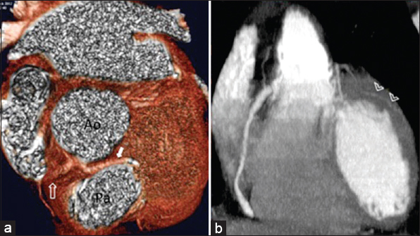
- 56-year-old male patient admitted with chest pain diagnosed with three coronary arteries originating separately from the right coronary sinus. (a) Volume-rendered three-dimensional image (VR-3D) shows the left anterior descending artery (LAD) originating from the right coronary sinus (short white arrow) and passing between the aorta (Ao) and the pulmonary artery (Pa) (open arrow: right coronary artery). (b) MDCT image shows LAD diagonal branch with myocardial bridging (arrowheads).
DISCUSSION
Anomalous origin of all the three coronary arteries originating from the right coronary sinus is a very rare anomaly; only a few cases are reported in the medical literature.[69] The course of the arteries that originate from the right coronary sinus is variable. The RCA follows the usual pathway to the right atrioventricular sulcus. The LCx typically courses posterior to the aortic root, but passing anterior to the pulmonary artery or along the interventricular septum has been reported previously. The LAD may have an anterior path to the pulmonary artery or right ventricular outflow tract, septal or interarterial course.[6]
Most of the coronary artery anomalies are not clinically important, but potentially life-threatening types need to be identified early. Aberrant vessels that pass between the aorta and pulmonary trunk are at risk for sudden death, particularly if the vessel supplies the LCA distribution network.[345] For several decades, these anomalous coronary arteries were identified by coronary angiography. MDCT has been accepted as the ideal method for evaluation of patients with atypical chest pain due to its excellent temporal and spatial resolution.[1278] Among patients with coronary artery anomalies identified with MDCT, conventional angiography alone allowed correct identification of the anomalies in only 53% of cases.[2] Magnetic resonance coronary angiography is a noninvasive method that does not require the use of contrast agents or ionizing radiation, and thus is superior compared to cardiac CT angiography and conventional coronary angiography. Its disadvantages are lengthy acquisition time and lower spatial resolution.[10]
Coronary artery anomalies consist of anomalies of origin, course, or both. Coronary arteries may take origin from the opposite or noncoronary sinus and show an anomalous course. The RCA arising from the left coronary sinus, the LCA arising from the right coronary sinus, the LAD or LCx arising from the right coronary sinus, and the LCA or RCA arising from the non-coronary sinus are most widely-known patterns of an anomalous origin of a coronary artery from opposite or non-coronary sinus.[1234] A coronary artery originating from the opposite or non-coronary sinus courses one of the four pathways: Between the aorta and pulmonary artery (interarterial), retroaortic, prepulmonic, or subpulmonic (septal).[14] Sudden cardiac death can be seen in cases where the coronary artery runs in the interarterial course.[5678] The LCA arises from the right sinus of Valsalva separately or as a branch of a single coronary artery in 0.09–0.11% of patients who undergo angiography. An interarterial course may be detected in up to 75% of patients with this anomaly. The acute angle of the ostium, the stretching of the intramural segment, and the compression between the commissure of the right and left coronary cusps may cause sudden cardiac death in these patients. The LCx artery or the LAD artery may arise from the right coronary sinus. The LCx artery arising from right aortic sinus is the commonest anomaly detected in approximately 0.32–0.67% of the population.[134] This anomalous LCx artery passes behind the aortic root.[12] Fortunately, this anomaly is of little clinical significance and has not been associated with death. However, it is very important to detect this anomaly before valve surgery or bypass grafting of the coronary arteries is carried out. In patients with tetralogy of Fallot, double outlet ring ventricle, and transposition complexes, LAD artery may arise from the right coronary sinus. In patients with normal hearts this anomaly is rarely seen. It may either take an interarterial or a prepulmonic course.[18] In the case reported here, the LAD originating from the right coronary sinus showed interarterial course. There is still disagreement on the surgical treatment, but some guidelines recommend surgical coronary revascularization in patients with anomalous LCA arising from the right sinus of Valsalva and showing interarterial course.[7] Our patient was old and conservative approach was preferred.
Myocardial bridging is a band of myocardial muscle covering a segment of a coronary artery. The incidence of myocardial bridging in angiography is between 0.5 and 2.5%. However, it is more commonly detected during pathologic analysis (15–85%). It is most commonly seen in the middle segment of the LAD artery. Myocardial bridging is often asymptomatic. However, myocardial bridging can cause angina pectoris, myocardial infarction, life-threatening arrhythmias, or even death. MDCT clearly shows the intramyocardial location of the involved coronary arterial segment.[1] We detected a myocardial bridging affecting the diagonal branch of the LAD.
CONCLUSION
In conclusion, conventional coronary angiography has been used for diagnosis of coronary anomalies. Its wide availability, improved technical expertise with simultaneous therapeutic intervention are its advantages. This imaging technique has some limitations due to its projectional and invasive nature. The limitations of catheter angiography have been significantly overcome by MDCT angiography. Curved multiplanar reconstruction and 3D reformatting enables non-invasive visualization of the anatomy, variants, and anomalies of the coronary arteries. MDCT offers superior definition of the ostial origin and path of the anomalous coronary artery, compared with conventional angiography. Although congenital coronary artery anomalies are uncommon, knowledge of the CT appearances of various coronary artery anomalies and understanding the clinical significance of these anomalies is essential for accurate diagnosis and patient treatment.
Available FREE in open access from: http://www.clinicalimagingscience.org/text.asp?2015/5/1/25/156137
Source of Support: Nil
Conflict of Interest: The authors declare no conflict of interest. None of the authors have disclosed financial interests related to the material in the manuscript. The authors have obtained patient consent.
REFERENCES
- Coronary artery anomalies: Classification and ECG-gated multi-dedector row CT findings with angiographic correlation. Radiographics. 2006;26:317-34.
- [Google Scholar]
- Coronary artery anomalies in 126,595 patients undergoing coronary angiography. Cathet Cardiovasc Diag. 1990;21:28-48.
- [Google Scholar]
- Triggerring of sudden death from cardiac causes by vigorous exertion. N Engl J Med. 2000;343:1355-61.
- [Google Scholar]
- Current concepts in multi-detector row CT evaluation of the coronary arteries: Principles, techniques, and anatomy. Radiographics. 2003;23:S111-25.
- [Google Scholar]
- Anomalous origin of the left coronary artery from the right sinus of Valsalva, which presented as acute myocardial infarction. Korean Circulation J. 2006;36:817-9.
- [Google Scholar]
- Origin of all three major coronary arteries from the right sinus of Valsalva: Clinical, angiographic, and magnetic resonance imaging findings and incidence in a select referral population. Catheter Cardiovasc Interv. 2007;69:711-8.
- [Google Scholar]
- ACC/AHA 2008 guidelines for the management of adults with congenital heart disease: Executive summary: A report of the American College of Cardiology/American Heart Association Task Force on practice guidelines (writing committee to develop guidelines for the management of adults withcongenital heart disease) Circulation. 2008;118:2395-451.
- [Google Scholar]
- Anomalous origin of the three coronary arteries from the right aortic sinus Valsalva: Role of MDCT coronary angiography. Int J Cardiovasc Imaging. 2006;22:723-9.
- [Google Scholar]
- Anomalous origin of the entire coronary system by three separate ostia within the right coronary sinus- A rarely observed coronary anomaly. Can J Cardiol. 2010;26:206-8.
- [Google Scholar]
- Identification of anomalous coronary arteries and their anatomic course by magnetic resonance coronary angiography. Circulation. 1995;92:3158-62.
- [Google Scholar]


