
Translate this page into:
Severe Aortic Coarctation in an Adult Patient with Normal Brachial Blood Pressure
Address for correspondence: Mrs. Tina H Leetmaa, Department of Cardiology, Aarhus University Hospital, Skejby, Brendstrupsgaardsvej 100, Aarhus N-8200, Denmark. E-mail: tina.leetmaa@gmail.com
-
Received: ,
Accepted: ,
This is an open-access article distributed under the terms of the Creative Commons Attribution License, which permits unrestricted use, distribution, and reproduction in any medium, provided the original author and source are credited.
This article was originally published by Medknow Publications & Media Pvt Ltd and was migrated to Scientific Scholar after the change of Publisher.
Abstract
The present case shows that a normal brachial blood pressure (BP) does not exclude severe coarctation and should be considered in normotensive patients presenting with a systolic murmur and/or unexplained severe left ventricular hypertrophy. Congenital coarctation of the aorta is a narrowing of the descending aorta, usually located distal to the origin of the subclavian artery, causing hypertension in the upper part of the body. This condition may be undiagnosed until adult life where the clinical presentation most often is high BP in the upper extremities. A 57-year-old patient with severe aortic coarctation and left ventricular hypertrophy presented with normal brachial BP. However, standard suprasternal view by echocardiography indicated coarctation. Multislice computed tomographic (CT) angiography revealed an uncommon location of the aortic narrowing with the right and left subclavian arteries originating below the area of coarctation, explaining the equally low BP in both upper extremities.
Keywords
Aortic coarctation
left ventricular hypertrophy
multislice computed tomography
normal blood pressure

INTRODUCTION

Congenital coarctation of the aorta is a narrowing of the descending aorta which typically is located at the ligamentum arteriosum just distal to the left subclavian artery. This condition may be undiagnosed until adult life, when the clinical presentation most often is high blood pressure (BP) in both or more seldom in only one of the upper extremities.[12] Other typical clinical manifestations may include headache, fatigue on exertion, and bilateral lower limb claudication. Coarctation of the aorta occurs in 5-8% of cases of congenital heart defects. This condition may occur along with ventricular septal defect and other related heart defects, or may occur isolated. In rare cases, severe trauma and injury may lead to coarctation of the aorta. In extremely rare cases, severe atherosclerosis or inflammatory diseases of the aorta may cause narrowing of the artery leading to aortic coarctation.
CASE REPORT
A 57-year-old patient was referred to our outpatient clinic by his primary care physician because the 12-lead ECG demonstrated left ventricular (LV) hypertrophy. Twenty-two years earlier, the patient had been referred for cardiological examination due to a cardiac systolic murmur. At that time, his BP was 98/50 mmHg, and simultaneous and equal radial and femoral pulses were described. No echocardiography was performed and the murmur was interpreted as physiological. No medical or cardiovascular history or cardiovascular risk factors were present, and the patient had no signs of genetic disorders. At the present consultation, the patient confirmed the absence of any cardiovascular symptoms. However, femoral pulses were weak on both sides. The BP was 110/70 mmHg in both arms. No cardiac murmurs could be heard at auscultation. Transthoracic echocardiography showed a non-dilated, hypertrophic left ventricle [Figure 1a and b] with end-diastolic interventricular septal thickness of 21 mm, end-diastolic LV posterior wall thickness of 12 mm, and an estimated LV mass of 449 g (LV mass index 214 g/m2). The LV ejection fraction was 50%. Except for a mild aortic regurgitation (in a normally shaped tricuspid aortic valve) and a dilatation of the ascending aorta of 40 mm, no concomitant structural cardiac abnormalities were found. A continuous wave Doppler examination from the suprasternal notch showed a peak systolic pressure gradient in the thoracic descending aorta of 80 mmHg without diastolic run-off [Figure 1c and d], indicating a severe obstruction at the classical site of a coarctation. Multislice computed tomographic (CT) angiography confirmed the finding of severe coarctation of the aorta. The CT scan demonstrated that both subclavian arteries originated distal to the severe coarctation, explaining the normal BP in both arms [Figure 2]. The neck vessels originated proximal to the obstruction and were significantly dilated. Moreover, a CT scan of the cerebrum revealed the vessels in the circle of Willis giving rise to numerous collaterals in the brain circulation.
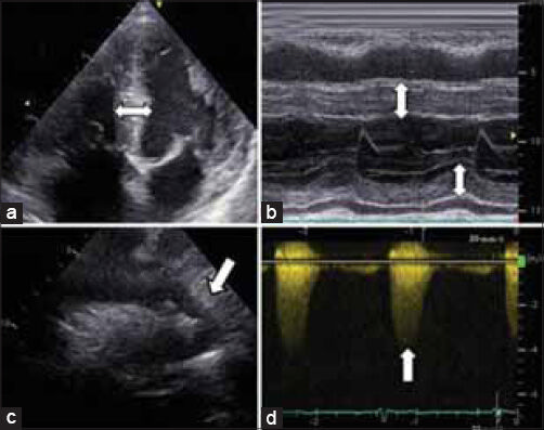
- 57-year-old male was referred to our outpatient clinic because the 12-lead ECG demonstrated left ventricular (LV) hypertrophy that was later diagnosed as due to congenital coarctation of the aorta. Transthoracic echocardiography (a) Apical four-chamber view and (b) M-mode show left ventricle hypertrophy (arrows); (c and d) suprasternal views show the narrowing in the thoracic descending aorta (arrow) and the continuous wave Doppler curve without diastolic run-off (arrow).

- 57-year-old male was referred to our outpatient clinic because the 12-lead ECG demonstrated left ventricular (LV) hypertrophy that was later diagnosed as due to congenital coarctation of the aorta. Multislice computed tomographic angiography and three-dimensional reconstruction show the aortic coarctation.
DISCUSSION
We report an uncommon case of congenital coarctation in a 57-year-old man without the clinical signs of coarctation. Because of the uncommon location of the aortic narrowing with both the right and left subclavian arteries originating distal to the area of coarctation, the BP was equally low in both upper extremities. The present case shows that a normal brachial BP does not rule out severe coarctation and should be considered in apparently normotensive patients presenting with a systolic murmur or target organ damage, in this case severe LV hypertrophy.
CONCLUSION
Uncorrected coarctation of the aorta in adults predisposes to congestive heart failure, aortic dissection and rupture, stroke, cerebral hemorrhage, and infective endocarditis. Therefore, an early diagnosis is important, and the present case emphasizes the use of suprasternal view as a part of a standard diagnostic echocardiography.
Treatment options include surgical repair or balloon angioplasty with or without stent implantation. Taking the atypical location, extent, and complexity of the lesion into account, it was decided to offer the patient surgery as the treatment option.
Available FREE in open access from: http://www.clinicalimagingscience.org/text.asp?2014/4/1/41/137835
Source of Support: Nil
Conflict of Interest: None declared.
REFERENCES
- Anomalous right subclavian artery arising distal to a coarctation of the aorta. Ann Surg. 1958;147:93-7.
- [Google Scholar]
- Coarctation of the aorta proximal of the left subclavian artery: Experience with six surgical cases. Ann Surg. 1957;146:145-51.
- [Google Scholar]


