Translate this page into:
Ellis Van Creveld Syndrome with Synpolydactyly, an Antenatal Diagnosis with Postnatal Correlation
Address for correspondence: Dr. Nischal G. Kundaragi, Consultant, Government Medical College, Nagpur and SRM University Medical College, Kanchipurum, Tamil Nadu, India. E-mail: drngk@yahoo.com
-
Received: ,
Accepted: ,
This is an open-access article distributed under the terms of the Creative Commons Attribution License, which permits unrestricted use, distribution, and reproduction in any medium, provided the original author and source are credited.
This article was originally published by Medknow Publications & Media Pvt Ltd and was migrated to Scientific Scholar after the change of Publisher.
Abstract
Ellis Van Creveld syndrome (EVC), also known as chondroectodermal dysplasia, presents at birth with short limbs accompanied by postaxial polydactyly, nail dysplasia, and dental anomalies. Other manifestations of EVC include atrial septum defects and other congenital heart diseases. We report a case of the EVC syndrome with postaxial polydactyly (Synpolydactyly with seven fingers on the right side and hexadactyly on the left side) and a partial atrioventricular canal defect diagnosed antenatally. This variation of EVS has not been reported in English literature till date.
Keywords
Antenatal diagnosis
cardiac malformation
ellis van creveld syndrome
synpolydactyly
ultrasonography
INTRODUCTION

Ellis Van Creveld syndrome (EVC) leads to babies born with short limbs, especially the middle and distal segments, accompanied by postaxial polydactyly of the hands and sometimes the feet. Nail dysplasia and dental anomalies (including absent and premature loss of teeth and upper lip defects) constitute the ectodermal dysplasia. Other manifestations also include congenital cardiac malformations.[1]
We report a case of the EVC syndrome with atypical postaxial synpolydactyly, with seven fingers on the right hand and hexadactyly on the left hand, with a partial atrioventricular canal defect, diagnosed antenatally. Small, hypoplastic and dystrophic nails were also noted in all the fingers.
CASE REPORT
A 23-year-old multipara on a routine antenatal check-up in the third trimester, revealed the present anomaly of EVS. Her past history revealed a still born first child with six fingers on both hands and a normal second child, two years of age. Marriage was of a non-consanguineous type and the height of both the parents was normal.
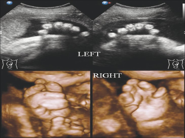
Antenatal examination demonstrated the following: Head circumference (HC) and abdominal circumference (AC) were corresponding to 37 weeks of gestation. Disproportionate shortening of the limbs was noted. Postaxial polydactyly was found on both the hands. Synpolydactyly with seven fingers on the right hand and hexadactyly on the left hand was detected. Right fifth finger duplication with complete soft tissue syndactyly of the fifth and sixth fingers, and partial soft tissue syndactyly of fourth and fifth fingers were noted, without syndactyly on the left side. A 3-D ultrasound was done for better demonstration and confirmation of polydactyly [Figure 1]. No polydactyly was noted in the fetal toes.

- Prenatal 2D and 3D ultrasound image showing right synpolydactyly with seven fingers and left hexadactyly.
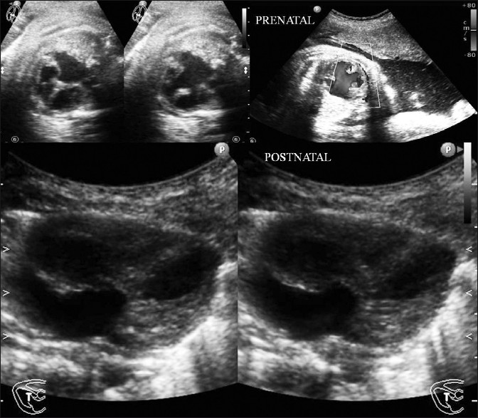
The fetal pre- and postnatal echocardiographic examination revealed a partial atrioventricular canal defect [Figure 2]. A narrow thorax with a disproportionate abdominothoracic ratio was noted. The abdominal viscera, brain, and spine were normal.

- Antenatal and postnatal fetal echocardiographic images showing partial atrioventricular canal defect.
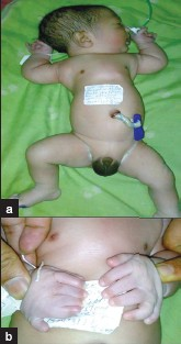
Pregnancy was induced and a live neonate was delivered. Postnatal infant examination revealed disproportionate short limb dwarfism, with a narrow thorax. Six fingers were noted on the right side with a wide fifth finger, representing the fused fifth and sixth fingers, and a partial soft tissue syndactyly of the fourth and fifth fingers was noted, without syndactyly on the left side. Polydactyly was not present in the foot. Nails of all the fingers and toes were small, hypoplastic, and dystrophic [Figures 3a and 3b]. Scalp hair was sparse.

- (a and b) Post natal infant examination shows narrow thorax, disproportionate dwarfism, polydactyly and dystrophic small nails in all fingers.
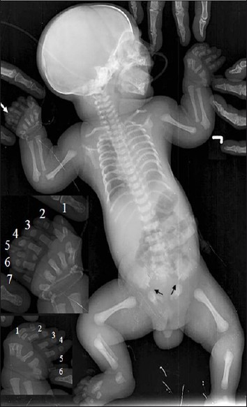
An infantogram revealed a narrow thorax with short ribs, disproportionate dwarfism, postaxial right synpolydactyly, and left hexadactyly, clubbed lower ends of both femurs and proximal ends of both the tibia and ulna, high placement of both clavicles, pelvic dysplasia with low iliac wings, and spur-like downward projections at the medial and lateral aspects of the acetabula. A radiographic examination also showed six metacarpals in both the hands, with few of the fingers having a hypoplastic proximal phalanx and absent or small terminal phalanges [Figure 4]. Right sixth metacarpal was wide and seen articulating with duplicated fifth and sixth fingers.[2]

- Post natal infantogram showing narrow thorax with short ribs, disproportionate dwarfism, postaxial synpolydactyly on right side and hexadactyly on left side (white arrow and inner small images), clubbed lower ends of both femur and proximal ends of both tibia and ulna, high placed both the clavicles, pelvic dysplasia with low iliac wings and spur like downward projections (small black arrows) at the medial and lateral aspects of the acetabulae.
Based on antenatal ultrasound features a diagnosis of EVS was offered and was confirmed on postnatal imaging features. The baby died after two days because of cardiac problems. The parents refused to get an autopsy done or undergo genetic testing.
DISCUSSION
In 1940, Ellis and van Creveld (EVC) described a syndrome, characterized by ectodermal dysplasia, polydactyly, chondrodysplasia, and congenital heart disease. EVC syndrome, also called chondroectodermal dysplasia, has autosomal recessive inheritance secondary to mutations in a novel gene, EVC, which is mapped to chromosome 4p16.1 or caused by mutations in a second gene, called EVC2, which gives rise to the same phenotype of the syndrome. EVC may be allelic to Weyers acrodental dysostosis (also called Weyers acrodental dysostosis), which is an autosomal dominant condition with dental anomalies, nail dystrophy, postaxial polydactyly, and mild short stature.[34]
The basic defect in EVC is generalized dysplasia of endochondral ossification, with a delay in appearance of the primary centers and premature appearance of the secondary centers, which leads to a short stature, with progressive distal shortening of the extremities.[5] The EVC syndrome comprises of a tetrad of clinical manifestations of chondrodystrophy, polydactyly, ectodermal dysplasia, and cardiac defects. Chondrodystrophy is the most common feature affecting the tubular bones, causing disproportionate dwarfism, with progressive distal limb shortening, symmetrically affecting the forearms and lower legs. Bone age is often less than the chronological age. Short stature, which is a common finding due to shortness of lower legs, is present at birth and becomes more prominent as the child grows. Genu valgus curvature of the humerus, talipes equinovarus (club foot), talipes calcaneovalgus, and pectus carinatum, with a long narrow chest, and respiratory difficulties are the other features.
Polydactyly, which is always present, is typically seen as postaxial hexadactyly of the hands, or in a few cases, the feet (about 20% of the cases). Polydactyly may be just extra soft tissue not adherent to the skeleton and devoid of bone, cartilage, joint or tendon, or there may be a complete digit formation or the digit, which may show duplication, with components like bifid metacarpals.
Syncarpalism (that is fusion of the hamate and capitate), synmetacarpalism (that is fusion of the metacarpals), polycarply (that is extra or ninth carpal bone appearing in the distal row) and polymetacarpalism (that is extra metacarpals) are frequently present. Carpal development is delayed.[6]
Oral manifestations of the syndrome include fusion of the upper lip to the maxillary gingival margin,[7] absence of the mucobuccal fold or the sulcus anteriorly, notching of the alveolar ridge, congenitally missing teeth in the mandibular region, natal teeth (teeth present at birth), erupted teeth having small crowns, and irregular spaces between teeth. In the present case, inconclusive oral manifestations were present.
Cardiac anomalies occur in 50 – 60% of the patients; the most common anomaly is a common atrium (40%). Cardiac anomalies such as the AV canal, ventricular septal defect, atrial septal defect, and patent ductus arteriosus have also been reported. The life expectancy of these patients is mainly reduced due to cardiac anomalies.
A common atrium exists when the entire atrial septum is absent. A partial or incomplete AV canal defect has separate mitral and tricuspid orifices, and may have atrial or ventricular septal defects or both, and clefts in the mitral and tricuspid valve leaflets. About two-third of all patients with the AV canal defect have the partial form. The most common partial form is an ostium primum atrial septal defect, which involves the central part of the atria inferior to that of the ostium secondum type, below the fossa ovalis, and adjacent to the AV valves, and cleft in the anterior (septal) leaflet of the mitral valve. The central part of the defect extends to the annuli of the atrioventricular valves, such that, no intervening tissue is present between the hinge point of the valve leaflets and atrial septal defect.[8]
Differential diagnosis of the EVC syndrome include short rib polydactyly syndrome type I (Sadino Noonan), these are frequently associated intestinal and genital malformations. In type II (Majewski), there is usually a preaxial polydactyly with orofacial clefts. Type III (Verma NaumoV) is similar to type I, but there are less malformations of the inner organs. In type IV (Beemer Langer) there is usually an omphalocele, but polydactyly is rare. Other differentials include achondroplasia, chondrodysplasia punctata, asphyxiating thoracic dystrophy, and Morquio syndrome.
Jeune dystrophy, McKusick-Kaufman syndrome, and Weyers syndrome are the postnatal differential diagnoses for the EVC syndrome. The McKusick-Kaufman and Ellis-Van Creveld syndromes are both recessively inherited disorders, and share postaxial polydactyly and congenital heart defect. The distinguishing characteristics of osteochondrodysplasia and ectodermal anomalies are seen in the EVC Syndrome, and the McKusick-Kaufman syndrome has hydrometrocolpos. McKusick-Kaufman is caused by mutations in a gene on chromosome 20p12, encoding a protein similar to the members of the chaperonin family, whereas, Ellis-Van Creveld Syndrome is caused by mutation in the EVC gene.[9] Congenital heart disease, supernumerary digits, and ectodermal dysplasia will almost always support the diagnosis of the EVC syndrome.
Synpolydactyly with seven fingers and partial atrioventricular canal defect has been reported in a few cases,[10] but antenatal diagnosis has not been made in any of them.
An antenatal demonstration of the spectrum of findings in the Ellis Van Creveld syndrome, with unreported type of synpolydactyly, will definitely add to the scientific progress in the antenatal syndromic approach.
Source of Support: Nil
Conflict of Interest: None declared.
Available FREE in open access from: http://www.clinicalimagingscience.org/text.asp?2011/1/1/59/91132
REFERENCES
- Kliegman: Nelson Textbook of Pediatrics. Disorders for which defects are poorly understood or unknown. (18th ed). Chapter 698. URL: http:/das/book/0/view/1608/1624.html
- [Google Scholar]
- McGlamry's comprehensive textbook of foot and ankle surgery. (3rd ed). Philadelphia: Lippincott Williams and Wilkins; 2001.
- [Google Scholar]
- Sequencing EVC and EVC2 identifies mutations in two-thirds of Ellis-van Creveld syndrome patients. Hum Genet. 2007;120:663-70.
- [Google Scholar]
- Mutations in a new gene in Ellis-van Creveld syndrome and Weyers acrodental dysostosis. Nature Genet. 2000;24:283-6.
- [Google Scholar]
- Atlas of genetic diagnosis and counseling; Chapter Ellis-van Creveld Syndrome. Totowa, New Jersey: Human Press; 2006. p. :350-5.
- [Google Scholar]
- Polycarpyly and other abnormalities of wrist in chondroectodermal dysplasia: The Ellis-Van Crevald syndrome. Radiology. 1984;151:393-6.
- [Google Scholar]
- Oral and dental anomalies in Ellis van Creveld syndrome (chondroectodermal dysplasia): report of a case. Int J Paediatr Dent. 1998;8:153-7.
- [Google Scholar]
- Cardiac Imaging: The Requisites. In: Chapter 8. Congenital Heart Disease (2nd ed). Maryland Heights, Missouri: Elsevier Mosby; 2005. p. :326-31.
- [Google Scholar]
- Chondroectodermal Dysplasia (Ellis-Van Creveld Syndrome): A case report. Internet J Pediatrics Neonatol. 2009;10:1.
- [Google Scholar]


