Translate this page into:
Contrast-enhanced Computed Tomography Imaging of Splenic Artery Aneurysms and Pseudoaneurysms: A Single-center Experience
Address for correspondence: Dr. Hirenkumar Kamleshkumar Panwala, Room 23, Department of Radiology, Christian Medical College, Vellore, Tamil Nadu, India. E-mail: drhiren1187@gmail.com
-
Received: ,
Accepted: ,
This is an open access journal, and articles are distributed under the terms of the Creative Commons Attribution-NonCommercial-ShareAlike 4.0 License, which allows others to remix, tweak, and build upon the work non-commercially, as long as appropriate credit is given and the new creations are licensed under the identical terms.
This article was originally published by Medknow Publications & Media Pvt Ltd and was migrated to Scientific Scholar after the change of Publisher.
Abstract
Aim:
The aim of our study was to evaluate the computed tomography (CT) imaging features of splenic artery aneurysm and pseudoaneurysm and to identify the disease conditions related to the same. We also wanted to ascertain any relationship between these associated disease conditions and the imaging features of the aneurysms.
Materials and Methods:
This retrospective study included patients diagnosed to have splenic artery aneurysms on contrast-enhanced CT examination between January 2001 and January 2016. Data were obtained from the picture archiving and communication system. The size, number, location, morphology, the presence of thrombosis, calcification, and rupture of the aneurysms were evaluated.
Results:
A total of 45 patients were identified with a mean age of 45 years. Splenic artery aneurysms were idiopathic in 12 (26.6%) patients. In the remaining patients, the main associated disease conditions included pancreatitis 15 (33%), chronic liver disease with portal hypertension 8 (18%), and extrahepatic portal vein obstruction (EHPVO) 6 (13%). Statistically significant findings included the relationship between EHPVO and multiple aneurysms (P = 0.002), chronic liver disease and fusiform aneurysm (P = 0.008), and smaller size of idiopathic aneurysms (P < 0.001).
Conclusion:
Based on this study, splenic artery aneurysms were associated with a variety of etiologies. The characteristics of the aneurysms such as size, location, and morphology vary with the associated disease conditions. These variations may have implications for the management.
Keywords
Aneurysms
computed tomography
splenic artery

INTRODUCTION
Splenic artery aneurysm is the most common type of splanchnic aneurysm.[1] Splanchnic aneurysms are important because of the high incidence of mortality related to rupture of the aneurysm.[1] The true prevalence of splenic artery aneurysms is unknown, with estimates varying widely, from 0.2% to as high as 10.4%.[23] The detection of splenic and other splanchnic artery aneurysms has become easier in recent years because of the use of multidetector computed tomography (CT). Multiplanar imaging and submillimeter sections enable detection of even small incidental lesions.
Our goal was to identify the disease conditions related to splenic artery aneurysm and to evaluate the common imaging findings of these aneurysms.
MATERIALS AND METHODS
This retrospective study was conducted in a tertiary care institution after approval from the Institutional Review Board.
We obtained the list of patients with a documented splenic artery aneurysm or pseudoaneurysm from the picture archiving and communication system (GE Healthcare, Milwaukee, Wisconsin) in Christian Medical College, Vellore based on contrast CT examination. Forty-five patients were identified between January 2001 and January 2016. The patient demographics, coexisting disease conditions, imaging characteristics of the aneurysm on CT, and treatment details were evaluated.
Multidetector computed tomography protocol
All CT scans were performed on Siemens (Emotion 16-slice Siemens Medical system, Erlangen, Germany), Philips (Brilliance 6-slice Philips Medical System, Best, the Netherlands), or GE (Discovery CT 750 HD 64-slice, GE Healthcare, Milwaukee, Wisconsin) scanners. Twenty-three studies were performed on the Philips Brilliance 6-slice CT, 12 studies on the Siemens emotion 16-slice CT, and 10 studies on GE Discovery 64-slice CT. Bolus tracking was used for determining scan delay, and biphasic scanning in the arterial and portal venous phases was performed. Intravenous contrast medium (low osmolar, nonionic, and 300 mg/mL iodine content) was used routinely at a dose of 1 ml/kg administered by pressure injector at a rate of 4 ml/s.
Image interpretation
All the CT examinations were reevaluated by a radiologist with over 7 years of experience in interpreting abdominal CT. Each CT study was evaluated for number, size, and location of splenic artery aneurysm. In patients with multiple aneurysms, the size of the largest aneurysm was recorded. Based on size, aneurysms were divided into less or >20 mm. A cutoff of 20 mm was selected since size larger than 20 mm is considered as an indication for treatment in asymptomatic patients.[1] For assessment of the location, the splenic artery was divided into three parts: proximal, middle, and distal third based on the length of the splenic artery. The presence of calcification and thrombosis was documented. Thrombosis was considered partial or complete according to the extent of involvement. All aneurysms were divided into saccular or fusiform according to morphology. Fusiform aneurysms involve the entire circumference while saccular aneurysms involve only a portion of the vessel wall. True aneurysms and pseudoaneurysms were distinguished by evaluating the underlying cause of the aneurysm and the presence of inflammatory changes or hematoma surrounding the lesion in the case of a pseudoaneurysm. Association with pancreatitis, iatrogenic injury, mycotic infection, and trauma was considered in favor of pseudoaneurysm. Associated disease conditions were recorded based on the imaging findings and data in the patient's electronic medical chart. Aneurysms were considered idiopathic when there were no associated conditions related to the aneurysm.
Statistical analysis
Frequency and percentage were used for analysis of categorical variables. Chi-square with continuity correction and Fisher's exact probability test was used to test the association of etiological with morphological parameters. P < 0.05 was considered statistically significant. The analysis was performed using SPSS (version 16.0, Chicago, IL, USA).
RESULTS
Demographics
Our study group comprised 25 males and 20 females. The mean age of the patients in our study was 44.8 years with a range of 17–72 years. The most common age groups involved were 50–59 years and 40–49 years with 24% each. The average age for patients with pancreatitis was 44 years, while patients with the chronic liver disease were aged 42 years. Extrahepatic portal vein obstruction (EHPVO) patients were relatively younger with an average age of 30 years, while the idiopathic group was aged 55.13 years out of 15 patients with pancreatitis were male (P = 0.003).
Associated conditions
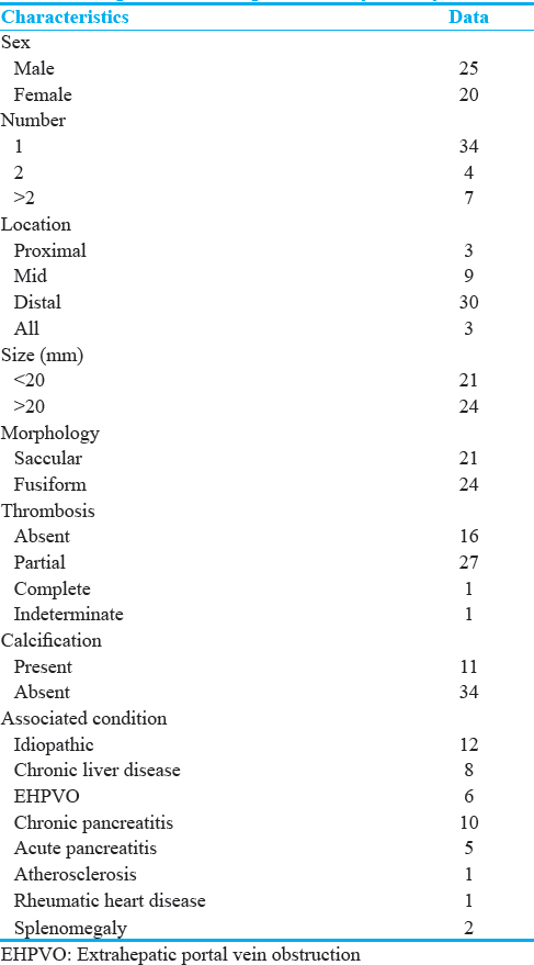
Pseudoaneurysms associated with pancreatitis formed the largest group with 15 (33%) patients out of which 10 were the chronic type. Incidentally, detected idiopathic aneurysms were the second largest group with 12 (26.6%) patients. Eight (17.8%) patients had chronic liver disease with portal hypertension, and 6 (13%) had EHPVO [Table 1].
Imaging findings
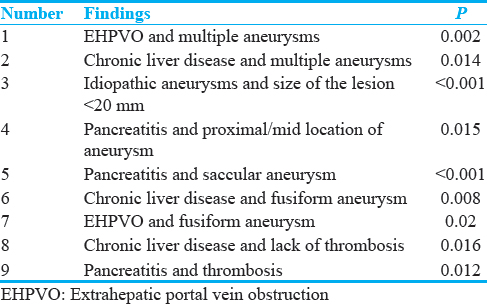
Eleven patients had multiple aneurysms out of which four patients had >2 aneurysms. Rest of the patients (34/45) had only one aneurysm. Ten out of 11 patients with multiple aneurysms were associated with chronic liver disease or EHPVO [Figures 1 and 2]. Conversely, 5 out of 6 patients with EHPVO and 5 out of 8 patients with chronic liver disease had multiple aneurysms. Only one patient had multiple idiopathic aneurysms. The relationship between both chronic liver disease (P = 0.014) and EHPVO (P = 0.002) with multiple aneurysms was statistically significant [Table 2].
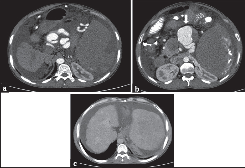
- A 56-year-old male with chronic liver disease and portal hypertension presenting with hematemesis (a and b). Contrast-enhanced computed tomography of the upper abdomen shows multiple fusiform aneurysms arising from the splenic artery with some showing peripheral calcification (arrow). (c) Computed tomography image shows irregular liver with enlarged portal vein and spleen, s/o chronic liver disease with portal hypertension.
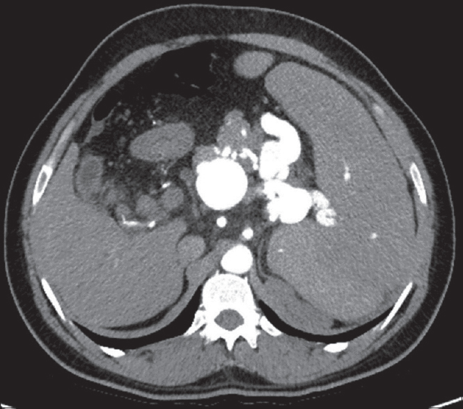
- A 26-year-old female with extrahepatic portal vein obstruction presenting with abdominal distension. Postcontrast computed tomography of upper abdomen shows multiple fusiform splenic artery aneurysms.
Twenty-one out of 45 (46.7%) patients had aneurysms of size <20 mm while the remaining 24 had size ≥20 mm. All incidentally detected idiopathic aneurysms measured <20 mm size (P < 0.001) [Figure 3]. Five out of 6 patients with EHPVO, 8 out of 15 patients with pancreatitis, and 7 out of 8 patients with chronic liver disease had aneurysms >20 mm size. Greater than 20-mm aneurysms in chronic liver disease and EHPVO taken together were statistically significant (P = 0.004).
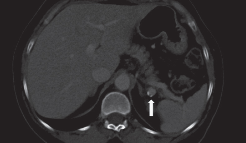
- A 50-year-old male presenting with altered bowel habit. Contrast-enhanced computed tomography of upper abdomen shows incidental splenic artery aneurysm with peripheral calcification (arrow).
The majority of the lesions were in the distal third of the splenic artery, 29 out of 45 (64%). In three patients, lesions were in the proximal third, while it was in the middle third in nine patients. In four patients with multiple aneurysms, lesions were seen in more than one segment. Statistical analysis was done after exclusion of these four patients. All the patients with the lesion in the proximal third had associated chronic pancreatitis. In the middle and distal third lesions, no distinct pattern in relation to the associated condition was noted. While 84.6% (22/26) of the nonpancreatitis-related lesions were in the distal third, only 7/15 (46%) of cases of pancreatitis showed distal involvement (P = 0.015).
Twenty-four (53%) patients had a fusiform aneurysm, while the remaining 21 (47%) had a saccular morphology. Fifteen (71%) patients with a saccular aneurysm had pancreatitis as an associated condition, and conversely, all the patients with pancreatitis showed saccular aneurysms (P < 0.001) [Figure 4]. Pseudoaneurysms, mostly associated with pancreatitis, were saccular in morphology as they involved only part of the arterial wall. Furthermore, none of the patients with chronic liver disease (P = 0.0084) or EHPVO (P = 0.02) showed saccular morphology.
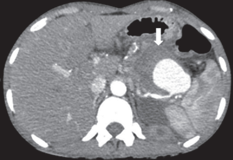
- A 40-year-old male with chronic pancreatitis presenting with abdominal pain. Contrast-enhanced computed tomography of upper abdomen shows partially thrombosed (arrow) saccular aneurysm arising from the distal third of the splenic artery.
Sixteen (35%) patients showed partial thrombosis of the aneurysm, while only one patient showed complete thrombosis. The presence of thrombosis could not be determined in one patient. Remaining 27 (60%) showed no evidence of thrombosis. Out of 15 patients with pancreatitis, 10 showed partial thrombosis (P = 0.012). Similarly, 6 out of 12 patients with idiopathic lesions showed thrombosis. On the other hand, none of the patients with chronic liver disease or EHPVO showed signs of thrombosis. The chronic liver disease patients showed a statistically significant relationship with lack of thrombosis (P = 0.016).
Eleven (24%) patients showed peripheral wall calcifications while the remaining 34 showed no evidence of calcification. The patients with chronic liver disease (2/8), EHPVO (2/6), and pancreatitis (1/15) (P = 0.03) showed a low incidence of calcifications. Five out of twelve idiopathic lesions showed calcifications.
One of the 45 patients presented with imaging findings of acute rupture in the form of active contrast extravasation.
Treatment
Twenty-four (53%) patients were conservatively managed, while 21 (47%) patients underwent surgical (2 patients) or endovascular interventional (19 patients) treatment [Figure 5]. Fifteen patients underwent coiling while three patients underwent a combination of coiling and polyvinyl alcohol embolization. One patient was managed using a covered stent under interventional guidance. All the idiopathic aneurysms were managed conservatively, while all the patients with pancreatitis-related lesions were managed by interventional techniques.
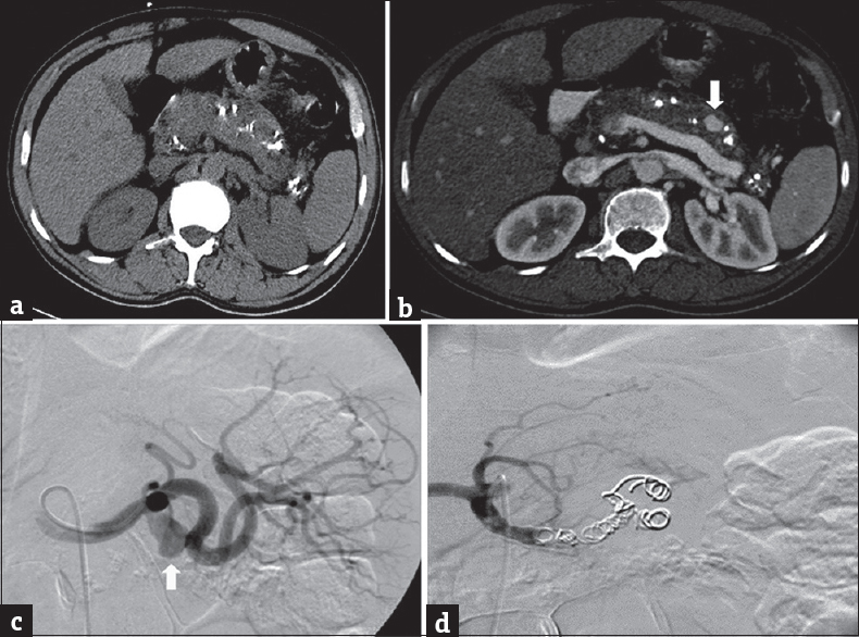
- A 46-year-old male with chronic pancreatitis presenting with abdominal pain and distension. (a) Plain computed tomography shows features of chronic calcific pancreatitis with multiple calcifications in the pancreatic parenchyma. (b and c) Arterial phase postcontrast computed tomography and angiogram shows pseudoaneurysm arising from the middle third of the splenic artery (arrow). (d) Postcoiling angiogram with no opacification of the aneurysm.
DISCUSSION
The first reported case of splenic artery aneurysm was by Beaussier in 1770 at autopsy.[2] Since then there have been many published clinical, operative, and autopsy studies of splenic artery aneurysms.[456] Before the advent of cross-sectional CT imaging, splenic artery aneurysms were considered relatively rare. However, with widespread availability of high-resolution multiplanar imaging, small incidental aneurysms are commonly encountered.
The causes of splenic artery aneurysm are idiopathic, portal hypertension, cirrhosis, pancreatitis, hypertension, and pregnancy.[78910] Less common causes include liver transplantation, arterial fibrodysplasia, arteritis, collagen vascular disease, α1-antitrypsin deficiency, systemic lupus erythematosus, and septic emboli.[1211121314] In our study, the largest group of true aneurysms consisted of the incidentally detected idiopathic lesions confirming the ability of multidetector CT in identifying them. Chronic liver disease with portal hypertension and EHPVO was the other main associated conditions in our study. However, after the inclusion of pseudoaneurysms, pancreatitis was the single largest associated condition.
The significant association with EHPVO (six cases) was unique to our study even though few isolated cases have been reported.[1516] This could be due to EHPVO being the most common cause of portal hypertension in the pediatric age group at our center.[17] Chronic cases of EHPVO can have associated liver parenchymal atrophy and lobulated contours which can mimic chronic liver disease.[1819] However, none of our cases of EHPVO had significant liver parenchymal changes. We did not have any case related to pregnancy as CT abdomen is usually not performed due to possible radiation hazards during pregnancy.
Splenic artery aneurysms are more common in females, but our study showed a male preponderance.[12] This could be attributed to the larger number of chronic pancreatitis in our study which is usually seen in males in our population, due to a higher prevalence of alcoholism in males. The average age at diagnosis of splenic artery aneurysm was 44 years in our study compared to about 60 years in previously published studies.[5] The difference probably reflects the larger proportion of pancreatitis, EHPVO, and chronic liver disease in our study population, all of which may present at an earlier age.
About 28% of patients in our study had multiple aneurysms which are similar to previous studies.[15] There was a statistically significant association of multiple aneurysms with chronic liver disease and EHPVO, which contributed to 90% of multiple aneurysms in our study. The relationship between multiple aneurysms and chronic liver disease has been reported by Ulu et al.[20] However, the association of multiple aneurysms with EHPVO has not been described earlier.
Most of the splenic artery aneurysms involved the distal third of the artery, similar to previous studies.[15] However, in our study proximal and mid part of the splenic artery involvement were also seen in patients having pancreatitis. The close anatomical relation of the pancreas with these splenic artery segments can explain the significant association between pancreatitis-related aneurysms and proximal/mid segments.
Using the size cutoff of 20 mm, none of the incidentally detected idiopathic lesions in our study required intervention. On the other hand, most of the patients with pancreatitis and chronic liver disease had lesions larger than 20 mm mandating active management.
Unlike the previous literature, our study had more number of aneurysms with fusiform than saccular morphology.[15] This is likely due to the larger number of chronic liver disease and EHPVO cases, both which had statistically significant association with fusiform aneurysms.
In our study, about 31% of the patients showed partial peripheral thrombosis with only one lesion showing complete thrombosis. The lack of thrombosis in chronic liver disease and the presence of thrombosis in pancreatitis were found to be statistically significant. However, no similar published studies are available for comparison of the incidence of thrombosis within the aneurysms.
Earlier studies suggest that nearly 70%–80% of patients with splenic artery aneurysm show peripheral calcification, but in our study, it was only 31%.[221] The patients with the chronic liver disease, EHPVO, and pancreatitis showed a low incidence of calcifications. This may be an indicator that these lesions develop in a shorter duration compared to the idiopathic lesions. However, there was no statistically significant association between calcification and varied etiologies.
Only one patient in our group presented with evidence of active extravasation suggesting acute rupture. Initial reports of splenic artery aneurysm suggested that the risk of rupture is 10%.[22] However, more recent data suggest rupture rates closer to 2%–3%.[2324]
The limitations of this study include the retrospective nature of the study and the lack of plain CT images. The retrospective evaluation of the CT reports may not reflect the actual number of splenic artery aneurysms as many small aneurysms can be missed on routine reporting. Plain CT sections were not taken as it is not a part of the routine protocol in our center. Hence, hematoma in the region of the lesion could not be adequately evaluated and this limited the ability to diagnose pseudoaneurysms definitively. Furthermore, since many cases were incidentally detected while evaluating for other conditions such as chronic liver disease, optimum imaging protocol for the evaluation of aneurysms could not be followed.
CONCLUSION
This study showed the etiologic factors associated with splenic artery aneurysms and the utility of multidetector CT in their diagnosis and evaluation. It brought out the differences in morphological features of the aneurysms based on their etiology. The aneurysms related to portal hypertension due to chronic liver disease and EHPVO tended to be multiple, larger, fusiform, and without thrombosis or calcification. The lesions associated with pancreatitis were saccular, large, single, more proximal in location, partially thrombosed, and without calcification. The incidentally detected idiopathic aneurysms were smaller than 20 mm, single, with or without thrombosis and showed peripheral calcifications.
Financial support and sponsorship
Nil.
Conflicts of interest
There are no conflicts of interest.
Acknowledgment
We would like to acknowledge Dr. Prateek Malik (PG final year, CMC Vellore) for editorial support, and Ms. Thenmozhi (Department of biostatistics, CMC)Vellore for statistical aid to complete this project.
Available FREE in open access from: http://www.clinicalimagingscience.org/text.asp?2018/8/1/37/239702
REFERENCES
- Splenic artery aneurysms and pseudoaneurysms: Clinical distinctions and CT appearances. AJR Am J Roentgenol. 2007;188:992-9.
- [Google Scholar]
- Clinical features and management of splenic artery pseudoaneurysm: Case series and cumulative review of literature. J Vasc Surg. 2003;38:969-74.
- [Google Scholar]
- The diagnosis and management of splenic artery aneurysms. J R Soc Med. 1988;81:387-8.
- [Google Scholar]
- Haemorrhagic complications of pancreatitis: Presentation, diagnosis and management. Ann R Coll Surg Engl. 1998;80:316-25.
- [Google Scholar]
- Management of splenic artery aneurysms: The significance of portal and essential hypertension. J Am Coll Surg. 1999;189:483-90.
- [Google Scholar]
- Review: Spontaneous rupture of splenic artery aneurysm in pregnancy. Eur J Obstet Gynecol Reprod Biol. 2003;109:124-7.
- [Google Scholar]
- Splenic artery aneurysms in portal hypertension. J Cardiovasc Surg (Torino). 1982;23:490-3.
- [Google Scholar]
- Splenic artery aneurysm associated with systemic lupus erythematosus: Report of a case. Surg Today. 1999;29:76-9.
- [Google Scholar]
- Splenic artery aneurysms in liver transplant patients. Transplantation. 1988;45:386-9.
- [Google Scholar]
- Splenic artery aneurysms: Two decades experience at mayo clinic. Ann Vasc Surg. 2002;16:442-9.
- [Google Scholar]
- Giant splenic artery mycotic aneurysm presenting with massive hematemesis. Indian J Gastroenterol. 2003;22:147-8.
- [Google Scholar]
- Splenic artery aneurysm presenting as extrahepatic portal vein obstruction: A case report. Case Rep Gastrointest Med 2011 2011 908529
- [Google Scholar]
- Aetiology of paediatric portal hypertension – Experience of a tertiary care centre in South India. Trop Doct. 2009;39:42-4.
- [Google Scholar]
- Imaging and radiological interventions in extra-hepatic portal vein obstruction. World J Radiol. 2016;8:556-70.
- [Google Scholar]
- Multimodality imaging of primary extrahepatic portal vein obstruction (EHPVO): What every radiologist should know. Br J Radiol. 2015;88:20150008.
- [Google Scholar]
- Multidetector CT findings of splenic artery aneurysm in children with chronic liver disease. Pediatr Radiol. 2008;38:1095-8.
- [Google Scholar]
- The diagnosis and management of visceral artery aneurysms. Surgery. 1980;88:619-24.
- [Google Scholar]
- Clinical importance and management of splanchnic artery aneurysms. J Vasc Surg. 1986;3:836-40.
- [Google Scholar]


