
Translate this page into:
Bilateral Fracture of the Handle Malleus: A Case Report and Review of the Literature
Address for correspondence: Dr. Ilson Sepulveda, Department of Radiology, ENT Head and Neck Surgery Service, General Hospital of Concepcion, San Martin Av. 1436, Concepción, Chile. E-mail: isepulvedaa@uft.edu
-
Received: ,
Accepted: ,
This is an open access journal, and articles are distributed under the terms of the Creative Commons Attribution-NonCommercial-ShareAlike 4.0 License, which allows others to remix, tweak, and build upon the work non-commercially, as long as appropriate credit is given and the new creations are licensed under the identical terms.
This article was originally published by Medknow Publications & Media Pvt Ltd and was migrated to Scientific Scholar after the change of Publisher.
Abstract
Malleus fracture is a rare condition. Usually, the handle of the malleus is involved, and we do not find reports in the literature of this condition in the bilateral presentation. It is present as sudden conductive hearing loss commonly after digital manipulation of the external auditory canal. The diagnosis is based principally on clinical examination by otomicroscopy and audiometry. Cone-beam computed tomography emerging as a powerful tool in the field of otolaryngology, especially for explorations of paranasal sinuses and temporal bone, due to imaging with a high resolution and few artifacts with lower dose radiation in comparison with multislice computed tomography.
Keywords
Bilateral
cone-beam computed tomography
fracture
handle
malleus


INTRODUCTION
The isolated malleus handle fracture is a rare entity, and its diagnosis is mostly achieved by clinic-radiological examinations. The first case was described by Meniere in 1855,[12] a very rare case of bilateral isolated fracture of the handle malleus, and our review of the literature does not reveal cases reported of a condition like this.
CASE REPORT
A 56-year-old female, teacher, presented without medical antecedents of relevance and clinical history of fluctuating hearing loss in the right ear and left sense of aural fullness after finger manipulations of the external auditory canal (ECA) while taking a shower. She also reported that 15 days ago, due to right ear itch and after finger introduction in ECA, she heard a sudden pop and experienced slight otalgia, with remission after the use of analgesics. Otomicroscopic examination showed a left intact tympanic membrane. The right tympanic membrane revealed an irregularly shaped malleus handle with excessive movement of the distal part of the malleus at Valsalva maneuver.
An audiogram showed conductive hearing loss with 18 dB air–bone gap (ABG) on the right ear [Figure 1].
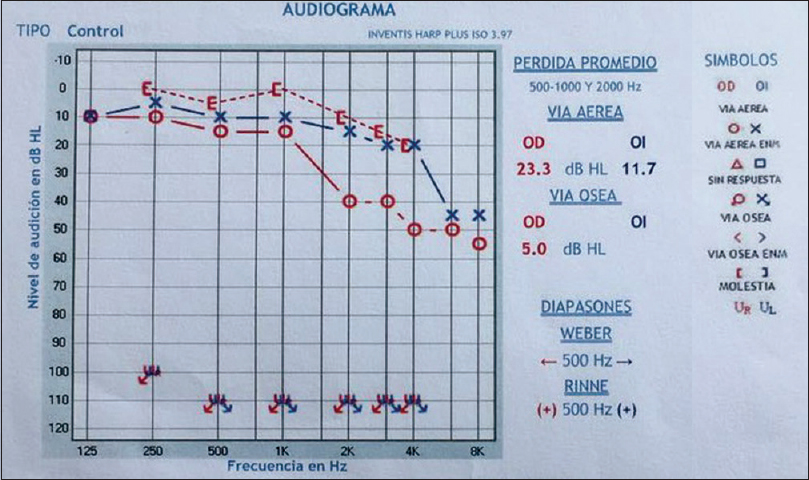
- A 56-year-old female with fracture of the handle malleus who presented with right conductive hearing loss. Audiogram: Shows a conductive hearing loss on the right ear, especially at the high frequencies (air–bone gap) 18 dB.
Temporal bone high-resolution cone-beam computed tomography (CBCT) with multiplanar reconstruction was performed and confirmed bilateral fracture of the handle malleus [Figures 2 and 3].
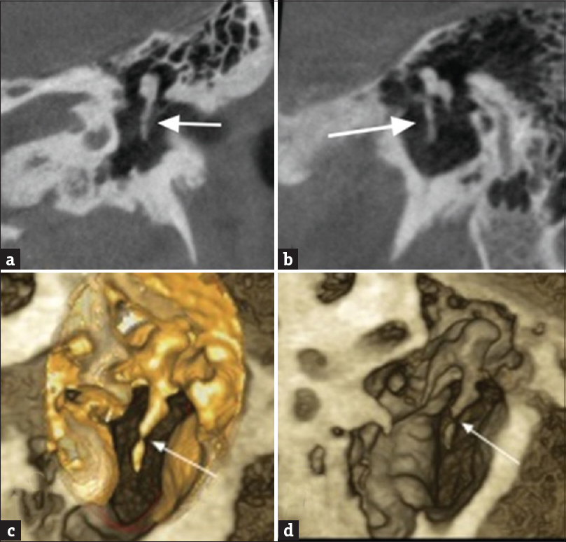
- A 56-year-old female with fracture of the handle malleus who presented with right conductive hearing loss. Cone-beam computed tomography of temporal bone shows isolated left fracture of the handle malleus (white arrows). Coronal (a). Oblique sagittal (b). Osseous volume rendering (c and d).
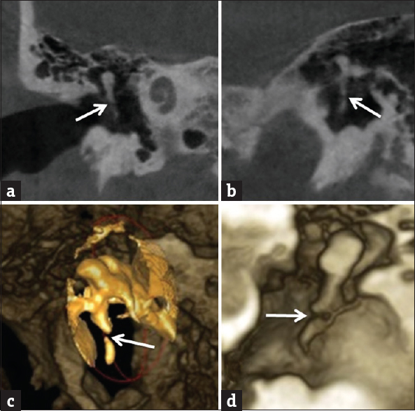
- A 56-year-old female with fracture of the handle malleus who presented with right conductive hearing loss. Cone-beam computed tomography of temporal bone shows an isolated right fracture of the handle malleus (white arrows). Coronal (a). Oblique sagittal (b). Osseous volume rendering (c and d).
A transcanal exploratory tympanotomy, under general anesthesia, was performed. Intraoperatively, the malleus handle fracture was visualized [Figure 4], reduced, and fixed with glass ionomer cement (GIC). Due do not presented hearing loss in the left fracture, conservative management was advised.

- A 56-year-old female with a fracture of the handle malleus who presented with right conductive hearing loss. Exploratory tympanostomy showed a right handle malleolus fracture (yellow arrows).
DISCUSSION
Isolated fracture of the handle malleus is a rare condition that can be overlooked or misdiagnosed.[1] First reports date from the second half of the 19th century and the first quarter of the 20th century.[3] The first case was described in 1855 by Meniere that healed spontaneously without any special treatment.[2]
Pedersen described that ossicular fracture is less common, and the incus is frequently exposed to luxation or fracture. Vulnerability of the incus is reasonable because the malleus is well stabilized by the tympanic membrane and the tensor tympani tendon. On the other hand, the stapes is firmly secured by the annular ligament and the stapedial tendon.[4]
Iurato and Quaranta performed an exhaustive review of the literature from 1855 to 1999 and found a total of about 43 cases of handle malleus fracture. In previous papers, the number of individual cases reported ranged from one to four.[5]
Common causes are digital manipulation of the ECA, direct instrumentation, head trauma, and barotrauma. The most common cause is a negative pressure by the suction-type mechanism when removing one's finger from the ear canal.[6] The patients describe the sudden-onset hearing loss, generally fluctuating with autoinflation, otalgia, distortion of sound, and tinnitus. Vertigo was not described.[7]
Delrue et al., presume that a patulous eustachian tube is a predisposing condition for a malleus handle fracture. They supposed that the combination of a negative pressure in the ECA and the atmospheric pressure in the middle ear could lead to lateral movement and hence fracturing of the malleus handle.[3]
This entity can be easily overlooked due to the normal appearance of the tympanic membrane. Careful otoscopy examination can reveal an irregularly shaped malleus handle.[7] Pneumatic insufflation, in some cases, may demonstrate hypermobility of the manubrium relative to the lateral process.[8]
Pure tone audiometry generally shows a mild conductive hearing loss, with normal sound transmission at low frequencies and an ABG in the higher frequencies due to major shunting of sound energy away from the ossicular chain at the fracture site.[26]
Evaluation of the temporal bone region requires high-resolution imaging as demonstrated in this particular case. High-resolution imaging should be used in the periodic assessment of patients with a history of temporal bone fractures. Several studies have been published on the use of CBCT technology for imaging of the temporal bone, specifically the middle- and inner-ear structures.[9] CBCT units use image intensifiers or flat panel detectors for fast imaging acquisition, lower radiation exposure to patients, and fewer artifacts.[10] This imaging technique has higher resolution than the conventional CT, due to the voxels are isotropic with a spatial resolution that varies from 0.07 to 0.25 mm depending on the manufacturer; the ability to detect small structures of the temporal bone in the preoperative and intraoperative is very useful in otosurgery.[9] The computed tomographic dose index of a multiple detector CT scan of the middle ear is around 170 mGy, compared to 15–30 mGy from CBCT imaging. This makes CBCT the ideal technique of choice for ENT.[11]
Conservative treatment is recommended initially, but if a significant ABG persists or other symptoms are particularly troublesome, the surgical repair can be considered. In the surgical approach, the use of endoscopes exclusively or as an adjunct to the microscope permits the surgeons to greatly enhance the visualization of the target pathology and landmarks.[12] The laser-assisted tympanostomy is ideal for patients with a narrow ECA. With the laser beam, a high-precision tympanostomy can be performed very easily, giving a good visualization of the middle-ear cavity.[13]
Various techniques have been described for surgical repair of the handle malleus fracture, including the use of the graft interposition in the fracture; other techniques require incudostapedial disarticulation for the realization of an ossiculoplasty placed between the stapes and the tympanic membrane with partial ossicular replacement prosthesis. Reconstruction of a malleus handle fracture with bone cement (calcium phosphate) is an elegant procedure, because it requires only minimal manipulation, with good results reported in the literature.[378]
Righini–Grunder et al., evaluated the use of GIC in middle-ear surgery in 444 patients and concluded that application of small amounts of GIC is suitable and useful in various areas of middle-ear surgery when used adequately, demonstrating few complications and no major risk for the patients.[14]
CONCLUSION
Isolated handle malleus is a very rare condition which may present as sudden-onset hearing loss, usually after digital manipulation of the ECA. We reported the first bilateral case. The diagnosis is not obvious, and the CBCT imaging plays a highly important role for diagnosis and the surgeon when planning surgical intervention in the temporal bone which may be required either because of complications due to the underlying disease or for other reasons.
Financial support and sponsorship
Nil.
Conflicts of interest
There are no conflicts of interest.
Available FREE in open access from: http://www.clinicalimagingscience.org/text.asp?2018/8/1/49/245526
REFERENCES
- Isolated malleus handle fracture: Two case reports. Egypt J Ear Nose Throat Allied Sci 2017:111-113. [Doi: org/10.1016/j.ejenta. 2017.02.001]
- [Google Scholar]
- Handling an isolated malleus handle fracture: Current diagnostic work-up and treatment options. Ann Otol Rhinol Laryngol. 2015;124:244-9.
- [Google Scholar]
- Traumatic middle ear lesions. Fracture of the malleus handle, aetiology, diagnosis and treatment. J Laryngol Otol. 1989;103:901-3.
- [Google Scholar]
- Malleus-handle fracture: Historical review and three new cases. Am J Otol. 1999;20:19-25.
- [Google Scholar]
- Isolated fracture of the malleus handle. Otolaryngol Head Neck Surg. 2009;141:653-4.
- [Google Scholar]
- Isolated fracture of the manubrium of the malleus. J Laryngol Otol. 2008;122:898-904.
- [Google Scholar]
- Classification and volumetric analysis of temporal bone pneumatization using cone beam computed tomography. Oral Surg Oral Med Oral Pathol Oral Radiol. 2014;117:376-84.
- [Google Scholar]
- Use of cone beam computed tomography in otolaryngologic treatments. Eur Arch Otorhinolaryngol. 2012;269:711-20.
- [Google Scholar]
- Cone-beam imaging: Applications in ENT. Eur Ann Otorhinolaryngol Head Neck Dis. 2011;128:65-78.
- [Google Scholar]
- Endoscopic and minimally-invasive ear surgery: A path to better outcomes. World J Otorhinolaryngol Head Neck Surg. 2017;3:129-35.
- [Google Scholar]
- Implications of laser assisted tympanostomy in adults. Otol Neurotol. 2005;26:361-3.
- [Google Scholar]
- Glass ionomer cement in otological microsurgery: Experience over 16 years. Eur Arch Otorhinolaryngol. 2015;272:2749-54.
- [Google Scholar]


