More results...

Generic selectors
Exact matches only
Search in title
Search in content
Post Type Selectors
Filter by Categories
Abdominal Radiology, Original Research
Abdominal Radiology, Pictorial Essay
Anthropology and Dental Radiology, Original Research
Breast Imaging, Case Report
Breast Imaging, Original Research
Breast Imaging, Pictorial Essay
Breast Imaging, Review Article
Cardiopulmonary Imaging, Case Report
Cardiopulmonary Imaging, Case Series
Cardiopulmonary Imaging, Education
Cardiopulmonary Imaging, Original Research
Cardiopulmonary Imaging, Pictorial Essay
Cardiopulmonary Imaging, Review Article
Case Report
Case Report, Breast Imaging
Case Report, Cardiopulmonary Imaging
Case Report, Diagnostic Radiology
Case Report, Neuroradiology/Head and Neck Imaging
Case Report, Vascular and Interventional Radiology
Case Series
Case Series, Diagnostic Radiology
Case Series, Vascular and Interventional Radiology
DENTAL ARTICLE
Dental Radiology, Case Report
Dental Radiology, Case Series
Dental Radiology, Original Research
Diagnostic Radiology, Case Report
Diagnostic Radiology, Case Series
Diagnostic Radiology, Original Research
Diagnostic Radiology, Pictorial Essay
Diagnostic Radiology, Review Article
Editorial
Education, Original Research
Emergency, Original Research
Erratum
Gastrointestinal Imaging, Case Report
Gastrointestinal Imaging, Case Series
Gastrointestinal Imaging, Original Research
Gastrointestinal Imaging, Pictorial Essay
General and Emergency Radiology, Original Research
General and Emergency Radiology, Review Article
Genitourinary and Gynecologic Imaging, Case Report
Genitourinary and Gynecologic Imaging, Original Research
Imaging Science, Original Research
Interventional Radiology, Original Research
Letter to Editor
Musculoskeletal Imaging, Case Report
Musculoskeletal Imaging, Original Article
Musculoskeletal Imaging, Original Research
Musculoskeletal Imaging, Pictorial Essay
Musculoskeletal Imaging, Review Article
Neuroradiology , Review Article
Neuroradiology Head and Neck Imaging, Pictorial Essay
Neuroradiology, Case Report
Neuroradiology, Pictorial Essay
Neuroradiology/Head and Neck Imaging, Case Report
Neuroradiology/Head and Neck Imaging, Case Series
Neuroradiology/Head and Neck Imaging, Original Research
Neuroradiology/Head and Neck Imaging, Review Article
Neuroradiology/Spine Imaging, Original Research
Nuclear Medicine, Case Report
Nuclear Medicine, Original Research
Nuclear Medicine, Pictorial Essay
Original Article
Original Article, Neuroradiology
Original Research
Original Research Article
Original Research, Dental Radiology
Original Research, Descriptive Study
Original Research, Diagnostic Radiology
Original Research, Gastrointestinal Imaging
Original Research, Genitourinary and Gynecologic Imaging
Original Research, Musculoskeletal Imaging
Original Research, Neuroradiology/Head and Neck Imaging
Original Research, Nuclear Medicine
Original Research, Vascular and Interventional Radiology
Pediatric Imaging, Case Report
Pediatric Imaging, Pictorial Essay
PICTORIAL ESSAY
PICTORIAL REVIEW
Radiologic-Pathologic Correlation
RADIOLOGICAL-PATHOLOGICAL CORRELATION
Radiology Business, Original Research
Research Article
Review Article
Review Article, Diagnostic Radiology
Review Article, Gastrointestinal imaging
Review Article, Musculoskeletal
Review Article, Vascular and Interventional Radiology
Technical Innovation
Technical Innovation, Gastrointestinal Imaging
Ultrasound, Case Report
Ultrasound, Original Research
Ultrasound, Review Article
Vascular and International Radiology, Case Report
Vascular and Interventional Radiology, Case Report
Vascular and Interventional Radiology, Case Series
Vascular and Interventional Radiology, Original Research
Vascular and Interventional Radiology, Pictorial Essay
Vascular and Interventional Radiology, Review Article
Vascular and Interventional Radiology, Short Communication
Vascular and Interventional, Original Research
View/Download PDF

Translate this page into:

Ultrasound
Case Report
2022
:12;
23
doi:
10.25259/JCIS_219_2021

Isolated congenital absence of bilateral femur: A rare case report with antenatal diagnosis and postnatal follow-up

Department of Radiology, City X-Ray and Scan Clinic Pvt. Ltd., New Delhi, Delhi, India
Sir Gangaram Hospital, New Delhi, Delhi, India
Corresponding author: Tushar Kapoor, Department of Radiology, City X-Ray and Scan Clinic Pvt. Ltd., New Delhi, Delhi, India. tusharkapoor2307@gmail.com
Licence
This is an open-access article distributed under the terms of the Creative Commons Attribution-Non Commercial-Share Alike 4.0 License, which allows others to remix, transform, and build upon the work non-commercially, as long as the author is credited and the new creations are licensed under the identical terms.

How to cite this article: Kapoor A, Kapoor T, Kapoor A, Kapoor A, Kapoor R, Arora V, et al. Isolated congenital absence of bilateral femur: A rare case report with antenatal diagnosis and postnatal follow-up. Journal of Clinical Imaging Science 2022;12:23.

Abstract

We report a rare case of isolated congenital absence of the bilateral femur diagnosed antenatally in an 18-19 weeks fetus on a level II scan. The bilateral femur bones were not visualized with normal bilateral tibia and fibula. The fetus was followed with a routine growth scan at 32-33 weeks along with a fetal MRI, which showed similar findings. The antenatal findings were confirmed clinically as well as with a postnatal follow-up X-Ray (infantogram) of the baby. Trio whole-exome sequencing was performed for the child as well as both the parents, which did not reveal any clinically significant variant that could explain the patient’s phenotype.

Keywords

Antenatal
Femur
Congenital

INTRODUCTION

Fetal growth and development of the lower extremities are guided by a multitude of growth factors. These are expressed in a particular sequence and at various concentrations during embryo development. Embryogenesis of the extremities occurs between 4 and 8 weeks in utero. Most limb deficiencies occur in this period of time.[1,2] Congenital defects of the femur vary from simple hypoplasia of the bone to near-complete or complete absence. The clinical distinction between the various types of femoral defect is important as a guide to the prognosis of limb development as well as further management of the patient.[3]

CASE REPORT

A 41-year-old multigravida patient was referred for a level II scan at 18-20 weeks. There was no history of consanguinity in the parents. No congenital abnormality was detected in the previous pregnancy of the patient or in any of the family members before. There was no history of any known teratogenic exposure. The scan revealed nonvisualization of the bilateral femur bones [Figure 1]. The bilateral tibia and fibula were within normal limits as per gestational age [Figure 2]. 3D and 4D ultrasounds also clearly demonstrated the same abnormality [Figure 3]. Associated club foot was seen on the right side. There were no other cranio-spinal or cardiovascular abnormalities. The patient was followed up at 32-33 weeks of gestation. Bilateral femur bones were still not seen. Also, the rest of the long bones were 5-6 weeks shorter than the gestational age. Fetal MRI was performed, which also showed similar findings [Figure 4]. The baby (female) was delivered by cesarean section at 38 weeks of gestation. The APGAR score was 7. The height of the baby was 37 cm (<1st percentile), weight was 2.35 kg (<3 percentile), OFC was 32 cm. The antenatal findings were confirmed postnatally both clinically [Figure 5] as well as with an X-ray infantogram [Figure 6]. The X-ray showed absent bilateral femur bones. The bilateral tibia was forming a pseudoarthrosis with the acetabulum. The acetabulum itself appeared shallow and dysplastic with a vertical orientation. The right fibula was visualized but the left fibula was not seen clearly (likely non-ossified). A routine transcranial and whole abdominal ultrasound were performed after birth, which was within normal limits.

Figure 1:
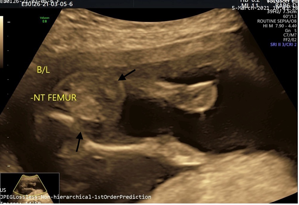
Antenatal ultrasound scan of the fetus at 18-20 weeks at the level of the lower abdomen and pelvis shows nonvisualization of bilateral femur bones.
Figure 2a:
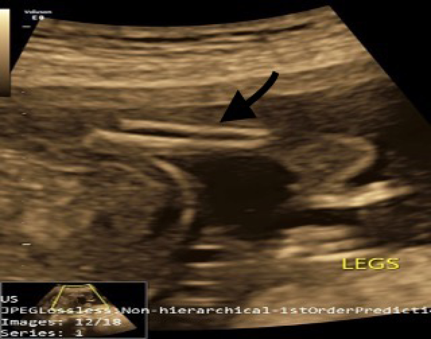
Antenatal ultrasound at 18-20 weeks at the level of the legs shows normal-appearing bilateral tibia and fibula of the right side (arrow).
Figure 2b:
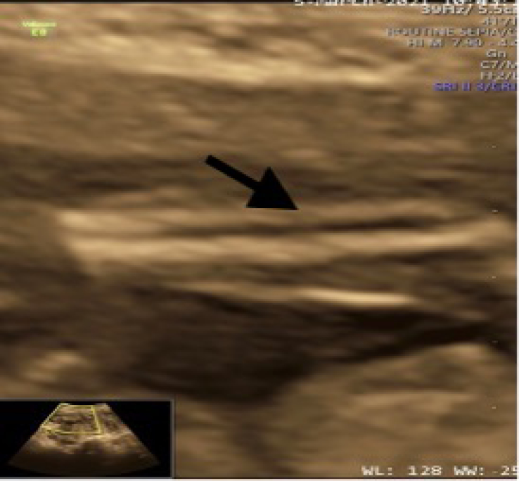
Antenatal ultrasound at 18-20 weeks at the level of the legs shows a normal-appearing bilateral tibia and fibula of the left side (arrow).
Figure 3a:
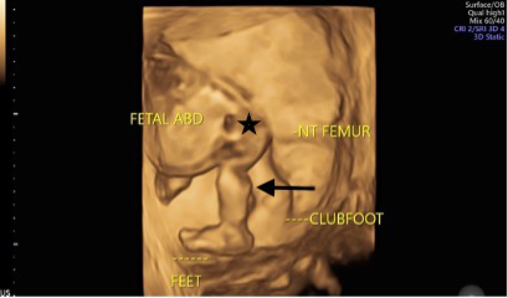
3D ultrasound image at the level of lower limbs clearly demonstrates nonvisualized thigh (asterisk) with a normally developed leg (arrow).
Figure 3b:
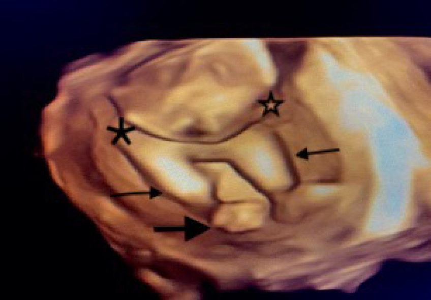
Another 3D ultrasound image at the level of lower limbs demonstrates normally developed both legs (tibia and fibula, denoted by thin arrows) directly articulating with the pelvis (asterisks) with nonvisualization of bilateral thighs (femurs). Note is made of right-sided club foot (bold arrow).
Figure 4:
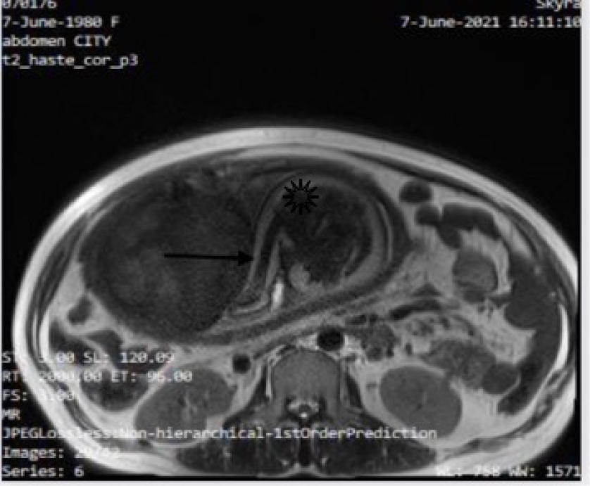
Fetal MRI has done at 32-33 weeks at the level of the fetal pelvis, shows nonvisualization of femur with the tibia and fibula (arrow) in close proximity with the acetabulum (asterisk).

Trio whole-exome sequencing (WES) was performed for the child and both the parents using blood as the tissue. No clinically significant variant was identified that can explain the patient’s phenotype: short long bone, aplasia/hypoplasia involving the skeletal musculature, abnormality of femur morphology, abnormality of the skeletal system. Microarray or karyotyping could not be performed; however, copy number variation in the WES data was looked for.

Figure 5:
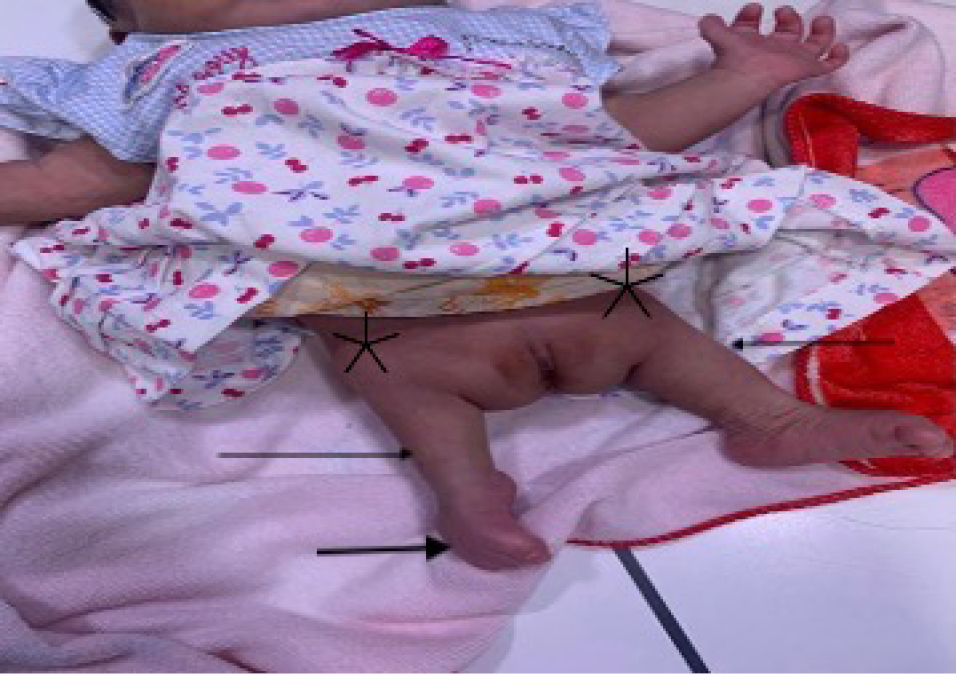
Postnatal clinical photograph of the baby demonstrates bilateral absent thighs with a right-sided club foot. This photograph is exactly the same as shown in the antenatal 3D ultrasound images shown in Figure 3b.
Figure 6:
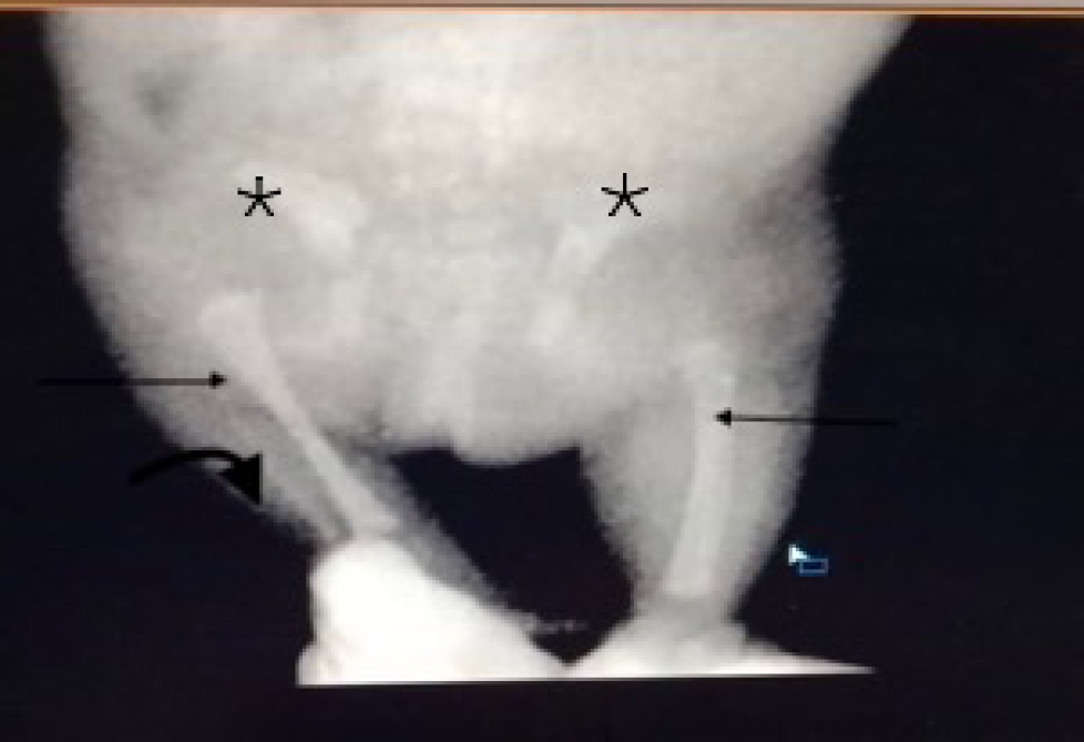
Postnatal X-ray infantogram confirms the prenatal observations. Bilateral femur bones are not seen. Bilateral proximal tibia (arrows) appears femur-like in shape and tibia is seen forming pseudoarthrosis with the acetabulum (asterisks). The acetabulum appears shallow and dysplastic (asterisks). The right fibula is visualized (bold arrow) but the left fibula is not seen clearly (likely non-ossified).

DISCUSSION

Bilateral absence of femur is a rare congenital anomaly and is at the extreme end of the congenital femur deficiency spectrum. These anomalies can occur isolated or in association with other anomalies, e.g., fibular hemimelia (most common), clubfoot, foot array abnormality, and acetabular dysplasia.[1] In our case, the acetabulum was shallow and dysplastic as well as there was a right-sided club foot. No other associated abnormality was seen. The exact cause of congenital femoral deficiencies or absence is not known in most cases. Some of the abnormalities may be caused by a genetic base while others may be caused by an indirect effect of teratogen.[4,5] The classification by Aitken for femoral deficiency has been widely used for many years. This classification is based on the severity of the hip and femur deficiency on radiographic findings. There are four classes in Aitken classification: class A- the presence of femoral head with varus deformity; class B- the presence of femoral head but with delayed ossification; varus, mild acetabular dysplasia, and pseudarthrosis may occur; class C- the absence of femoral head as well as acetabular dysplasia and shortening of femur; and class D- the absence of femoral head with severe dysplastic and severely shortened femur.

The case presented here does not fit into any of the classifications above. There is a complete absence of femur and tibia-fibula complex that appear to be connected to the acetabulum. The proximal part of the tibia appears femur-like in shape.

Trio WES did not reveal any variant which could explain the phenotype. Several variants have been prioritized according to the EVIDENCE, a software that calculates comprehensive scores for phenotypic similarity to the disease and pathogenicity of the variants based on the American College of Medical Genetics guideline.[6] We did not detect any variants associated with the patient’s phenotype, although we paid special attention to all detected variants to select relevant variants to the patient’s clinical information. However, the possibility of missed detection of the pathogenic variants cannot be excluded, because the coverage of the exons may not be complete due to the technical limitations of the method (nondetection of uniparental disomy, low-level mosaicism, copy number variations, and intronic variants). Further whole-genome sequencing may be worthwhile to look for novel genes.

CONCLUSION

Bilateral complete absence of femur is a very rare anomaly. To the best of our knowledge, it is the first reported antenatal case with postnatal follow-up in the world literature. This case is interesting as it does not fit in any category of the femoral deficiency classification which was given by Aitken, and is the most commonly used classification in clinical practice to guide patient management. More cases like these might need a newer classification, which covers this abnormality and to guide better management for these patients.

Declaration of patient consent

Patient’s consent not required as patient’s identity is not disclosed or compromised.

Financial support and sponsorship

Nil.

Conflicts of interest

There are no conflicts of interest.

References

  1. , . Congenital femoral deficiency reconstruction and lengthening surgery. In: , ed. Pediatric Lower Limb Deformities. Switzerland: Springer International Publishing; . p. :361.:425.
    [CrossRef] [Google Scholar]
  2. . Limb deficiencies. In: , ed. Tachdjian’s Pediatric Orthopaedics Vol 1. (5th ed.). Philadelphia, PA: WB. Saunders; . p. :951.:74.
    [Google Scholar]
  3. . Congenital abnormalities of the femur. Arch Dis Child. 1961;36:410-7.
    [CrossRef] [PubMed] [PubMed Central] [Google Scholar]
  4. , , . Congenital femoral deficiency: A rare case report. Int J Contemp Pediatr. 2017;4:1118-21.
    [CrossRef] [Google Scholar]
  5. , , , . The child with a limb deficiency. In: , , eds. Lovell and Winter’s Pediatric Orthopaedics Vol 2. (7th ed.). Philadelphia, PA: Lippincott Williams and Wilkins; . p. :1526.:95.
    [Google Scholar]
  6. , , , , , , et al. Standards and guidelines for the interpretation of sequence variants: A joint consensus recommendation of the American College of Medical Genetics and Genomics and the Association for Molecular Pathology. Genet Med. 2015;17:405-24.
    [CrossRef] [PubMed] [PubMed Central] [Google Scholar]

Fulltext Views
5,240

PDF downloads
4,665
View/Download PDF
Download Citations
BibTeX
RIS
Show Sections