More results...

Generic selectors
Exact matches only
Search in title
Search in content
Post Type Selectors
Filter by Categories
Abdominal Radiology, Original Research
Abdominal Radiology, Pictorial Essay
Anthropology and Dental Radiology, Original Research
Breast Imaging, Case Report
Breast Imaging, Original Research
Breast Imaging, Pictorial Essay
Breast Imaging, Review Article
Cardiopulmonary Imaging, Case Report
Cardiopulmonary Imaging, Case Series
Cardiopulmonary Imaging, Education
Cardiopulmonary Imaging, Original Research
Cardiopulmonary Imaging, Pictorial Essay
Cardiopulmonary Imaging, Review Article
Case Report
Case Report, Breast Imaging
Case Report, Cardiopulmonary Imaging
Case Report, Diagnostic Radiology
Case Report, Neuroradiology/Head and Neck Imaging
Case Report, Vascular and Interventional Radiology
Case Series
Case Series, Diagnostic Radiology
Case Series, Vascular and Interventional Radiology
DENTAL ARTICLE
Dental Radiology, Case Report
Dental Radiology, Case Series
Dental Radiology, Original Research
Diagnostic Radiology, Case Report
Diagnostic Radiology, Case Series
Diagnostic Radiology, Original Research
Diagnostic Radiology, Pictorial Essay
Diagnostic Radiology, Review Article
Editorial
Education, Original Research
Emergency, Original Research
Erratum
Gastrointestinal Imaging, Case Report
Gastrointestinal Imaging, Case Series
Gastrointestinal Imaging, Original Research
Gastrointestinal Imaging, Pictorial Essay
General and Emergency Radiology, Original Research
General and Emergency Radiology, Review Article
Genitourinary and Gynecologic Imaging, Case Report
Genitourinary and Gynecologic Imaging, Original Research
Imaging Science, Original Research
Interventional Radiology, Original Research
Letter to Editor
Musculoskeletal Imaging, Case Report
Musculoskeletal Imaging, Original Article
Musculoskeletal Imaging, Original Research
Musculoskeletal Imaging, Pictorial Essay
Musculoskeletal Imaging, Review Article
Neuroradiology , Review Article
Neuroradiology Head and Neck Imaging, Pictorial Essay
Neuroradiology, Case Report
Neuroradiology, Pictorial Essay
Neuroradiology/Head and Neck Imaging, Case Report
Neuroradiology/Head and Neck Imaging, Case Series
Neuroradiology/Head and Neck Imaging, Original Research
Neuroradiology/Head and Neck Imaging, Review Article
Neuroradiology/Spine Imaging, Original Research
Nuclear Medicine, Case Report
Nuclear Medicine, Original Research
Nuclear Medicine, Pictorial Essay
Original Article
Original Article, Neuroradiology
Original Research
Original Research Article
Original Research, Dental Radiology
Original Research, Descriptive Study
Original Research, Diagnostic Radiology
Original Research, Gastrointestinal Imaging
Original Research, Genitourinary and Gynecologic Imaging
Original Research, Musculoskeletal Imaging
Original Research, Neuroradiology/Head and Neck Imaging
Original Research, Nuclear Medicine
Original Research, Vascular and Interventional Radiology
Pediatric Imaging, Case Report
Pediatric Imaging, Pictorial Essay
PICTORIAL ESSAY
PICTORIAL REVIEW
Radiologic-Pathologic Correlation
RADIOLOGICAL-PATHOLOGICAL CORRELATION
Radiology Business, Original Research
Research Article
Review Article
Review Article, Diagnostic Radiology
Review Article, Gastrointestinal imaging
Review Article, Musculoskeletal
Review Article, Vascular and Interventional Radiology
Technical Innovation
Technical Innovation, Gastrointestinal Imaging
Ultrasound, Case Report
Ultrasound, Original Research
Ultrasound, Review Article
Vascular and International Radiology, Case Report
Vascular and Interventional Radiology, Case Report
Vascular and Interventional Radiology, Case Series
Vascular and Interventional Radiology, Original Research
Vascular and Interventional Radiology, Pictorial Essay
Vascular and Interventional Radiology, Review Article
Vascular and Interventional Radiology, Short Communication
Vascular and Interventional, Original Research
View/Download PDF

Translate this page into:

CASE REPORT
2012
:2;
1
doi:
10.4103/2156-7514.92363

Arachnoid Pit and Extensive Sinus Pnematization as the Cause of Spontaneous Lateral Intrasphenoidal Encephalocele

Department of Radiology, King Saud Medical City, Riyadh, Kingdom of Saudi Arabia
Department of Otolaryngology, King Saud Medical City, Riyadh, Kingdom of Saudi Arabia
Address for correspondence: Dr. Bandar Al Qahtani, King Saud Medical City, Riyadh, Saudi Arabia. E-mail: Khamsi20@yahoo.com
Licence

This is an open-access article distributed under the terms of the Creative Commons Attribution License, which permits unrestricted use, distribution, and reproduction in any medium, provided the original author and source are credited.

Disclaimer:
This article was originally published by Medknow Publications & Media Pvt Ltd and was migrated to Scientific Scholar after the change of Publisher.

Abstract

Lateral sphenoid encephalocele, especially within the lateral aspect of the sphenoid sinus, when the sphenoid sinus has pneumatized extensively into the pterygoid recess, are considered exceedingly rare. We report a rare case of lateral intrasphenoidal encephalocele with spontaneous cerebral spinal fluid (CSF) rhinorrhea. Computed tomography demonstrated bilateral arachnoid pit, extensive sphenoid sinus pneumatization, and a defect in the superior wall of the left lateral recess of the sphenoid sinus. Magnetic resonance imaging demonstrated anteromedial temporal lobe herniating through the bony defect.

Keywords

Arachnoid pit
lateral recess
sphenoid sinus
spontaneous CSF-leaks
spontaneous encephalocele

INTRODUCTION

An encephalocele is the protrusion of intracranial contents, including brain matter and meninges through a defect in the cranium or skull base. Abiko et al. noted two types of transsphenoidal meningoencephaloceles: The intrasphenoidal and the true transsphenoidal. Intrasphenoidal encephaloceles describe those extending into the sphenoid sinus but confined by its floor.[1] Intrasphenoidal encephaloceles are subdivided by location into medial perisellar and lateral sphenoid recess types.[2] Perisellar encephaloceles within the sphenoid sinus are considered more common, whereas basal encephaloceles limited to the lateral sphenoid sinus are rare. Temporal lobe encephalocele protruding through a middle fossa defect is rare and has few distinctive features, so is difficult to identify clinically.[3] In a study of 15 patients, the mean delay between onset of symptoms such as CSF rhinorrhea and the diagnosis was 13.1 months.[4] The clinical manifestations included CSF rhinorrhea, chronic headache, and a history of meningitis.[5]

Arachnoid pits are smooth, lobulated bony defects that are related to aberrant arachnoid granulations. CSF pulsation and its pressure also play an important role in the creation of these defects. This role of the CSF is evident from the pattern of the bony remodeling, with outward concave orientation of adjacent bones.[6]

CASE REPORT

A 40-year-old male patient presented to the ENT clinic with watery nasal discharge that had persisted for 10 months. This patient had a history of headache associated with nasal obstruction that had been diagnosed by another doctor as hypertrophied turbinate and was advised surgery. But the patient refused to have surgery.

For the past 2 years, the patient had started to experience anosmia. Though gradual in onset and not associated with any watery discharge initially, it was found to be associated with nasal obstruction. For the past 10 months he was having watery nasal discharge which increased when he bent forward or coughed. The discharge trickled into the nasopharynx, though he could not taste any of this discharge it was associated with headache. The patient has no past history of any nasal trauma or surgery and no sign of meningitis.

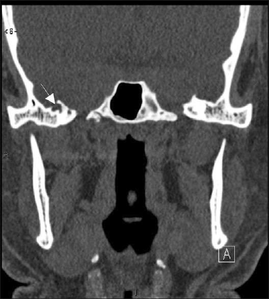
Computed tomography of brain and paranasal sinuses demonstrated bilateral arachnoid pit lateral to sphenoid sinus and osseous erosion lateral to the foramen rotundum, lateral sphenoid sinus recess pneumatization, and an approximately 9-mm size defect in the left anterolateral wall of the sphenoid sinus just lateral to foramen rotundum, with a soft tissue density mass filling the adjacent part of the sphenoid sinus [Figures 14]. Magnetic resonance imaging demonstrated left temporal lobe herniation through a wide defect in the left inferiolateral wall of sphenoid sinus surrounded by a cerebrospinal fluid filled meningeal sac which contains the herniated brain parenchyma [Figures 5, 6].

Figure 1
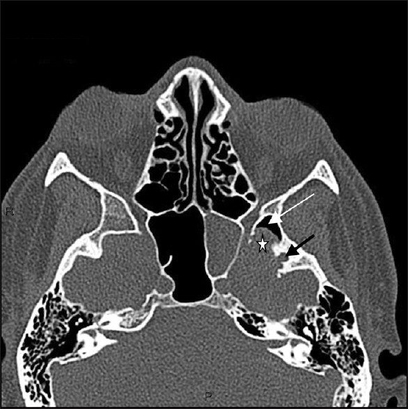
Axial CT scan demonstrates arachnoid pit lateral to sphenoid sinus (black arrow). An approximately 9-mm size defect in the left superolateral wall of the sphenoid sinus (white asterisk) just lateral to foramen rotundum, with a soft tissue density mass filling the adjacent part of the sphenoid sinus. Notice the pneumatization of most far lateral recesses of sphenoid sinus (white arrow).
Figure 2
Coronal-reformatted CT scan demonstrates osseous erosion lateral to the left foramen rotundum and lateral sphenoid sinus recess pnematization. An approximately 9-mm size defect in the left superolateral wall of the sphenoid sinus (white asterisk) just lateral to foramen rotundum (black arrow), with a soft tissue density mass filling the sphenoid sinus (curved arrow).
Figure 3
Arachnoid pit lateral to the right sphenoid sinus is noted (arrow).
Figure 4
Coronal-reformatted CT scan demonstrates arachnoid pits (white arrows) lateral to sphenoid sinus. A soft tissue density mass filling the sphenoid sinus is noted (black arrow).
Figure 5
Coronal IR-FSE T2-weighted image demonstrates left temporal lobe herniation through a wide defect in the left inferiolateral wall of sphenoid sinus surrounded by a cerebrospinal fluid filled meningeal sac.
Figure 6
Axial T2-weighted spin-echo imaging demonstrate the left temporal lobe herniation through a wide defect in the left inferiolateral wall of sphenoid sinus surrounded by a cerebrospinal fluid filled meningeal sac.

DISCUSSION

The most common site of CSF leakage is through the floor of the anterior fossa, which communicates with the ethmoid or frontal sinuses, or with the nasal fossa. The sphenoid sinus is rarely implicated as a source of spontaneous CSF fistula.[6] Communication with the middle cranial fossa and CSF fistula is more likely if the sphenoid sinus is laterally pneumatized.[6]

CSF pressures and the hydrostatic pulsative forces may lead to the development of pit holes on the middle fossa at the sites of arachnoid villi with herniation of dura/arachnoid or brain tissue.[6] If such defects are located over the underlying lateral extension of the sphenoid sinus, encephalocele can develop and lead to CSF leakage into the sinus. Therefore, erosion of bone by arachnoid granulations is only significant when it affects a pneumatized part of the skull.[7]

In a study on a subgroup of 11 patients with spontaneous sphenoid sinus fistulas, the authors concluded that the constellation of empty sella, arachnoid pits, and extensive pneumatization plays a role in the etiopathogenesis of sphenoid sinus fistulas.[67]

A persisting Sternberg's canal should be considered the source of spontaneous CSF-leaks with or without meningoencephaloceles in sphenoid sinuses with extensive lateral pneumatisation, especially when located laterally and below the maxillary nerve.[8] However, in another study, the conclusion was Sternberg's canal, as historically defined, is not nearly as prevalent as previously reported. Furthermore, the presence of arachnoid pits in all sphenoid CSF leaks and the predominant leak location lateral to the sites of fusion of ossification centers suggests that the leaks are acquired. Contributing factors may include arachnoid pits/weaknesses in the skull base and intracranial hypertension.[9]

The leaks related to arachnoid granulations along the lateral sphenoid were significantly larger than those along the ethmoid and midline sphenoid osteodural defects with an average size of 3.5 ± 0.80 mm.[7] In this case, the defect was approximately 9 mm and considered one of the largest acquired defect.

Plain high-resolution CT can give indirect evidence of the CSF fistula by revealing bone defect and opacification of sinuses or air cells. MR cisternography can non-invasively reveal CSF leakage in multiple planes without the disadvantage of ionizing radiation. On the T2-weighted spin-echo sequence, the bright signal emanating from the CSF column is well visualized against the paranasal sinuses background of air.[10] MRI is essential to confirm the extension of the lesion and any associated abnormalities. MRI is able to provide mulitplanar images of the encephalocele that is useful for both diagnosis and surgical planning.

Without repair of the defect, patients with CSF leakage have increased risk of developing ascending infection. Landreneau et al. in a study of four patients, conclude that fistulas involving lateral extension of the sphenoid sinus require a transcranial approach for direct visualization and obliteration of the defect, whereas fistulas involving the central portion of the sinus may be successfully obliterated transsphenoidally.[11] Transcranial multilayered closure of the defect is safe and reliable, particularly for large CSF fistula at the far lateral sphenoid sinus.[3] The patient refused the transcranial approach.

Endoscopic management is technically challenging, nevertheless its advantages are a good view of the surgical field while being less traumatic than transcranial approaches.[8]

Determination of the ideal approach is based on various factors, including the degree of lateral sphenoid pneumatization, location, and size of the meningoencephalocele, and ability to perform an adequate skull base repair through a given exposure.[5]

Source of Support: Nil

Conflict of Interest: None declared.

REFERENCES

  1. , , , . Intrasphenoidal encephalocele. Report of a case. Neurosurgery. 1988;22:933-6.
    [Google Scholar]
  2. , , , . Sphenoid encephaloceles: disease management and identification of lesions within the lateral recess of the sphenoid sinus. Laryngoscope. 2002;112:1800-5.
    [Google Scholar]
  3. , , , , , , . Spontaneous Cerebrospinal Fluid Rhinorrhea Associated With a Far Lateral Temporal Encephalocele. Neurol Med Chir (Tokyo). 2010;50:243-5.
    [Google Scholar]
  4. , , , , , , . Endonasal endoscopic repair of Sternberg's canal cerebrospinal fluid leaks. Laryngoscope. 2007;117:345-9.
    [Google Scholar]
  5. , , , , , , . Endoscopic management of spontaneous meningoencephalocele of the lateral sphenoid sinus. J Neurosurg. 2010;112:1070-7.
    [Google Scholar]
  6. , , , , , . A retrospective analysis of spontaneous sphenoid sinus fistula: MR and CT findings. AJNR Am J Neuroradiol. 2000;21:337-42.
    [Google Scholar]
  7. , , , , . Nontraumatic skull base defects with spontaneous csf rhinorrhea and arachnoid herniation: Imaging findings and correlation with endoscopic sinus surgery in 27 patient. AJNR Am J Neuroradiol. 2008;29:542-9.
    [Google Scholar]
  8. , , . Spontaneous CSF-leaks and meningoencephaloceles in sphenoid sinus by persisting Sternberg's canal. Rhinology. 2009;47:369-74.
    [Google Scholar]
  9. , , , , . Sternberg's canal: Fact or fiction? Am J Rhinol Allergy. 2009;23:167-71.
    [Google Scholar]
  10. , , , . Evaluation of high-resolution CT and MR cisternography in the diagnosis of cerebrospinal fluid fistula. AJNR Am J Neuroradiol. 1998;19:633-9.
    [Google Scholar]
  11. , , , . Surgical treatment of cerebrospinal fluid fistula involving lateral extension of the sphe noid sinus. Neurosurgery. 1998;42:1101-5.
    [Google Scholar]

Fulltext Views
10,971

PDF downloads
3,137
View/Download PDF
Download Citations
BibTeX
RIS
Show Sections