More results...

Generic selectors
Exact matches only
Search in title
Search in content
Post Type Selectors
Filter by Categories
Abdominal Radiology, Original Research
Abdominal Radiology, Pictorial Essay
Anthropology and Dental Radiology, Original Research
Breast Imaging, Case Report
Breast Imaging, Original Research
Breast Imaging, Pictorial Essay
Breast Imaging, Review Article
Cardiopulmonary Imaging, Case Report
Cardiopulmonary Imaging, Case Series
Cardiopulmonary Imaging, Education
Cardiopulmonary Imaging, Original Research
Cardiopulmonary Imaging, Pictorial Essay
Cardiopulmonary Imaging, Review Article
Case Report
Case Report, Breast Imaging
Case Report, Cardiopulmonary Imaging
Case Report, Diagnostic Radiology
Case Report, Neuroradiology/Head and Neck Imaging
Case Report, Vascular and Interventional Radiology
Case Series
Case Series, Diagnostic Radiology
Case Series, Vascular and Interventional Radiology
DENTAL ARTICLE
Dental Radiology, Case Report
Dental Radiology, Case Series
Dental Radiology, Original Research
Diagnostic Radiology, Case Report
Diagnostic Radiology, Case Series
Diagnostic Radiology, Original Research
Diagnostic Radiology, Pictorial Essay
Diagnostic Radiology, Review Article
Editorial
Education, Original Research
Emergency, Original Research
Erratum
Gastrointestinal Imaging, Case Report
Gastrointestinal Imaging, Case Series
Gastrointestinal Imaging, Original Research
Gastrointestinal Imaging, Pictorial Essay
General and Emergency Radiology, Original Research
General and Emergency Radiology, Review Article
Genitourinary and Gynecologic Imaging, Case Report
Genitourinary and Gynecologic Imaging, Original Research
Imaging Science, Original Research
Interventional Radiology, Original Research
Letter to Editor
Musculoskeletal Imaging, Case Report
Musculoskeletal Imaging, Original Article
Musculoskeletal Imaging, Original Research
Musculoskeletal Imaging, Pictorial Essay
Musculoskeletal Imaging, Review Article
Neuroradiology , Review Article
Neuroradiology Head and Neck Imaging, Pictorial Essay
Neuroradiology, Case Report
Neuroradiology, Pictorial Essay
Neuroradiology/Head and Neck Imaging, Case Report
Neuroradiology/Head and Neck Imaging, Case Series
Neuroradiology/Head and Neck Imaging, Original Research
Neuroradiology/Head and Neck Imaging, Review Article
Neuroradiology/Spine Imaging, Original Research
Nuclear Medicine, Case Report
Nuclear Medicine, Original Research
Nuclear Medicine, Pictorial Essay
Original Article
Original Article, Neuroradiology
Original Research
Original Research Article
Original Research, Dental Radiology
Original Research, Descriptive Study
Original Research, Diagnostic Radiology
Original Research, Gastrointestinal Imaging
Original Research, Genitourinary and Gynecologic Imaging
Original Research, Musculoskeletal Imaging
Original Research, Neuroradiology/Head and Neck Imaging
Original Research, Nuclear Medicine
Original Research, Vascular and Interventional Radiology
Pediatric Imaging, Case Report
Pediatric Imaging, Pictorial Essay
PICTORIAL ESSAY
PICTORIAL REVIEW
Radiologic-Pathologic Correlation
RADIOLOGICAL-PATHOLOGICAL CORRELATION
Radiology Business, Original Research
Research Article
Review Article
Review Article, Diagnostic Radiology
Review Article, Gastrointestinal imaging
Review Article, Musculoskeletal
Review Article, Vascular and Interventional Radiology
Technical Innovation
Technical Innovation, Gastrointestinal Imaging
Ultrasound, Case Report
Ultrasound, Original Research
Ultrasound, Review Article
Vascular and International Radiology, Case Report
Vascular and Interventional Radiology, Case Report
Vascular and Interventional Radiology, Case Series
Vascular and Interventional Radiology, Original Research
Vascular and Interventional Radiology, Pictorial Essay
Vascular and Interventional Radiology, Review Article
Vascular and Interventional Radiology, Short Communication
Vascular and Interventional, Original Research
View/Download PDF

Translate this page into:

RADIOLOGICAL-PATHOLOGICAL CORRELATION
2011
:1;
65
doi:
10.4103/2156-7514.92187

Gardner's Syndrome

Department of Oral Medicine and Radiology, Institute of Dental Studies and Technologies, Kadrabad, Modinagar, Ghaziabad, Uttar Pradesh, India
Department of Oral Medicine and Radiology, Kles V.K Institute of Dental Sciences, Nehru Nagar, Belgaum, Karnataka, India
Department of Conservative Dentistry and Endodontics, Institute of Dental Studies and Technologies, Kadrabad, Modinagar, Ghaziabad, Uttar Pradesh, India
Address for correspondence: Dr. Sapna Panjwani, Department of Oral Medicine and Radiology, Institute of Dental Studies and Technologies, Kadrabad, Modinagar, Ghaziabad, Uttar Pradesh, India. E-mail: dr.sapna28@gmail.com
Licence

This is an open-access article distributed under the terms of the Creative Commons Attribution License, which permits unrestricted use, distribution, and reproduction in any medium, provided the original author and source are credited.

Disclaimer:
This article was originally published by Medknow Publications & Media Pvt Ltd and was migrated to Scientific Scholar after the change of Publisher.

Abstract

Gardner's syndrome is an autosomal dominant disease and is a subtype of familial adenomatous polyposis. It is characterized by adenomatous intestinal polyps, multiple osteomas in the skull, maxillae, mandible, and multiple cutaneous and subcutaneous masses (epidermoids and desmoid). Intestinal polyps, if not treated, have 100% chance of becoming malignant. We report a case of a 25-year-old female patient with Gardner's syndrome, with clinical manifestations including impacted supernumerary teeth, odontomes, sebaceous cyst on the scalp, and osteomas. It is important for the general dental practitioners to be aware of the clinical and radiological characteristics of Gardner's syndrome.

Keywords

Familial intestinal polyposis
gardner's syndrome
impacted teeth
osteoma

INTRODUCTION

In 1951, Eldon J. Gardner (1909 – 1989), a college teacher of genetics, first described the syndrome, as a rare autosomal, dominant, inherited disorder with a high degree of penetrance, characterized by a triad of multiple osteomas, colonic polyposis, and mesenchymal tumors of the skin and soft tissues.[1] It is a variant of the familial adenomatous polyposis syndrome, known to be caused by the mutation in the Adenomatous Polyposis Coli (APC) gene located on chromosome 5q21 (band q21 on chromosome 5).[2]

The incidence of Gardner's syndrome in the general population has been estimated as one in 14,025 live births.[3] Dental anomalies including odontoma and multiple unerupted supernumerary and permanent teeth have been reported.[4] Dental anamolies are seen in 30% of patients with Gardner's syndrome.

Presence of osteomas is required to make the diagnosis of Gardner's syndrome. The mandible is the most common location, however, osteomas may occur in the skull, paranasal sinuses, and the long bones.[5] Classic location of osteomas in mandible is the mandibular angle and inferior surface. Osteomas precede the clinical and radiographic evidence of colonic polyposis or Gardner's syndrome, therefore, may be insightful markers for the disease. It is also proposed that radiography of the jaws may serve as a valuable tool for the early detection of carriers of Gardner's syndrome.

The epidermoid cyst is the most common cutaneous finding in Gardner's syndrome and is present in multiple forms in 50 – 60% of the patients and may occur on the face, extremities, and / or the scalp.[6]

Desmoid tumor is benign fibrous proliferation that occurs under the skin predominantly in the abdominal wall. This tumors has high rate of recurrence after excision. It is seen in 8.6% of cases of Gardner syndrome.

Our patient was a 25-year-old female with a history of multiple swellings in the lower jaw that had developed over a period of 11 years. The swelling was not associated with pain, discharge, or numbness. An extraoral examination revealed gross facial asymmetry, because of a diffuse swelling on the left side of the mandible measuring about 3 cm × 4 cm in size, extending from the parasymphysis to the posterior border of the mandible [Figure 1]. Similar swellings, three in number, were present on the inferior aspect on the right side of the mandible. The skin over the swellings appeared normal. On palpation, all the swellings were bony hard in consistency, non-tender, and were fixed to the mandible.

Figure 1
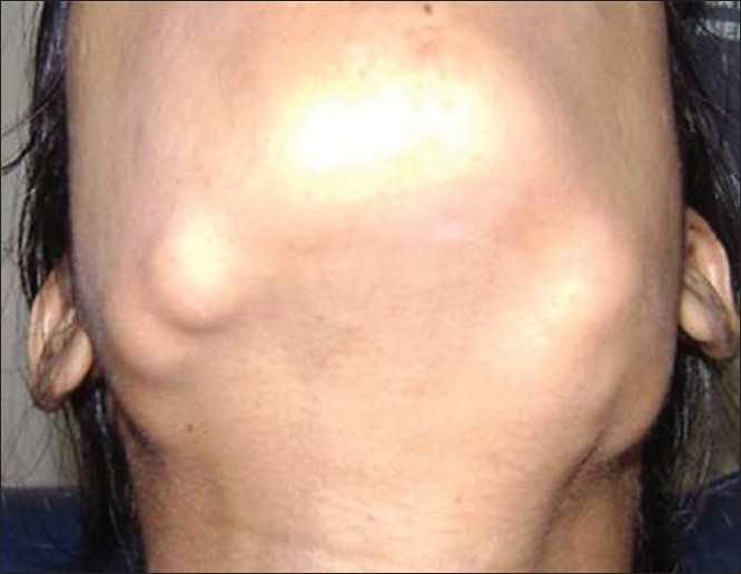
Extraoral osteomas. A 25-year-old female patient with diffuse bilateral swellings on the lower third of face along the mandibular border.

Intraoral examination revealed a diffuse swelling, obliterating the right buccal vestibule, measuring about 2 cm × 2 cm in size, and extending from the mesial aspect of tooth 46 to the distal aspect of tooth 48 anteroposteriorly. The overlying mucosa was intact, with no ulceration or sinus discharge. On palpation, all the swellings were bony hard in consistency and non-tender. There were clinically missing teeth 21 and 23 (no history of extraction revealed) and over-retained tooth 63. Other dental findings were a carious tooth 47. Patient had a small swelling measuring about 1 cm × 1 cm on the scalp, which was fluctuant and cystic in nature (epidermoid / sebaceous cyst).

The patient did not have any symptoms of rectal bleeding, cramping abdominal pain, weight loss, diarrhea or small bowel or colonic obstruction, and was reluctant to get a colonoscopy done to rule out the presence of polyps in the intestine, colon, and rectum. The clinical and radiographic findings, of osteomas, odontomes, impacted teeth, and sebaceous cyst suggested a diagnosis of Gardner's syndrome.

Our patient's need was aesthetic improvement, for which the osteomas were surgically excised and sent for histopathological examination. The patient was followed up for one year and has remained asymptomatic, with no relapse of the lesions [Figure 2].

Figure 2
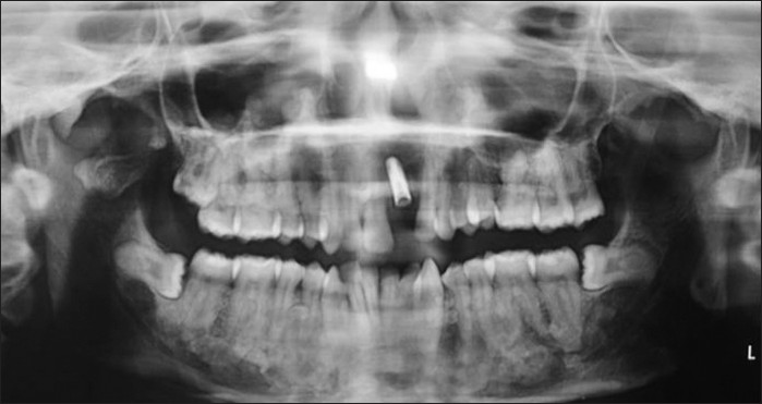
Postoperative radiograph after removal of osteomas, impacted teeth, and implant placement, with respect to tooth 21.

RADIOLOGICAL FEATURES

The patient underwent maxillary and mandibular cross-sectional occlusal radiograph, orthopantomograph (OPG), and posteroanterior radiograph [Figure 3]. The OPG revealed impacted teeth 21 and 23 and homogenous radiopaque masses over the right and left inferior aspect of the mandible, suggestive of osteomas [Figure 4]. Also, multiple small, homogeneous radiopaque masses surrounded by a radiolucent halo could be seen throughout the maxilla and mandible, suggestive of complex odontomes. Diffuse sclerotic masses were present throughout the body of the mandible giving it a mottled appearance. Impacted teeth 21 and 23 and the complex odontomes in the near vicinity were better visualized in the maxillary cross-sectional occlusal radiograph [Figure 5].

Figure 3
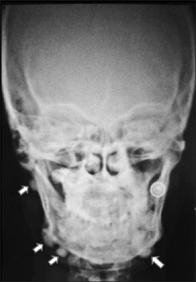
Posteroanterior radiograph shows extra-oral osteomas along the mandible (white arrows).
Figure 4
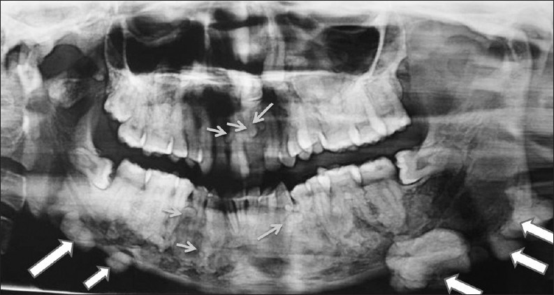
OPG demonstrates impacted teeth 21 and 23, homogenous radiopaque masses over the right and left inferior aspect of the mandible suggestive of osteomas (large arrows). Also multiple small, homogeneous radiopaque masses surrounded by a radiolucent halo could be seen throughout the maxilla and mandible suggestive of complex odontomes (small arrows).Diffuse sclerotic masses are present throughout the body of mandible giving it a mottled appearance
Figure 5
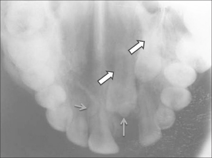
Maxillary occlusal radiograph shows impacted teeth 21, 23 (large arrows) and odontomes (small arrows) in the near vicinity.

PATHOLOGICAL FEATURES

The histopathological report revealed the presence of compact lamellar bone with a haversian canal, lacunae, histiocytes, and reversal and resting lines suggestive of osteoma [Figure 6].

Figure 6
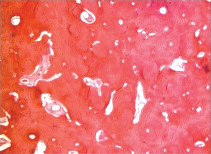
Photomicrograph shows the presence of compact lamellar bone with haversian canal, lacunae, histiocytes and reversal and resting lines suggestive of osteoma. (Hematoxylin-Eosin stain 10×).

DISCUSSION

Gardner's syndrome is a disease that affects multiple systems. It is an autosomal dominant condition with an incidence between 1 in 8300 and 1 in 14,025 live births, affecting both genders equally, with a uniform worldwide distribution.[7] Symptoms are usually present by the end of the second decade of life, but they may present anytime between two months and 70 years. As the clinical and radiological features in the maxillofacial region, such as osteomas, skin cysts, atypical skin pigmentation, and abnormal dental findings or radiographical lesions can precede frequent asymptomatic intestinal polyps for many years, dental practitioners and radiologists should be familiar with the manifestations of this syndrome.

Unlike most patients with Gardner's syndrome who present with gastrointestinal symptoms such as bloody diarrhea and pain in the abdomen, our patient presented with dental and facial cosmetic issues. Our case showed the classical clinical features associated with the syndrome: Multiple osteomas and odontomes, epidermoid cyst, and impacted teeth.

Skeletal abnormalities appear in approximately 90% of the patients with Gardner's syndrome, and osteoma is considered to be the most common.[8] Supernumerary teeth, compound odontomes, and / or impacted teeth are seen in 30% of the patients with this disease.

The gastrointestinal manifestations of Gardner's syndrome include colonic adenomatous polyps (tubular, villous, tubulovillous), gastric and small intestinal adenomatous polyps (12% of the patients), and periampullary carcinomas (2% of the patients).[9] In our patient there were no gastrointestinal symtoms and she had refused colonoscopy.

Patients may also suffer from congenital hypertrophy of the retinal pigment epithelium,[7] although the finding was absent in our case.

Panoramic radiography by the dentist can be useful for early detection of Gardner's syndrome, because the components of this entity, like osteoma, odontoma, supernumerary, and impacted teeth, can be detected. However, panoramic radiography is of limited value in the evaluation, localization, and extension of the tumor mass, considering the superimposition of the bony structures and the two-dimensional image.[10]

The maxillofacial features of the syndrome can appear many years before the intestinal polyposis.[10] Therefore, dental professionals should be familiar with the significance of the syndrome as a pre-cancerous condition. Dental management includes removal of the impacted teeth, cysts of the jaw or face, as well as, resection of osteomas for functional or cosmetic reasons. Tooth extraction can be difficult because of generalized increased alveolar bone density and complete absence of periodontal space caused by hypercementosis.[7]

CONCLUSION

Osteomas of the skull, angle of the mandible and its inferior surface are classic bony features, in addition to intestinal ployps, in patients with Gardner syndrome. A practicing dental surgeon should be aware of this entity in which intestinal polyps have 100% chance of malignant transformation.

ACKNOWLEDGEMENT

The authors would like to thank Dr Zameera Naik for her contribution to this manuscript.

Source of Support: Nil

Conflict of Interest: None declared.

REFERENCES

  1. , . “A genetic and clinical study of intestinal polyposis, a predisposing factor for carcinoma of the colon and rectum”. Am J Hum Genet. 1951;3:167-76.
    [Google Scholar]
  2. , , , , . Diagnosis and treatment of Gardner's syndrome with gastric polyposis: A case report and review of literature. World J Gastroenterol. 2008;14:2121-3.
    [Google Scholar]
  3. , , , , , . A Genetic study of Gardner syndrome and Congenital Hypertrophy of the Retinal Pigment Epithelium. Am J Hum Genet. 1988;42:290-6.
    [Google Scholar]
  4. , . Gardner's syndrome: A case report. Br J Oral Surg. 1970;8:51-7.
    [Google Scholar]
  5. , , , . Syndromes of the Head and Neck (2nd ed). New York: McGraw-Hill; . p. :324-8.
  6. , , , . Cutaneous symptoms of Gardner's syndrome. J Eur Acad Dermatol Venereol. 1999;12:80-1.
    [Google Scholar]
  7. , , , , , . The role of Cone beam CT in the evaluation and management of a family with Gardners syndrome. J Craniomaxillofac Surg. 2009;37:461-8.
    [Google Scholar]
  8. , , , , . Osteomas of the condyle associated with Gardner's syndrome causing limited mandibular movement. J Oral Maxillofac Surg. 1999;57:1004-9.
    [Google Scholar]
  9. , , , , , , . Gardner's syndrome: A case report and review of the literature. World J Gastroenterol. 2005;11:5408-11.
    [Google Scholar]
  10. , , , , , , . Radiographic assessment of Gardners syndrome. Dentomaxillofac Radiol. 2007;36:121-4.
    [Google Scholar]
Show Sections