More results...

Generic selectors
Exact matches only
Search in title
Search in content
Post Type Selectors
Filter by Categories
Abdominal Radiology, Original Research
Abdominal Radiology, Pictorial Essay
Anthropology and Dental Radiology, Original Research
Breast Imaging, Case Report
Breast Imaging, Original Research
Breast Imaging, Pictorial Essay
Breast Imaging, Review Article
Cardiopulmonary Imaging, Case Report
Cardiopulmonary Imaging, Case Series
Cardiopulmonary Imaging, Education
Cardiopulmonary Imaging, Original Research
Cardiopulmonary Imaging, Pictorial Essay
Cardiopulmonary Imaging, Review Article
Case Report
Case Report, Breast Imaging
Case Report, Cardiopulmonary Imaging
Case Report, Diagnostic Radiology
Case Report, Gastrointestinal Imaging
Case Report, Genitourinary and Gynecologic Imaging
Case Report, Neuroradiology/Head and Neck Imaging
Case Report, Vascular and Interventional Radiology
Case Series
Case Series, Abdominal Radiology
Case Series, Diagnostic Radiology
Case Series, Musculoskeletal Imaging
Case Series, Vascular and Interventional Radiology
DENTAL ARTICLE
Dental Radiology, Case Report
Dental Radiology, Case Series
Dental Radiology, Original Research
Diagnostic Radiology, Case Report
Diagnostic Radiology, Case Series
Diagnostic Radiology, Original Research
Diagnostic Radiology, Pictorial Essay
Diagnostic Radiology, Review Article
Editorial
Education
Education, Diagnostic Radiology
Education, Imaging science
Education, Original Research
Emergency, Original Research
Erratum
Gastrointestinal Imaging, Case Report
Gastrointestinal Imaging, Case Series
Gastrointestinal Imaging, Original Research
Gastrointestinal Imaging, Pictorial Essay
General and Emergency Radiology, Original Research
General and Emergency Radiology, Review Article
Genitourinary and Gynecologic Imaging, Case Report
Genitourinary and Gynecologic Imaging, Original Research
Imaging Science, Original Research
Interventional Radiology, Original Research
Letter to Editor
Musculoskeletal Imaging, Case Report
Musculoskeletal Imaging, Original Article
Musculoskeletal Imaging, Original Research
Musculoskeletal Imaging, Pictorial Essay
Musculoskeletal Imaging, Review Article
Neuroradiology , Review Article
Neuroradiology Head and Neck Imaging, Pictorial Essay
Neuroradiology, Case Report
Neuroradiology, Pictorial Essay
Neuroradiology/Head and Neck Imaging, Case Report
Neuroradiology/Head and Neck Imaging, Case Series
Neuroradiology/Head and Neck Imaging, Original Research
Neuroradiology/Head and Neck Imaging, Review Article
Neuroradiology/Spine Imaging, Original Research
Notice of Retraction
Nuclear Medicine, Case Report
Nuclear Medicine, Original Research
Nuclear Medicine, Pictorial Essay
Original Article
Original Article, Neuroradiology
Original Research
Original Research Article
Original Research, Cardiopulmonary Imaging
Original Research, Dental Radiology
Original Research, Descriptive Study
Original Research, Diagnostic Radiology
Original Research, Gastrointestinal Imaging
Original Research, Genitourinary and Gynecologic Imaging
Original Research, Musculoskeletal Imaging
Original Research, Neuroradiology/Head and Neck Imaging
Original Research, Nuclear Medicine
Original Research, Vascular and Interventional Radiology
Pediatric Imaging, Case Report
Pediatric Imaging, Pictorial Essay
PICTORIAL ESSAY
Pictorial essay, Musculoskeletal Imaging
Pictorial essay, Neuroradiology/Head and Neck Imaging
PICTORIAL REVIEW
Radiologic-Pathologic Correlation
RADIOLOGICAL-PATHOLOGICAL CORRELATION
Radiology Business, Original Research
Research Article
Review Article
Review Article, Diagnostic Radiology
Review Article, Gastrointestinal imaging
Review Article, Musculoskeletal
Review Article, Musculoskeletal Imaging
Review Article, Vascular and Interventional Radiology
Technical Innovation
Technical Innovation, Cardiopulmonary Imaging
Technical Innovation, Gastrointestinal Imaging
Ultrasound, Case Report
Ultrasound, Original Research
Ultrasound, Review Article
Vascular and International Radiology, Case Report
Vascular and Interventional Radiology, Case Report
Vascular and Interventional Radiology, Case Series
Vascular and Interventional Radiology, Original Research
Vascular and Interventional Radiology, Pictorial Essay
Vascular and Interventional Radiology, Review Article
Vascular and Interventional Radiology, Short Communication
Vascular and Interventional, Original Research
View/Download PDF

Translate this page into:

CASE REPORT
2014
:4;
60
doi:
10.4103/2156-7514.143732

Aneurysmal Bone Cyst of the Calcaneus

Department of Radiology, Ankara Keçiören Training and Research Hospital, Ankara, Turkey
Department of Radiology, Ankara Numune Training and Research Hospital, TR-06100, Ankara, Turkey
Department of Radiology, Diskapi Yildirim Beyazit Training and Research Hospital, Ankara, Turkey

Address for correspondence: Dr. Veysel Kaplanoğlu, Department of Radiology, Ankara Keçiören Training and Research Hospital, TR-06100, Ankara, Turkey. E-mail: hatice.altnkaynak@yahoo.com.tr

Licence

This is an open-access article distributed under the terms of the Creative Commons Attribution License, which permits unrestricted use, distribution, and reproduction in any medium, provided the original author and source are credited.

Disclaimer:
This article was originally published by Medknow Publications & Media Pvt Ltd and was migrated to Scientific Scholar after the change of Publisher.

Abstract

Aneurysmal bone cysts (ABCs) are benign, non-neoplastic, expansile, vascular, locally destructive lesions. The lesion may arise de novo (65%) or secondarily (35%) in pre-existing benign or malignant lesions (giant cell tumor, osteoblastoma, chondroblastoma, angioma, and others). The calcaneus is a rare localization for ABC, comprising only 1.6% of the cases. In this paper, we present a case of a female patient with a 3-month history of heel pain that got worse and was accompanied by swelling and difficulty in walking. The magnetic resonance images of the postero-lateral calcaneus showed a contrast-enhanced cystic lesion located in the medullary cavity; exophytic portion of the tumor extended into the soft tissue causing distinctive cortical thinning. Heterogeneous hyperintense septae formations and blood level components were also detected. After correlation with pathology results, the lesion was diagnosed as an ABC. Since an ABC of the calcaneus is a rarely seen phenomenon, we present the radiologic findings in this case and a review of the literature.

Keywords

Aneurysmal bone cyst
benign bone lesions
calcaneus

INTRODUCTION

Aneurysmal bone cysts (ABCs) account for 1% of primary bone tumors. They are more common in women than in men (1.04:1).[1] Eighty-five percent of the cases are younger than 20 years, very rarely being under the age of 5 years or over 50 years of age.[2] The average age of calcaneal ABC is 24 years.[3] ABCs are non-malignant vascular lesions.[1] The lesion may arise as a primary lesion (65%) or secondary (35%) to pre-existing benign or malignant lesions (giant cell tumor, osteoblastoma, angioma, and frequently in the chondroblastoma of the calcaneus, but in rare cases in fibrous dysplasia, chondromyxoid fibroma, simple bone cyst, fibrous histiocytoma, eosinophilic granuloma, radiation osteitis, osteosarcoma, trauma, and fibrosarcoma).

Despite being reported in almost every bone of the body, it is most commonly encountered on the metaphyses of the long bones, especially on the distal part of the femur, proximal part of the tibia, and the vertebrae.[24] Calcaneus is a rare localization, comprising 1.6% of ABCs.[4] Therefore, we present our case where the lesion was diagnosed as ABC and provide radiography, ultrasonography (US), computerized tomography (CT), and magnetic resonance imaging (MRI) findings.

CASE REPORT

A 17-year-old female patient presented with a 3-month history of pain in her left heel. However, the pain had increased in the preceding 2-3 weeks. She had developed edema, and found it difficult to walk. The patient did not have any history of trauma or infection. On the lateral foot X-ray, there was an expanding radiolucent mass on the postero-lateral part of the calcaneus [Figure 1]. CT scan showed a lytic lesion with soft tissue density leading to erosion and expansion of the bone cortex, located on the postero-lateral part of the calcaneus [Figure 2]. MRI showed a cystic mass lesion on the postero-lateral part of the calcaneus. The lesion was causing prominent cortical thinning and destruction. It was located in the medullary cavity, and on the T2-weighted (T2W) sequence, a significant hypointense sclerotic rim surrounding the medullar component was visible. The T1-weighted (T1W) sequence showed heterogeneous hyperintense septae formations. T2W MRI also revealed heterogeneous hyperintense septae formations, blood level components, with contrast enhancement on the surrounding tissue and septae [Figure 3]. A surgical curettage and bone grafting was performed. On microscopic evaluation, cavernous cavities of different dimensions filled with blood without endothelial sequence and separated by collagenous tissue were seen throughout the specimen. The cavernous cavities contained fibroblasts, macrophages, and osteoclastic giant cells. The patient was diagnosed with ABC.
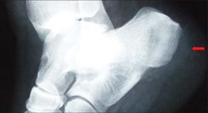
17-year-old female with pain in the left heel diagnosed as due to an aneurysmal bone cyst. Lateral X-ray of the left foot shows radiolucent lesion (arrow) leading to expansion on the postero-lateral part of the calcaneus.
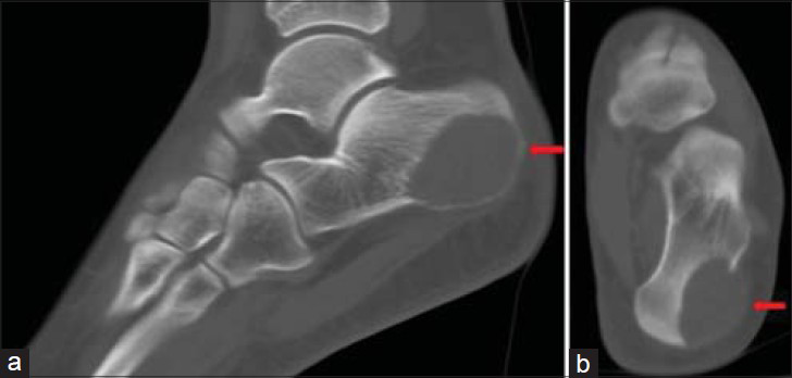
Figure 1
17-year-old female with pain in the left heel diagnosed as due to an aneurysmal bone cyst. Lateral X-ray of the left foot shows radiolucent lesion (arrow) leading to expansion on the postero-lateral part of the calcaneus.
17-year-old female with pain in the left heel diagnosed as due to an aneurysmal bone cyst. CT scans of the left foot a) sagittal and b) coronal views show lytic mass lesion (arrow), 35 × 25 mm in dimension, localized postero-laterally, leading to cortical bone erosion, expansion, and destruction.
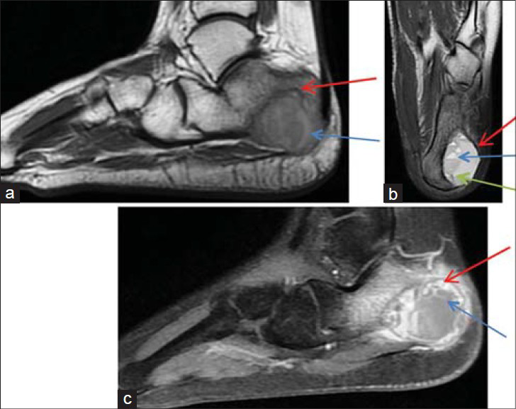
Figure 2
17-year-old female with pain in the left heel diagnosed as due to an aneurysmal bone cyst. CT scans of the left foot a) sagittal and b) coronal views show lytic mass lesion (arrow), 35 × 25 mm in dimension, localized postero-laterally, leading to cortical bone erosion, expansion, and destruction.
17-year-old female with pain in the left heel diagnosed due an aneurysmal bone cyst. MRI on the postero-lateral part of the calcaneus shows a cystic mass lesion in the medullary cavity. a) T1W and b) T2W sequences show heterogeneous hyperintense sclerotic rim (red arrow) surrounding the medullar component and central heterogeneous hyperintense septae formations (green arrow). T2W sequence shows blood level components (blue arrow). c) Image following intravenous contrast media administration shows contrast enhancement surrounding the lesion (red arrow) and in the septae formations (blue arrow).
Figure 3
17-year-old female with pain in the left heel diagnosed due an aneurysmal bone cyst. MRI on the postero-lateral part of the calcaneus shows a cystic mass lesion in the medullary cavity. a) T1W and b) T2W sequences show heterogeneous hyperintense sclerotic rim (red arrow) surrounding the medullar component and central heterogeneous hyperintense septae formations (green arrow). T2W sequence shows blood level components (blue arrow). c) Image following intravenous contrast media administration shows contrast enhancement surrounding the lesion (red arrow) and in the septae formations (blue arrow).

DISCUSSION

An ABC was first described in 1893, with the first histological description given in 1925. The term “aneurysmal bone cyst” was first used in 1942 by Jaffe and Lichtenstein on ABC lesions seen on direct radiographic images.[3] An ABC is classified as a primary lesion or a secondary lesion. Pre-existing arteriovenous malformations are of the primary type and are seen in children. A history of accompanying lesion or trauma is not present in children. However, in adults, a history of trauma is present. The secondary type is accompanied by cyst, tumor, and degeneration of fibro-osseous lesion.[2] Eighty-five percent of the cases are under 20 years of age. It is very rarely seen in patients younger than 5 years or older than 50 years of age, because it appears in bones that continue growing. Primary ABC (65%) is seen in children and adolescents.[3] According to the literature, the mean age of ABC of the calcaneus is 24 years.[3] It is more common in females than in males (1.04:1).[1]

Despite being reported in almost every bone of the body, it is commonly encountered in the metaphyses of the long bones, especially the distal part of the femur, proximal part of the tibia, and vertebrae.[24] Sixty percent of the cases originate from metaphyses, while 28% originate from the meta-epiphyseal and subchondral parts.[3] ABC comprises about 1% of primary bone tumors.[125] According to reports in the literature, ABC is rarely seen on the calcaneus, which comprises approximately 1.6% of all ABCs.[4] An ABC located on the calcaneus is often symptomatic with patients mostly presenting with heel pain after minor trauma, walking discomfort, and sometimes swelling. Heel pain is often of a chronic nature and worsens with time following the trauma. Fractures of the calcaneus are rare. Nonspecific clinical findings give rise to a suspicion of ABC and the diagnosis is primarily made radiologically.[4]

On X-ray, ABC is seen as an expansile osteolytic lesion causing cortical thinning or destruction of the calcaneus, as in our case.[4] It is an eccentrically placed foamy-shaped lesion, widening the periosteum with the border of the thin cortical roof usually being preserved.[14] CT is useful to display the anatomical integrity of the lesion on the calcaneus. An ABC is usually seen as cortical erosion; however, in our case, no cortical erosion was seen on X-ray, but it was evident on the CT. The lesion density helps differentiate the cavity. Densities over 70 Hounsfield Units (HU) may represent solid lesions such as fibrous dysplasia. The image of blood and serum as a layer of fluid-fluid level is also defined as a feature of ABC. The densities of these layers vary between 16 and 47 HU.[1] The MRI images of ABC on T1 and T2 typically show a non-homogenous cystic lesion and multiple fluid-fluid levels surrounded by a rim with low signal.[4] The fluid-fluid levels are better evaluated on a T2W MRI. In the current case, the fluid-fluid levels and internal septae were also visualized better on MRI. Fluid-fluid levels may also be seen in giant cell tumors, telangiectatic osteosarcomas, and chondroblastomas. The patient's age, localization of the lesion, and radiological images of the lesion can be of help in differential diagnosis. Ultrasound can be of help when showing fluid-fluid levels.[1] In the case reported here, a complex lesion containing fluid-fluid levels, formation of septae, and solid fields were seen.

In the differential diagnoses of calcaneal ABC, simple bone cyst, chondroblastoma, and rarely, osteosarcoma are considered. Simple bone cysts that are not seen often in the body are very commonly encountered as benign tumors/tumor-like lesions of the calcaneus, comprising 41% of these lesions. They are asymptomatic and incidentally encountered on X-ray images.[4] Chondroblastomas are rarely encountered benign lesions, comprising 15% of the secondary ABC cases. This relation is important because the relapse of chondroblastoma in the presence of ABC is approximately 100%. Chondroblastoma is more often seen in the long bones and only 7% of the cases are calcaneal. Pain is a common feature. On imaging, lesion with good margins surrounded by reactive bone and calcification is seen.[3] Osteosarcoma is rarely encountered, and this is important in differential diagnosis. It is found in similar age group as ABC and the symptoms are also similar. On X-ray, in particular, telangiectatic osteosarcoma may imitate ABC and biopsy is crucial for definite diagnosis since this condition is much more aggressive and has a progressive course.[4]

CONCLUSION

Even though the calcaneus is a rare localization for an aneurysmal bone cyst, it should be considered in the differential diagnosis. For an appropriate diagnosis, correlation of the clinical presentation, anatomical localization, radiological profile, and histological evaluation are required.

Source of Support: Nil

Conflict of Interest: None declared.

REFERENCES

  1. , , . “Soap bubble” in the calcaneus. CMAJ. 2011;183:1171.
    [Google Scholar]
  2. , , , , . Costa originated giant aneurysmal bone cyst. Erciyes Med J. 2010;32:295-300.
    [Google Scholar]
  3. , , . Aneurysmal bone cyst: Concept, controversy, clinical presentation, and imaging. AJR Am J Roentgenol. 1995;164:573-80.
    [Google Scholar]
  4. , , , , , . Pathologic fracture of a calcaneal aneurysmal bone cyst. J Foot Ankle Surg. 2011;50:727-32.
    [Google Scholar]
  5. , , , , , , . Treatment of pelvic aneurysmal bone cysts in two children: Selective arterial embolization as an adjunct to curettage and bone grafting. Diagn Interv Radiol. 2007;13:49-52.
    [Google Scholar]

Fulltext Views
13,154

PDF downloads
3,484
View/Download PDF
Download Citations
BibTeX
RIS
Show Sections