Translate this page into:
Can Patient’s Body Weight Represent Body Diameter for Pediatric Size-Specific Dose Estimate in Thoracic and Abdominal Computed Tomography?

*Corresponding author: Supika Kritsaneepaiboon, Department of Radiology, Faculty of Medicine, Prince of Songkla University, Hat Yai, 90110, Thailand. supikak@yahoo.com
-
Received: ,
Accepted: ,
Abstract
Objective: The objective of the study was to determine whether body weight (BW) can be substituted for body diameters to calculate size-specific dose estimate (SSDE) in the children.
Materials and Methods: A total of 196 torso computed tomography (CT) studies were retrospectively reviewed. Anteroposterior diameter (DAP) and lateral diameter (Dlat) were measured, and DAP+Dlat, effective diameter, SSDE diameter and SSDEBW were calculated. Correlation coefficients among body diameters, all SSDE types and percentage changes between CT dose index volumes and SSDEs were analyzed by BW and age subgroups.
Results: Overall BW was more strongly correlated with body diameter (r = 0.919–0.960, P < 0.001) than was overall age (r = 0.852–0.898, P < 0.001). The relationship between CT dose index volume and each of the SSDE types (r = 0.934–0.953, P < 0.001), between SSDEBW and all SSDE diameters (r = 0.934–0.953, P < 0.001), and among SSDE diameters (r = 0.950–0.989, P < 0.001) overall had strong correlations with statistical significance. The lowest magnitude difference was SSDEBW−SSDEeff.
Conclusion: BW can be used instead of body diameter to calculate all SSDE types, with our suggested best accuracy for SSDEeff and the least variation in age < four years and BW < 20 kg.
Key Messages: Size-specific dose estimate (SSDE) is a new and accurate dose-estimating parameter for the individual patient which is based on the actual size or body diameter of the patient. BW can be an important alternative for all body diameters to estimate size-specific dose or calculate SSDE in children.
Keywords
Body diameter
Body weight
Computed tomography dose index volume
Size-specific dose estimate
Torso
INTRODUCTION
The use of pediatric computed tomography (CT) has grown dramatically in the past decade and the risk of radiation-induced cancers in children is of more concern than in adults. The most commonly used CT parameters for calculating CT radiation dosage are CT dose index volume (CTDIvol) and dose length product (DLP).[1-3] However, the CTDIvol is delivered from a specific standard phantom size and does not indicate the actual radiation dose applied to the individual patient, leading to underestimation of the total received radiation dose to children or adults with small body size.[1-2,4-8]
Size-specific dose estimate (SSDE) is a new parameter for individual specific patients which was developed by the American Association of Physicists in Medicine (AAPM Report 204).[9] The SSDE is the patient dose estimate with corrections based on the actual size or body diameter of the patient.[4,9-10] There have been several reports examining SSDE in children[11-15] and the combination of measurements (sum of body diameters or effective diameters (Deff) is recommended to determine the appropriate SSDE correction.[11] Achieving a patient’s body diameters to calculate SSDE is more difficult than obtaining a patient’s body weight (BW) in routine work, which would make SSDE calculation more simple and rapid. However, only one report has examined conversion factors for pediatric SSDEBW.[16] The purposes of this study were to determine whether SSDE based on BW could be substituted for other SSDE values and to compare all SSDE values with CTDIvol among pediatric patients who underwent chest and abdominal CT.
METHODS
Patients and study design
The study was approved by our Human Research Ethics Committee. We retrospectively reviewed the imaging records of pediatric patients (<18 years) who underwent intravenous contrast chest or abdominal CT alone or contiguous chest and abdominal CT examinations from October 2011 to October 2016. Of the 2340 studies, 198 were randomly selected by computer, and two studies were excluded due to incorrect CT dose protocols.[17] Finally, 196 studies were reviewed. The demographic data, age, BW, and gender of the patients were collected from the hospital medical records. The patients were categorized into age and BW subgroups. The age subgroups were 0–<5years (n = 71), 5–<10 years (n = 39), 10–<15 years (n = 31), and 15–<18 years (n = 55). The BW subgroups were classified according to our institutional practice CT protocol: 4–9 kg (n = 19), 10–19 kg (n = 70), 20–29 kg (n = 22), 30–39 kg (n = 15), 40–49 kg (n = 26), and 50–64 kg (n = 26), >64 kg (n = 18).[18]
Definitions, dosimetry, and body diameter measurement
Anteroposterior diameter (DAP) was defined as the skin-to-skin thickness of the body part of the patient at the maximum thickness axial slice image [Figure 1]. Lateral diameter(Dlat) was defined as the skin-to-skin thickness of the body part of the patient at the maximum thickness axial slice image and/or anterior-posterior dimension localizer image.[19] Anteroposterior plus lateral diameter (DAP+lat) was defined as the diameter calculated as AP diameter plus Dlat. The Deff was calculated as the square root of the AP dimension multiplied by the lateral dimension.[9]

- Contiguous chest and abdominal computed tomography (CT) demonstrating the anteroposterior and lateral dimension measurements. A 4-month-old boy weighed 6.1 kg underwent CT scan for tumor surveillance in underlying Langerhans cell histiocytosis.
The CTDIvol (units: mGy) is the mean radiation absorbed dose to the patient at a given point of scan volume and is defined as weighted CTDIw/pitch. The CTDIvol was calibrated using a pencil-shaped ionization chamber with either a dedicated 16-cm or 32-cm diameter polymethylmethacrylate phantom representing the head or a body region, respectively. The DLP was defined as the CTDIvol x exposed scan length. These parameters were displayed on the CT scanner consoles and Picture Archiving and Communication System (PACS). In multiphase-scanning, the CTDIvol of the maximum DLP was used. Only CTDIvol based on 32-cm phantom was included in this study. SSDE were calculated as CTDIvol multiplied by the conversion factor in the table and depended on BW, AP, and lateral and Deff according to the AAPM Report 204 and Khawaja et al. study.[9,16] The exact conversion factor for each patient was calculated by the provided equations in the AAPM Report 204.[9]
Data collection
The two CT scanner models used during the study period were a 64-multislice Philips Brilliance CT scanner and a 160-slice Toshiba Aquilion Prime CT scanner. The images were retrieved from a PACS workstation. The body diameters were independently measured by one 13-year-experience pediatric radiologist and one-third year resident training in diagnostic radiology with consensus. The BW, age, dose indices (CTDIvol and SSDEBW, SSDEAP, SSDElat, SSDEAP+lat, and SSDEeff), and body diameters (AP, lateral, AP+lat, effective) for each patient were recorded into a spreadsheet (Microsoft Office Excel 2010; Microsoft Corporation, Redmond, WA, USA).
Statistical analysis
We presented the quantitative parameters involving BW and body diameters (AP, lateral, AP+lat, effective) using median ± interquartile range (IQR) due to non-normal distribution data. Percentage changes between CTDIvol and each SSDE type and the magnitude differences between the SSDEBW and SSDEdiameters were calculated.
Correlations among BW, age, dose indices, and body diameter measurements were established with Spearman Rank correlation coefficients (r) for the following: Correlations between each body diameter and BW and between each body diameter and age; and correlations among dose indices (CTDIvol, SSDEBW, SSDEAP, SSDElat, SSDEAP+lat, and SSDEeff) across BW and age subgroups. The power to determine sample size in BW and age subgroups for calculating correlation among dose indices was >0.8. Estimated relationships between median dose indices (CTDIvol and SSDE) and mean BWs were calculated by quantile regression analysis. Differences among the SSDE values were calculated by Wilcoxon Rank sum test. P = 0.05 or less was considered to indicate a statistically significant difference. Interobserver variations among the two reviewers were calculated using intraclass correlation coefficient (ICC) values.
RESULTS
Demographic data
This study included 196 CT studies from 196 patients, 112 male and 84 female, 72 contiguous chest and abdominal, 66 abdominal and 58 chest CTs. The median BW classified in BW and age subgroups are shown in Table 1. Males had a lower median BW (median [IQR], 18.50 [12.00–47.25 kg]) than females (median [IQR], 25.50 [13.15–46.23 kg]). The largest age subgroup was children 1 day–4 years (n = 71, 36.2%) and the 10–19 kg subgroup was the largest BW subgroup (n = 70, 35.7%).
| Parameters | BW† (kg) in median (IQR) | Diameter† (cm) in median (IQR) | |||||
|---|---|---|---|---|---|---|---|
| Total (n=196) | Male (n=112) | Female (n=84) | AP diameter | Dlat | AP+Dlat s | Deff | |
| BW subgroup | |||||||
| Overall | 20.40 | 18.50 | 25.50 | 15.42 | 20.54 | 35.93 | 17.81 |
| 4–9 kg (n=19) | (12.00–47.00) 5.27 | (12.00–47.25) 6.10 | (13.15–46.23) 4.70 | (13.19–19.05) 10.99 | (17.55–27.21) 14.04 | (31.17–46.66) 25.08 | (15.28–22.85) 12.42 |
| 10–19 kg (n=70) | (4.57–6.83) 13.35 | (4.25–7.87) 13.35 | (4.70–5.50) 13.55 | (10.19–12.53) 13.54 | (12.59–14.83) 18.05 | (23.09–27.60) 31.47 | (11.40–13.47) 15.63 |
| 20–29 kg (n=22) | (11.07–15.50) 21.75 | (11.20–15.87) 21.25 | (10.37–15.00) 25.00 | (12.81–14.15) 15.00 | (17.04–18.85) 20.89 | (29.97–32.91) 35.52 | (14.84–16.19) 17.56 |
| 30–39 kg (n=15) | (20.35–25.75) 33.00 | (20.45–23.52) 35.50 | (20.45–25.75) 32.60 | (14.64–15.61) 16.92 | (20.18–22.84) 24.33 | (35.06–38.08) 41.80 | (17.33–18.61) 20.30 |
| 40–49 kg (n=26) | (30.80–35.75) 45.50 | (32.45–36.40) 47.00 | (31.02–33.25) 45.00 | (15.87–17.94) 17.83 | (23.25–25.87) 27.13 | (39.90–43.05) 45.32 | (19.59–21.26) 22.20 |
| 50–64 kg (n=26) | (41.40–47.00) 56.80 | (44.50–47.50) 58.00 | (40.00–46.90) 55.9 | (17.18–19.37) 20.81 | (25.7–28.27) 29.33 | (43.36–47.01) 50.10 | (21.29–23.02) 24.65 |
| >64 kg (n=18) | (53.50–60.00) 73.45 (65.43–77.33) |
(54.00–60.00) 72.75 (65.18–74.33) |
(52.70–59.00) 79.15 (71.87–84.52) |
(20.06–21.54) 22.24 (21.37–24.04) |
(27.83–30.64) 32.42 (29.81–33.52) |
(48.05–51.80) 54.52 (52.28–57.43) |
(23.62–25.60) 26.62 (25.74–28.32) |
| Age subgroup | |||||||
| Overall | 20.40 (12.00–47.00) | 18.50 (12.00–47.25) | 25.50 (13.15–46.23) | 15.42 (13.19–19.05) | 20.54 (17.55–27.21) | 35.93 (31.17–46.66) | 17.81 (15.28–22.85) |
| 1 day–4 years (n=71) | 10.70 (8.75–13.00) | 11.20 (9.10–13.50) | 9.25 (5.57–11.92) | 12.84 (12.24–13.72) | 17.05 (15.54–18.14) | 30.10 (28.30–31.92) | 14.88 (13.93–15.75) |
| 5–9 years (n=39) | 18.70 (15.75–21.25) | 18.70 (17.00–20.85) | 17.65 (14.97–22.60) | 15.06 (13.76–15.64) | 20.35 (18.89–20.67) | 35.35 (32.45–36.42) | 17.40 (16.05–18.05) |
| 10–14 years (n=31) | 39.20 (30.25–55.95) | 37.00 (30.30–55.40) | 42.10 (30.75–56.17) | 18.25 (15.73–20.74) | 26.37 (23.85–28.67) | 45.70 (39.42–48.7) | 22.31 (19.25–24.08) |
| 15–18 years (n=55) | 50.00 (45.50–63.10) | 59.00 (47.50–65.15) | 47.05 (40.00–53.88) | 19.9617.64–21.30) | 28.43 (26.35–30.86) | 48.10 (44.65–51.75) | 23.44 (22.01–25.55) |
Dose metrics
The overall CTDIvol at 32 cm phantom size was 2.90 (2.88–5.84 mGy) (median [IQR]).
BW and body diameters across BW and age subgroups
The overall body diameters, AP, lat, AP+lat, and effective, were median (IQR), 15.42 (13.19–19.05), 20.54 (17.55–27.21), 35.93 (31.17–46.66), and 17.81 (15.28–22.85) cm, respectively. The median body diameters across BW and age subgroups are shown in Table 1. The Dlat was larger than the AP diameter in all BW and age subgroups. All of the body diameters were in ascending order in both BW and age subgroups. Interobserver agreement using ICC between the two reviewers was excellent (ICC = 0.99).
Correlation coefficients
Overall and subgroup correlations between body diameters and BW and between body diameters and age are shown in Table 2. The DAP, Dlat, DAP+lat, and Deff were strongly correlated to the overall BW (r = 0.919, 0.96, 0.935, and 0.943, respectively, P < 0.001). The correlations between body diameters and overall age were also strong but less than the body diameter – BW correlations (r = 0.852–0.898, P < 0.001) [Table 2].
| AP diameter | Dlat | AP+Dlat s | Deff | |||||
|---|---|---|---|---|---|---|---|---|
| Coefficient† | P | Coefficient† | P | Coefficient† | P | Coefficient† | P | |
| BW subgroup | ||||||||
| Overall | 0.919 | <0.001 | 0.960 | <0.001 | 0.935 | <0.001 | 0.943 | <0.001 |
| 4–9 kg (n=19) | 0.722 | <0.001 | 0.815 | <0.001 | 0.552 | <0.001 | 0.465 | 0.039 |
| 10–19 kg (n=70) | 0.638 | <0.001 | 0.718 | <0.001 | 0.77 | <0.001 | 0.706 | <0.001 |
| 20–29 kg (n=22) | 0.102 | 0.651 | 0.695 | 0.003 | 0.46 | 0.02 | 0.418 | 0.052 |
| 30–39 kg (n=15) | 0.522 | 0.046 | 0.073 | 0.795 | 0.39 | 0.147 | 0.450 | 0.092 |
| 40–49 kg (n=26) | 0.082 | 0.689 | 0.496 | 0.010 | 0.58 | 0.001 | 0.490 | 0.010 |
| 50–64 kg (n=26) | 0.340 | 0.097 | 0.259 | 0.212 | 0.34 | 0.06 | 0.382 | 0.059 |
| >64 kg (n=18) | 0.864 | <0.001 | 0.850 | <0.001 | 0.91 | <0.001 | 0.922 | <0.001 |
| Age subgroup | ||||||||
| Overall | 0.852 | <0.001 | 0.898 | <0.001 | 0.870 | <0.001 | 0.872 | <0.001 |
| 1 d–4 years (n=71) | 0.561 | <0.001 | 0.761 | <0.001 | 0.635 | <0.001 | 0.685 | <0.001 |
| 5–9 years (n=39) | 0.078 | 0.637 | 0.250 | 0.12 | 0.128 | 0.434 | 0.128 | 0.434 |
| 10–14 years (n=31) | 0.150 | 0.420 | 0.264 | 0.15 | 0.193 | 0.296 | 0.189 | 0.309 |
| 15–18 years (n=55) | 0.181 | 0.185 | 0.278 | 0.040 | 0.268 | 0.047 | 0.24 | 0.075 |
AP: Anteroposterior †Spearman’s rank correlation interpretation (r): r=1 perfectly positive, 0.8≤r<1 strongly positive, 0.5≤r<0.8 moderately positive, 0.1≤r<0.5 weakly positive, 0<r<0.1 lowest positive. BW: Body weight, Dlat: lateral diameter, Deff: Effective diameter
The correlations across the SSDEBW and SSDE body diameters in the BW and age subgroups were moderate to strong with statistical significance (r = 0.719–0.979, P < 0.001 in the BW subgroups and r = 0.758–0.965, P < 0.001 in the age subgroups) [Table 3]. The correlations across the SSDE body diameters in the BW and age subgroups were strong with statistical significance as shown in Table 4 (r = 0.862–1, P < 0.001 in the BW subgroup and r = 0.872–0.9991, P < 0.001 in the age subgroup).
| Parameter | SSDEBW−SSDEAP | SSDEBW−SSDElat | SSDEBW−SSDEAP+lat | SSDEBW−SSDEeff | ||||
|---|---|---|---|---|---|---|---|---|
| Coefficient† | P | Coefficient† | P | Coefficient† | P | Coefficient† | P | |
| BW subgroup | ||||||||
| Overall | 0.934 | <0.001 | 0.951 | <0.001 | 0.942 | <0.001 | 0.953 | <0.001 |
| 4–9 kg (n=19) | 0.926 | <0.001 | 0.938 | <0.001 | 0.837 | <0.001 | 0.860 | <0.001 |
| 10–19 kg (n=70) | 0.719 | <0.001 | 0.751 | <0.001 | 0.802 | <0.001 | 0.733 | <0.001 |
| 20–29 kg (n=22) | 0.899 | <0.001 | 0.933 | <0.001 | 0.940 | <0.001 | 0.935 | <0.001 |
| 30–39 kg (n=15) | 0.957 | <0.001 | 0.975 | <0.001 | 0.975 | <0.001 | 0.979 | <0.001 |
| 40–49 kg (n=26) | 0.908 | <0.001 | 0.946 | <0.001 | 0.960 | <0.001 | 0.955 | <0.001 |
| 50–64 kg (n=26) | 0.949 | <0.001 | 0.939 | <0.001 | 0.953 | <0.001 | 0.956 | <0.001 |
| >64 kg (n=18) | 0.976 | <0.001 | 0.938 | <0.001 | 0.968 | <0.001 | 0.962 | <0.001 |
| Age subgroup | ||||||||
| Overall | 0.934 | <0.001 | 0.951 | <0.001 | 0.942 | <0.001 | 0.953 | <0.001 |
| 1 d–4 years (n=71) | 0.807 | <0.001 | 0.824 | <0.001 | 0.758 | <0.001 | 0.823 | <0.001 |
| 5–9 years (n=39) | 0.921 | <0.001 | 0.944 | <0.001 | 0.945 | <0.001 | 0.945 | <0.001 |
| 10–14 years (n=31) | 0.927 | <0.001 | 0.865 | <0.001 | 0.922 | <0.001 | 0.899 | <0.001 |
| 15–18 years (n=55) | 0.950 | <0.001 | 0.942 | <0.001 | 0.965 | <0.001 | 0.962 | <0.001 |
SSDE: Size-specific dose estimate, BW: Body weight, AP: Anteroposterior diameter, Dlat: Lateral diameter, AP+lat: Anteroposterior plus Dlat, eff: Effective diameter. †Spearman’s rank correlation interpretation (r): r=1 perfectly positive, 0.8≤r<1 strongly positive, 0.5≤r<0.8 moderately positive, 0.1≤r<0.5 weakly positive, 0<r<0.1 lowest positive
| Parameter | SSDEAP-SSDElat | SSDEAP−SSDEAP+lat | SSDEAP−SSDEeff | SSDElat−SSDEAP+lat | SSDEAP+lat−SSDEeff | SSDElat−SSDEeff | P‡ |
|---|---|---|---|---|---|---|---|
| Coefficient† | Coefficient† | Coefficient† | Coefficient† | Coefficient† | Coefficient† | ||
| BW subgroup | |||||||
| Overall | 0.950 | 0.989 | 0.966 | 0.978 | 0.979 | 0.986 | <0.001 |
| 4–9 kg (n=19) | 0.989 | 0.938 | 0.895 | 0.997 | 0.898 | 0.881 | <0.001 |
| 10–19 kg (n=70) | 0.932 | 0.978 | 0.961 | 0.983 | 0.982 | 0.967 | <0.001 |
| 20–29 kg (n=22) | 0.891 | 0.969 | 0.973 | 0.961 | 0.997 | 0.952 | <0.001 |
| 30–39 kg (n=15) | 0.982 | 0.982 | 0.985 | 1.000 | 0.996 | 0.996 | <0.001 |
| 40–49 kg (n=26) | 0.862 | 0.950 | 0.956 | 0.962 | 0.998 | 0.959 | <0.001 |
| 50–64 kg (n=26) | 0.917 | 0.976 | 0.977 | 0.969 | 0.9992 | 0.964 | <0.001 |
| >64 kg (n=18) | 0.953 | 0.976 | 0.982 | 0.988 | 0.997 | 0.985 | <0.001 |
| Age subgroup | |||||||
| Overall | 0.950 | 0.989 | 0.966 | 0.978 | 0.979 | 0.986 | <0.001 |
| 1 d–4 years (n=71) | 0.880 | 0.982 | 0.895 | 0.941 | 0.937 | 0.973 | <0.001 |
| 5–9 years (n=39) | 0.971 | 0.986 | 0.990 | 0.993 | 0.9991 | 0.991 | <0.001 |
| 10–14 years (n=31) | 0.872 | 0.956 | 0.935 | 0.971 | 0.977 | 0.943 | <0.001 |
| 15–18 years (n=55) | 0.917 | 0.972 | 0.978 | 0.979 | 0.998 | 0.975 | <0.001 |
SSDE: Size-specific dose estimate, BW: Body weight, AP: Anteroposterior diameter, Dlat: lateral diameter, AP+lat: Anteroposterior plus Dlat, eff: Effective diameter. †Spearman’s rank correlation interpretation (r): r=1 perfectly positive, 0.8≤r<1 strongly positive, 0.5≤r<0.8 moderately positive, 0.1≤r<0.5 weakly positive, 0<r<0.1 lowest positive. ‡P values for Spearman’s rank correlation coefficient in SSDEAP-SSDElat, SSDEAP-SSDEAP+lat, SSDEAP-SSDEeff, SSDElat-SSDEAP+lat, SSDEAP+lat-SSDEeff and SSDElat-SSDEeff
Quantile regression analysis
Quantile regression analysis was used to generate and predict the trends of the median dose indices (CTDIvol, SSDEBW, and all SSDE body diameters) and BW. The trends of all SSDE values were higher than the CTDIvol. The equations to predict dose indices from BW were:
| CTDIvol = (0.09586 × BW) + 1.475231 | Equation 1 |
| SSDEBW = (0.104456 × BW) + 4.2285934 | Equation 2 |
| SSDEAP = (0.108038 × BW) + 4.465022 | Equation 3 |
| SSDElat = (0.104385 × BW) + 4.915016 | Equation 4 |
| SSDEAP+lat = (0.104802 × BW) + 4.753674 | Equation 5 |
| SSDEeff = (0.105634 × BW) + 4.698292 | Equation 6 |
Percentage change
The percentage change between CTDIvol and SSDE according to the BW and body diameters is shown in the box plot chart in Figure 2. Almost all SSDE values were greater than the CTDIvol values. There was only one patient (0.5%) in which SSDEBW was less than CTDIvol (6%) and this patient weighed >100 kg. In the SSDE diameter group, eight SSDE diameters (SSDEAP =1, SSDElat = 2, SSDEAP+lat = 2, and SSDEeff = 3) were less than CTDIvol, and all of them were maximum diameters in each SSDE diameter subgroup. The percentage change shown as median (IQR) was as follows: (SSDEBW−CTDIvol)/CTDIvol 88% (66–112%) and range −6–147%; (SSDEAP−CTDIvol)/CTDIvol 94% (61–119%) with range −39.82–171%; (SSDElal−CTDIvol)/CTDIvol 111% (67–99%) with range –27–172%; (SSDEAP+lal−CTDIvol)/CTDIvol 104% (62–96%) with range −3–176%, and (SSDEeff− CTDIvol)/CTDIvol 101% (62–94%) with range −5–181%.

- Boxplot percentage change between computed tomography DIvol and SSDE (BW and body diameters).
Differences between SSDEBW and SSDEdiameters
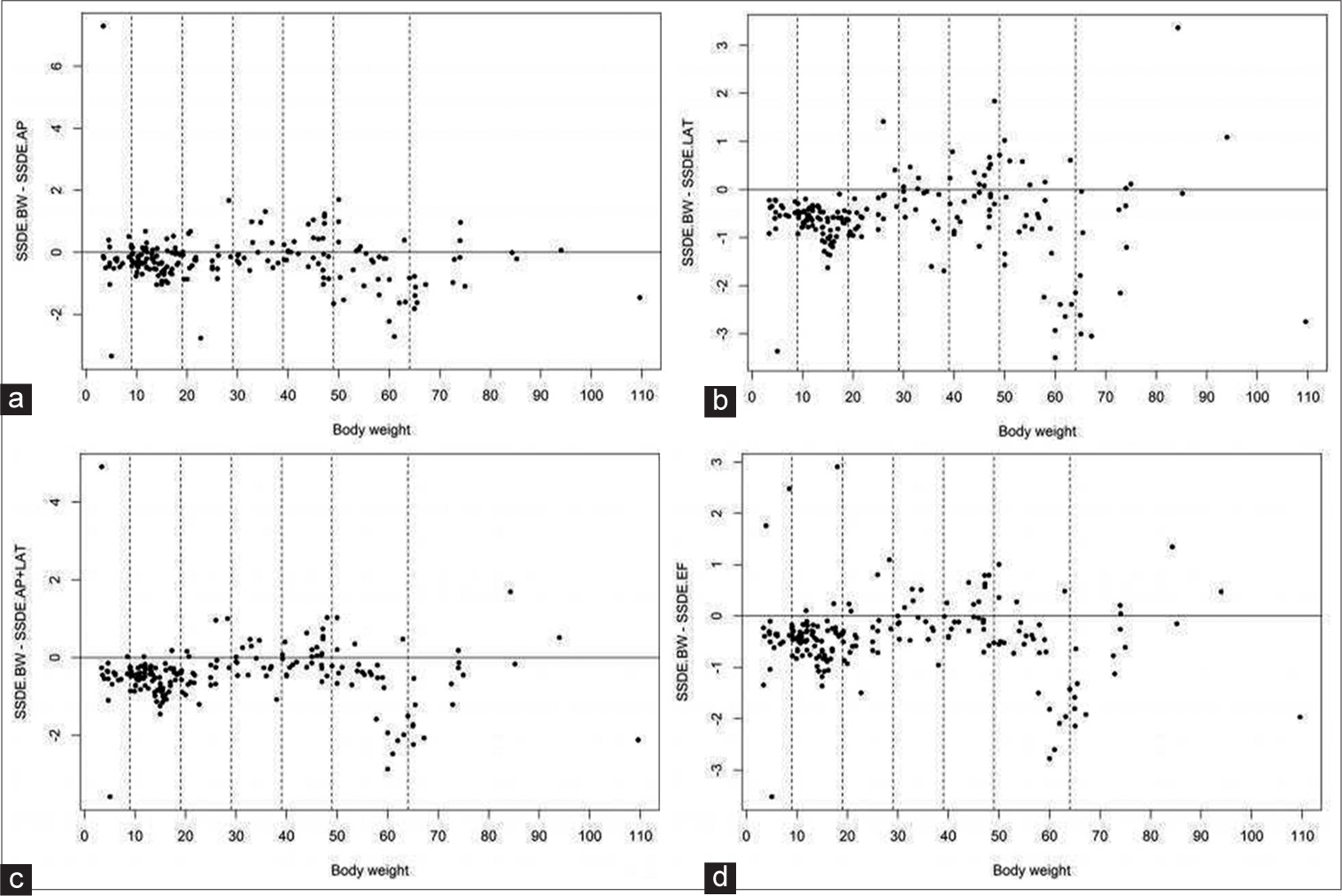
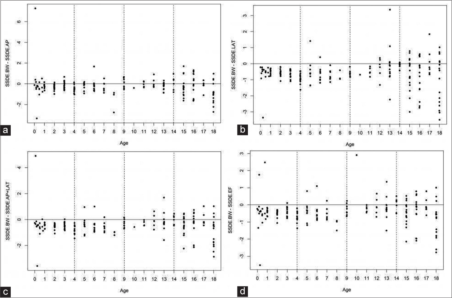
The difference between SSDEBW and all SSDE diameters of each patient was not statistically significant in SSDEBW−SSDEAP (P = 0.3854), and SSDEBW−SSDEAP+lat (P = 0.09188), and SSDEBW− SSDEeff (P = 0.1167) except in SSDEBW−SSDElat (P = 0.03113) by Wilcoxon Rank sum test. The SSDE magnitude differences between all SSDEBW and all SSDE diameters of each patient were plotted in graphs and categorized by age and BW subgroups [Figures 3 and 4]. The lowest magnitude was the difference between SSDEBW and SSDEeff. −4.22–2.91, while the highest magnitude was between SSDEBW and SSDEAP −4.18–7.3. The other magnitudes were −4.31–3.37 for SSDEBW–SSDElat and −4.23–4.91 for SSDEBW−SSDEAP+lat.

- Scatter plots of differences between SSDEBW and each SSDE body diameter by BW subgroups; SSDEBW–SSDEAP (a), SSDEBW–SSDElat (b), SSDEBW–SSDEAP+lat (c), and SSDEBW–SSDEeff (d).

- Scatter plots of differences between SSDEBW and each SSDE body diameter by age subgroups; SSDEBW–SSDEAP (a), SSDEBW–SSDElat (b), SSDEBW–SSDEAP+lat (c), and SSDEBW–SSDEeff (d).
DISCUSSION
Our study found that all body diameters and overall BW were strongly correlated (r = 0.919–0.960, P < 0.001). The Dlat, DAP+lat, and Deff in our study had higher correlations with overall BW than with DAP, which could be explained by understanding the general growth pattern of children, in which the child’s body grows in the Dlat more rapidly than in the AP diameter.[2] The correlations for all body diameters and overall age were also strong but not as high as the body diameter-BW relationships (r = 0.852–0.898, P < 0.001). However, the study by Kleinman et al. found that the predicted individual patient size was not correlated with age.[2]
All relationships of SSDEBW-all SSDE diameters (r = 0.934–0.953, P < 0.001) and among SSDE body diameters (r = 0.950–0.989, P < 0.001) with overall BW and with overall age showed strong and statistically significant correlations [Tables 3 and 4]. The strongest correlations were found in the 30–39 kg subgroup and the 5–9 years subgroup. Another previous study by Khawaja et al. reached the same conclusion as our study that BW could be substituted to estimate size-specific dose in children.[16] Another study by Parikh et al. also found that BW could be used to estimate SSDE with reasonable accuracy at body width >20 cm.[14]
In our study, we could predict the SSDE and CTDIvol using BW from Equations 1–6, while Christner et al. study concluded that only CTDIvol increased linearly with patient size (DAP + Dlat), while SSDE was independent of patient size.[10] Furthermore, almost all SSDE type values (n = 189/196, 96.4%) were higher than CTDIvol, except for large-sized patients and those weighing >100 kg. Therefore, emphasizing CTDIvol underestimates the radiation dose in most pediatric or small-sized patients and overestimates the radiation dose in large-sized patients.[2,8,14,20]
Although the SSDEBW had a statistically significant difference from SSDElat by Wilcoxon Rank sum test, the magnitude difference between SSDEBW and SSDElat was still in the acceptable range (within 7% of dose index in diagnostic radiology). The lowest magnitude difference was between SSDEBW and SSDEeff, while the highest magnitude difference was between SSDEBW and SSDEAP. These results could be explained by considering a study from Brady et al., which found that either an individual AP or Dlat measurement alone was less useful than a combination of AP and Dlat measurement for SSDE determination.[11] However, all SSDEBW–SSDEdiameters magnitude differences in our study were still in the acceptable range. The smallest variations of the SSDE differences in all subgroups by age and BW were in the lower BW ranges and younger age groups. In addition, most of the SSDEBW values tended to be lower than the SSDEdiameters. This implies that the SSDEBW can be substituted for SSDEdiameters, especially SSDEeff, but with caution as the SSDEBW tended to be lower than SSDEdiameters.
Our study had a few limitations. First, we could not statistically determine correlations between each body diameter and the BW <20 kg and >64 kg subgroups and between each body diameter and the age >4 year subgroups because the power of the sample size in those subgroups was <0.8. We suggest further research should be conducted with increased sample sizes in each subgroup if the study objective is to determine the correlation between body diameters and age or BW subgroups. Second, we did not calculate the SSDE from the water equivalent diameter (Dw), which is a physical parameter based on patient attenuation. In case of patients having high body attenuation, for example, those suffering from mediastinal or intra-abdominal tumors with low to normal BW, the SSDEDw is more accurate than SSDEdiameter to determine the correct patient dose.[21] We suggest further studies including SSDEDw and clinical indications. Finally, the findings of our study may not be applicable in institutions and hospitals that have automatic software to determine the body measurements and SSDE.
CONCLUSION
Accurate dose-estimating parameters and size-specific dose indices are important for calculating accurate radiation dosage in the pediatric population. Our study found that the body diameter-BW correlation was stronger than the body diameter – age relationship. This calculation is simple and rapid to perform, and BW can be an important alternative for all body diameters to estimate size-specific dose or calculate SSDE in children. Our findings indicate this method has the best accuracy for SSDEeff and the least variation in ages less than 4 years and BWs < 20 kg.
Financial support and sponsorship
Nil.
Conflicts of interest
There are no conflicts of interest.
References
- How accurate is size-specific dose estimate in pediatric body CT examinations? Pediatr Radiol. 2016;46:2016-46.
- [CrossRef] [PubMed] [Google Scholar]
- Patient size measured on CT images as a function of age at a tertiary care children’s hospital. AJR Am J Roentgenol. 2010;194:2010-194.
- [CrossRef] [PubMed] [Google Scholar]
- CT dosimetry: Comparison of measurement techniques and devices. Radiographics. 2008;28:2008-28.
- [CrossRef] [PubMed] [Google Scholar]
- Optimizing CT radiation dose based on patient size and image quality: The size-specific dose estimate method. Pediatr Radiol. 2014;44(Suppl 3):501-5.
- [CrossRef] [PubMed] [Google Scholar]
- CT dose index and patient dose: They are not the same thing. Radiology. 2011;259:2011-259.
- [CrossRef] [PubMed] [Google Scholar]
- CT dose: How to measure, how to reduce. Health Phys. 2008;95:2008-95.
- [CrossRef] [PubMed] [Google Scholar]
- Dose reduction and compliance with pediatric CT protocols adapted to patient size, clinical indication, and number of prior studies. Radiology. 2009;252:2009-252.
- [CrossRef] [PubMed] [Google Scholar]
- It is time to retire the computed tomography dose index (CTDI) for CT quality assurance and dose optimization. For the proposition. Med Phys. 2006;33:2006-33.
- [CrossRef] [PubMed] [Google Scholar]
- Size-Specific dose Estimates (SSDE) in Pediatric and Adult Body CT Examinations. 2018. The American Association of Physicists in Medicine. Report of AAPM Task Group. 204:2018-204. Available from: https://www.aapm.org/pubs/reports/RPT_204.pdf [Last cited on 2018 Nov 14]
- [Google Scholar]
- Size-specific dose estimates for adult patients at CT of the torso. Radiology. 2012;265:2012-265.
- [CrossRef] [PubMed] [Google Scholar]
- Investigation of american association of physicists in medicine report 204 size-specific dose estimates for pediatric CT implementation. Radiology. 2012;265:2012-265.
- [CrossRef] [PubMed] [Google Scholar]
- Diagnostic reference ranges for pediatric abdominal CT. Radiology. 2013;268:2013-268.
- [CrossRef] [PubMed] [Google Scholar]
- Pediatric chest CT diagnostic reference ranges: Development and application. Radiology. 2017;284:2017-284.
- [CrossRef] [PubMed] [Google Scholar]
- A comparison study of size-specific dose estimate calculation methods. Pediatr Radiol. 2018;48:2018-48.
- [CrossRef] [PubMed] [Google Scholar]
- Local diagnostic reference level based on size-specific dose estimates: Assessment of pediatric abdominal/pelvic computed tomography at a Japanese national children’s hospital. Pediatr Radiol. 2015;45:2015-45.
- [CrossRef] [PubMed] [Google Scholar]
- Simplifying size-specific radiation dose estimates in pediatric CT. AJR Am J Roentgenol. 2015;204:2015-204.
- [CrossRef] [PubMed] [Google Scholar]
- Comparing Means In: Chow SC, Jones B, Liu J, Peace KE, eds. Sample Size Calculations in Clinical Research (2nd ed.). Boca Raton, FL: Chapman and Hall/CRC; 2007. p. :49-82.
- [Google Scholar]
- Multidetector CT in children: Current concepts and dose reduction strategies. Pediatr Radiol. 2010;40:2010-40.
- [CrossRef] [PubMed] [Google Scholar]
- Automated size-specific CT dose monitoring program: Assessing variability in CT dose. Med Phys. 2012;39:2012-39.
- [CrossRef] [PubMed] [Google Scholar]
- Effective dose determination using an anthropomorphic phantom and metal oxide semiconductor field effect transistor technology for clinical adult body multidetector array computed tomography protocols. J Comput Assist Tomogr. 2007;31:2007-31.
- [CrossRef] [PubMed] [Google Scholar]
- Use of Water Equivalent Diameter for Calculating Patient Size and Size-Specific Dose Estimates (SSDE) in CT: The Report of AAPM Task Group 220. AAPM Rep. 2014;2014:6-23.
- [Google Scholar]


