Translate this page into:
Ureterovesical Opaque Densities after Ureterovesical Reflux Injection Therapy: Diagnostic and Therapeutic Dilemma
Address for correspondence: Dr. Hamed Akhavizadegan, Department of Urology, Baharloo hospital, Behdari Street, Rahahan square, Tehran, Iran. E-mail: h-akhavizadegan@tums.ac.ir
-
Received: ,
Accepted: ,
This is an open-access article distributed under the terms of the Creative Commons Attribution License, which permits unrestricted use, distribution, and reproduction in any medium, provided the original author and source are credited.
This article was originally published by Medknow Publications & Media Pvt Ltd and was migrated to Scientific Scholar after the change of Publisher.
Abstract
Primary vesicoureteral reflux can be treated by injection of a bulking agent into the wall of the ureterovesical junction. Over time, the bulking agent can get calcified. Radiological images of the area show findings that mimic those seen in ureterovesical junction calculi. In this report, we present the imaging findings of this phenomenon and discuss its challenging aspects.
Keywords
Bulking agent
calcification
collagen
ureterovesical stone
vesicoureteral junction
vesicoureteral reflux
INTRODUCTION

Bulking agent injection is now a standard treatment for patients with grade 1-3 primary vesicoureteral reflux (VUR). Patients with higher grades and secondary types of VUR show an unacceptably high failure rate when treated with injections of a bulking agent.[1] Collagen has been used for many years as a bulking agent in ureterovesical orifice and bladder neck to treat VUR and incontinence.[2] Collagen, however, can get calcified in the tissue,[2] a process that can cause a series of problems for patients. We present a case of a patient in whom calcified injected collagen showed imaging features mimicking ureteral stone.
CASE REPORT
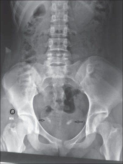
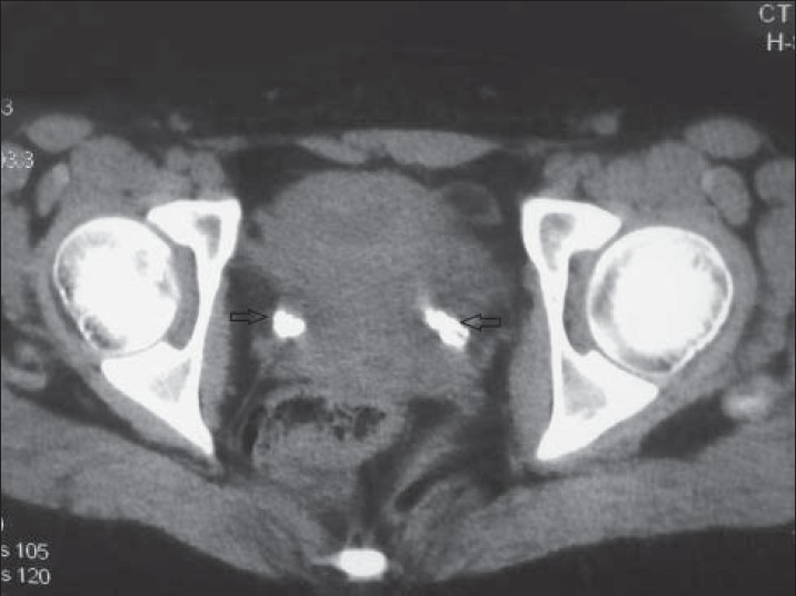
A 15-year-old girl presented with moderate degree of right flank pain for 2 weeks. Pain was localized on the right side, altered with change in position, and did not relate to any particular organ. Physical examination and urine analysis were normal. The patient had been treated successfully for bilateral grade 2 VUR when she was 3 years old. A kidney ureter bladder (KUB) radiography was requested and the report indicated presence of bilateral ureteral stone [Figure 1]. Ultrasonography showed bilateral ureterovesical junction (UVJ) stone and mild stasis in both kidneys, which was compatible with the findings of the non-enhanced spiral abdomino-pelvic computed tomography (CT) scan [Figure 2]. However, diethylene triamine pentaacaetic acid (DTPA) scan with furosemide injection and radionuclide cystography (RNC) scan ruled out obstruction and relapse of VUR. Calcification of bilateral intravesical collagen injected to treat her VUR 12 years earlier was responsible for the positive findings on KUB, ultrasonography, and spiral CT. The patient was referred to the physiotherapist for her low back pain. The pain improved with treatment.

- 15-year-old female with bilateral flank pain, which was suspected to be due to stones in bilateral ureterovesical junction, later diagnosed as due to calcification of collagen used 12 years earlier to treat vesicoureteral reflux. Kidney ureter bladder graphy shows bilateral UVJ calcifications (arrow).

- 15-year-old female with bilateral flank pain, which was suspected to be due to stones in bilateral ureterovesical junction, later diagnosed as due to calcification of collagen used 12 years earlier to treat vesicoureteral reflux. Spiral CT scan of pelvis without intravenous and oral contrast shows bilateral UVJ calcifications (arrows).
DISCUSSION
Endoscopic therapy for VUR has gained popularity because of elimination of invasive surgical procedures in children. In addition, it has an acceptable success rate and a low level of complications.[1] Although a transition has occurred in the material of choice from Teflon to collagen and now to hyaluronic acid copolymers. However, we do find patients who were treated with collagen now in the second decade of their life. Calcification of injected collagen has been described since 1994.[23] The injected material, being a foreign body, can induce inflammatory response that leads to tissue calcification.[4] This calcification has been reported to be symptomatic with some patients reporting episodes of stone passing, hematuria, and even obstruction.[4] This is the second case report of ureterovesical injection calcification presenting as a ureteral stone during differential diagnosis[4] and the first report of its imaging. Unlike the first case report where the patient presented with renal colic and passing of stone, our patient was completely asymptomatic, except for positional non-related flank pain. There is no difference between a ureteral stone and calcification of injected collagen, when viewed by different imaging modalities like KUB [Figure 1], CT scanning [Figure 2], and also ultrasound examination. This similarity is because of the place where the bulking agent is injected. In doubtful cases, as in our case, UVJ obstruction can be ruled out by DTPA nuclear scan in which glomerular filtration of injected nuclear radioisotope reveals any obstruction in the upper urinary tract. There are also other challenging issues. The diagnosis of UVJ stone in the presence of UVJ calcification, possibility of ureteral stone passing (in a patient with renal colic) after UVJ injection therapy, and the probability of recurrent VUR after ureteral intervention (in which calcified ureteral orifice will tolerate dilatation to pass the ureteroscope) remain to be studied. VUR can be assessed by RNC in which a radioisotope passing to the bladder via the urethral catheter reveals presence of VUR.
CONCLUSION
The standard treatment for grade 1 and 3 primary VUR is injection of a bulking agent into the UVJ. Clinicians should be aware that calcification of bulking agent like collagen can occur and they need to differentiate this from ureterovesical stones on imaging findings.
Available FREE in open access from: http://www.clinicalimagingscience.org/text.asp?2014/4/1/27/133261
Source of Support: Nil
Conflict of Interest: None declared.
REFERENCES
- Usefulness of concomitant autologous blood and dextranomer/hyaluronic acid copolymer injection to correct vesicoureteral reflux. J Urol. 2012;188:948-52.
- [Google Scholar]
- Physical and histological behavior of a new injectable collagen (GAX65) implanted into the submucosal space of the mini-pig bladder. J Urol. 1995;154:812-5.
- [Google Scholar]
- Histological behavior of glutaraldehyde cross-linked bovine collagen injected into the human bladder for the treatment of vesicoureteral reflux. J Urol. 1994;152:632-5.
- [Google Scholar]
- Symptomatic calcification of subureteral collagen ten years after injection. Urology. 2007;69:982.e1-2.
- [Google Scholar]


