
Translate this page into:
Ultrasound Findings of the Painful Ankle and Foot
Address for correspondence: Dr. Suheil Artul, Department of Radiology, EMMS Nazareth Hospital, Nazareth, Israel. E-mail: suheil_artul@hotmail.com
-
Received: ,
Accepted: ,
This is an open-access article distributed under the terms of the Creative Commons Attribution License, which permits unrestricted use, distribution, and reproduction in any medium, provided the original author and source are credited.
This article was originally published by Medknow Publications & Media Pvt Ltd and was migrated to Scientific Scholar after the change of Publisher.
Abstract
Objectives:
To document the prevalence and spectrum of musculoskeletal ultrasound (MSKUS) findings at different parts of the foot.
Materials and Methods:
All MSKUS studies conducted on the foot during a 2-year period (2012-2013) at the Department of Radiology were reviewed. Demographic parameters including age, gender, and MSKUS findings were documented.
Results:
Three hundred and sixty-four studies had been conducted in the 2-year period. Ninety-three MSKUS evaluations were done for the ankle, 30 studies for the heel, and 241 for the rest of the foot. The most common MSKUS finding at the ankle was tenosynovitis, mostly in female patients; at the heel it was Achilles tendonitis, also mostly in female patients; and for the rest of the foot it was fluid collection and presence of foreign body, mainly in male patients. The number of different MSKUS abnormalities that were reported was 9 at the ankle, 9 at the heel, and 21 on the rest of the foot.
Conclusions:
MSKUS has the potential for revealing a huge spectrum of abnormalities. The most common finding was collection/hematoma and foreign bodies at the foot, tenosynovitis at the ankle, and Achilles tendinitis at the heel.
Keywords
Abnormalities
foot
ultrasound

INTRODUCTION

Musculoskeletal ultrasound (MSKUS) imaging is becoming more and more popular in the evaluation and management of various problems in rheumatology. Its first clinical application in rheumatology was reported in 1974 with the differentiation of a Baker's cyst from thrombophlebitis.[1]
MSKUS has been shown to be helpful in the evaluation of joint, tendon, muscle, bursa, bone, and other soft tissue abnormalities.[23456] It has the ability to directly visualize, characterize, and quantify the earliest and smallest inflammatory and structural changes. These qualities can help also in directing appropriate treatment and monitoring response and disease progression.
MSKUS has several unique features that not only help in performing the above measurements but also confer significant advantages over other imaging techniques. It is safe, non-invasive, and uses no ionizing radiation. It is portable and can be made available in an out-patient setting, enabling rapid, “real-time,” and dynamic examination of one or more joints or areas.
The Doppler dimension of this modality adds important information regarding vascular components that ultimately help in identifying abnormalities like synovitis, hemangiomas, and tumors.[78] Contrast-enhanced ultrasound and 3D ultrasonography improve the sensitivity, specificity, and accuracy of this modality.[910]
Feet are rich in tendons, muscles, blood vessels, nerves, and joints, which can be the source of different complaints for the patient. Foot pain is a common orthopedic or rheumatologic problem.
In this retrospective study, we wanted to evaluate the results of all MSKUS studies of the feet that were done at the Nazareth hospital during the last 2 years.
MATERIALS AND METHODS
All MSKUS studies of the feet that were done from 2012 to 2013 at the Nazareth hospital were identified from the registry of MSKUS studies in the Department of Radiology. Epidemiologic and clinical parameters including age, gender, complaint, and MSKUS findings were documented. All the studies were done by an experienced radiologist in the field of MSKUS (first author). During this period, a Philips HD 11, Amsterdam (the Netherlands) ultrasound machine with Ph ilips (New York, USA) 3-12 MHz probes was used.
In case of more than one diagnosis given by the evaluating radiologist, we chose the first diagnosis from the list. Accidental findings were not included in our study, but the diagnosis regarding the main reason for which the patient was referred to the imaging investigation was only selected. Repeated studies of the same patient for the same clinical problem were considered as one study.
RESULTS
Three hundred and sixty-four studies of different patients were selected. The ankle was investigated in 93 patients, the heel in 30 patients, and the rest of the foot in 241 patients.
Table 1 summarizes the different findings of the ankles. Table 2 summarizes the different findings on the heel, and Table 3 summarizes the findings of the rest of the foot. Due to the fact that in the overwhelming majority of the referrals, the exact duration of symptoms was not included, no data regarding duration of symptoms/signs was included in the tables. Also, since the reasons for referrals were brief, being either pain or swelling, such data were not included.


DISCUSSION
The main finding of our study is the huge spectrum of findings that MSKUS is able to visualize. No other modality, except probably magnetic resonance imaging (MRI), can visualize such a spectrum of findings. However, the availability and the cost of MSKUS investigations make MSKUS much more favorable. The spectrum of findings that MSKUS could pick up that were unthinkable until a few years ago include metastasis [Figure 1], pigmented villunodular synovitis (PVNS) [Figure 2], osteomyelitis [Figure 3], and other entities. Even with MSKUS being in use for a long time, improvements in this technology are rapidly increasing the applicability of this modality.

- 55-year-old woman, usually healthy, was referred for ultrasound study of the ankle due to a history of pain for 2 months around the medial aspect of the ankle, with clinical suspicion of tendonitis. (a) Ultrasound image shows a lytic expansile lesion (white arrow) of distal tibia, with interruption of the cortex (blue arrow). (b) Color power ultrasound image shows neogenesis of vessels (red arrow) in the lesion, suggestive of a metastasis. Further systemic investigation revealed primary malignancy of breast.

- 44-year-old female with painful movement of the ankle. Ultrasound of the anterior part of the ankle shows intra-articular tibio-talar inflamed synovial pannus (white arrow) and echogenic small foci (red arrow) suggestive of pigmented villonodular synovitis later confirmed by surgery.
- 3-year-old-girl with fever and a painful subcutaneous mass anterior to the distal tibia. Ultrasound shows a 3-cm mass adherent to the distal tibia with elevation of the periosteum (arrows) consistent with osteomyelitis.
There are still a few reports about the detection of bone tumor or metastasis by ultrasound. Most of these studies deal with rib metastasis where findings of increased uptake on bone scintigraphy can impose a challenge to the radiologist in the differentiation of metastasis from rib fractures. Ultrasonography can help differentiate between the two entities.
In general, expansile lytic lesion, irregular interruption of the cortex, and neogenesis are highly suggestive of malignant tumor. In one study on 55 patients, a high-resolution ultrasonography picked up 94% of the patients with proven rib metastasis and 97% of the patients with proven rib fractures.[11] In another study, ultrasound had high accuracy rate in the diagnosis of bone cage metastasis and was a tool in ultrasound-guided tissue biopsy.[12] Ultrasonography was also found to be accurate and safe for obtaining core needle biopsy from bone tumors.[13]
PVNS is a rare benign proliferative tumor of the synovium. It can affect the synovium of the joint or tendon sheet. The knee is the most common joint involved, but any other joint, including even spine, could be affected. Usually one joint or tendon sheet is involved, but bilateral knee or shoulder PVNS have been reported. Hematic joint or sheet fluid with lack of trauma is supportive of PVNS. MRI is the most sensitive modality for the diagnosis of PVNS.[14] But there are a few reports in the literature utilizing ultrasonography in the diagnosis of PVNS, and the sonographic characteristics of PVNS are yet to be defined. We believe that demonstration of synovial pannus with effusion, small echogenic foci in the fluid, and irregularity of the affected articular bone are highly suggestive of PVNS.
The utilization of ultrasonography in the evaluation and management of osteomyelitis, especially in pediatrics, is becoming more and more popular.[15] Periosteal thickening and periosteal abscess can easily be visualized by ultrasonography. Vascular flow evaluated by color Doppler ultrasonography is an important indicator in patients with osteomyelitis. Such a flow within or around the infected periosteum correlates with advanced acute osteomyelitis requiring surgery in most of the patients. Lack of such a flow is usually indicative of early stages of osteomyelitis that can be managed by antibiotics only, in most of the patients. In addition, ultrasonography can provide guidance for therapeutic aspiration or obtaining tissue biopsy samples.[16]
MSKUS studies of the ankle revealed either tenosynovitis or arthritis of the ankle joint in more than 40% of the cases studied. More than two-thirds of the cases of tenosynovitis were females, and 50% of the cases of arthritis at the ankle joint were also females. Arthritis is usually associated clinically with swelling, while tenosynovitis could be a subtle finding making its diagnosis by MSKUS important as local steroid injection may significantly improve symptoms of tenosynovitis.
As expected, Achilles tendon problems, especially tendinitis and tears, were the most common findings in MSKUS of the heel [Figure 4]. Plantar fasciitis [Figure 5] was second in prevalence. Achilles tendon abnormalities and plantar fasciitis accounted for more than 90% of the findings of MSUKS of the heel in our study. Actually most of the findings at the heel area can be diagnosed clinically, and the role of MSKUS at this part of the body is more of confirming the suspected clinical diagnosis or diagnosing rare findings such as presence of a foreign body (FB) [Figure 6] and fibroma. This might explain the relatively small number of referrals for the evaluation of heel pain or swelling.

- 55-year-old male with thigh pain and a limp following a football match. Ultrasound panoramic view shows a total tear of Achilles tendon (blue arrow) 5.8 cm away from the insertion at the calcaneus (red arrow).

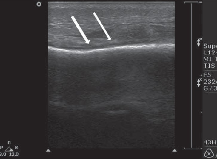
- 48-year-old female with heel pain while walking. Ultrasound shows a thick plantar fascia (white arrow) at the insertion to the calcaneus (red arrow) consisting of plantar fasciitis that improved after local corticosteroid injection.

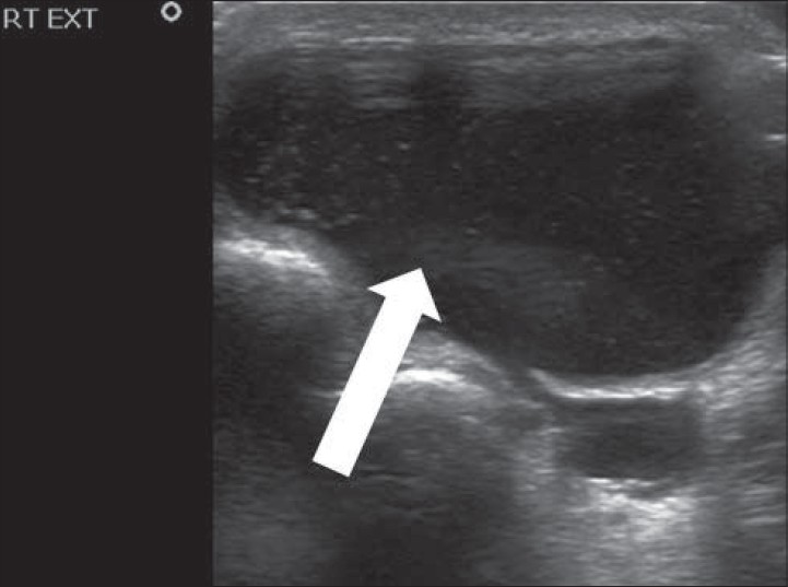
- 44-year-old male referred to ultrasound due to painful mass at the heel. Ultrasound shows a subcutaneous foreign body (blue arrow).
MSKUS can differentiate between micro-, partial-, or full-tear of Achilles tendon or tendinosis, with an accuracy of 92%.[17] It also can differentiate between acute, chronic, non-operatively, or operatively healing tendon tear.[18] Ultrasonographic signs of acute tear include markedly distorted fibrillar architecture, hyperemic edge of the torn tendon, hematoma, and gap at the rupture site. The signs of healed rupture include mild residual distortion of the normal fibrillar architecture, anterior bulging or irregularity of the healed tendon, and hypoechoic area at the rupture site.
Plantar fasciitis is the most common cause of inferior heel pain. It consists of inflammation of the plantar fascia that results from traction forces during weight bearing. It mainly affects middle-aged women and younger, predominantly male, runners. The plantar fascia, similar to the tendons elsewhere, is shown sonographically as a homogeneous echogenic band with internal linear interfaces on longitudinal sections. Diagnosis by ultrasound is easy and consists of showing thickened fascia of more than 4 mm.[19]
Sonography can differentiate the main area of plantar fasciitis as an insertion problem, distal fascia disease, atypical distal fascia disease, or a mixed lesion.[20] It can also be utilized for guidance toward either local corticosteroid injection or surgery.
The major bulk of the MSKUS studies were done for the mid and front portions of the foot [Table 3]. Most of the results of the studies were normal. Majority of the findings were non-inflammatory including hematoma/collection, presence of FBs, and ganglions [Figure 7]. Unusual findings included neurofibroma, hemangioma, xanthoma, arteriovenous malformation (AVM), metastasis, osteomyelitis, avulsion, and others. So, many of the findings here are not easily diagnosed clinically with or without X-ray films and MSKUS plays an important role in the diagnosis of many abnormalities in this part of the foot. MSKUS should be a first-line study in the evaluation of unexplained pain and/or uncertain observed/palpated abnormality of the mid or front portion of the foot.
- 48-year-old man referred to ultrasound due to painful mass at the external part of the ankle. Ultrasound shows a 3.5-cm cystic mass consisting of ganglion cyst (white arrow).
FBs could be a source of serious infection in the foot, especially after trauma. Radioopaque ones are usually picked up by conventional radiography; however, retained radiolucent FBs could be easily missed by conventional radiography, and ultrasound is the method of choice for detecting such bodies.[212223] Ultrasonography also better localizes the FB, especially when surgery is considered.
The typical sonographic appearance of FB consists of a small strong reflector surrounded by hypoechoic tissue. Even a very small-sized FB can be visualized by ultrasound. The suspected area of FB should be scanned in both axial and sagittal planes. Detection of FBs depends on echogenicity, posterior acoustic shadowing, reverberations, and development of a hypoechoic ring or granuloma around the FB. Such a hypoechoic ring usually develops after 24 h and represents an inflammatory reaction. False-negative ultrasound is usually due to perpendicular position of FB, or due to overlying subcutaneous gas.[24]
Ganglion cysts are usually the result of focal myxomatous degeneration of collagenous tissue or from a communication with a joint or tendon sheath. In fact, one useful sonographic criterion is showing the connection between the cysts with the articulation itself.
Ganglion cysts typically contain a viscous, gelatinous fluid that is surrounded by a wall composed of a dense, fibrous connective tissue. Lower extremity ganglion cysts account for an estimated 15-20% of all ganglion cysts. Unlike the wrist ganglia, septations are frequently observed in the ankle ganglia and were observed in 50% of cases in one study.[25]
The typical sonographic appearance of ganglion cyst ranges from round, completely anechoic masses to hypoechoic, multilobulated, multiseptated masses with dependent debris. Most of the ganglion cysts show posterior acoustic enhancement, except for very small cysts. The posterior enhancement could be also obscured when the cyst is too close to the bone.[26]
It should be mentioned here that findings such as tarsal tunnel impingement were not reported in our results, since such a diagnosis was not routinely looked for unless specifically it was asked for or suspected in the referral letter.
One of the main issues regarding MSKUS is the accuracy in the diagnosis of the different findings. This is dependent on many factors, including the person performing ultrasound, version of the machine, prevalence of the findings in a population, type of finding (For example, diagnosis of a ganglion cyst of an extensor tendon of the hand by MSKUS is more accurate than the diagnosis of bone tumor) and others. However, many studies have shown ultrasound to have high accuracy levels in evaluation, especially of the lesions of the foot.
CONCLUSIONS
A huge number of pathologies of the ankle and foot can be detected by MSKUS as it offers a clear advantage of easy availability, lack of radiation, lower cost, and guidance during intervention. Since its first application in 1974 for musculoskeletal use, MSKUS has become the popular method of choice in evaluating different pathologies of the foot with high accuracy, including ganglion cyst, Achilles tendon problems, plantar fasciitis, benign tumors of soft tissue, tenosynovitis, arthritis, collections, vascular abnormalities, and FB. It is also useful in differentiating other pathologies like osteomyelitis, while its use in identifying other pathologies like bone tumors is under study.
MSKUS should be the first modality of imaging in the evaluation of foot and ankle pain.
Available FREE in open access from: http://www.clinicalimagingscience.org/text.asp?2014/4/1/25/133257
Source of Support: Nil
Conflict of Interest: None declared.
REFERENCES
- Comparison of ultrasound and positive contrast arthrography in the diagnosis of popliteal and calf swellings. Ann Rheum Dis. 1974;33:408.
- [Google Scholar]
- Ultrasound and MRI of the peroneal tendons and associated pathology. Skeletal Radiol. 2013;42:1191-200.
- [Google Scholar]
- Traumatic injuries of thigh and calf muscles in athletes: Role and clinical relevance of MR imaging and ultrasound. Insights Imaging. 2012;3:591-601.
- [Google Scholar]
- Heel ultrasound can assess maintenance of bone mass in women with breast cancer. J Clin Densitom. 2012;15:290-4.
- [Google Scholar]
- Achilles enthesis ultrasound: The importance of the bursa in spondyloarthritis. Clin Exp Rheumatol. 2013;31:422-7.
- [Google Scholar]
- Correlation of Radiographic Progression with the Cumulative Activity of Synovitis Estimated by Power Doppler Ultrasound in Rheumatoid Arthritis: Difference Between Patients Treated with Methotrexate and Those Treated with Biological Agents. J Rheumatol. 2013;40:1967-76.
- [Google Scholar]
- Sonographic synovial vascularity of synovitis in rheumatoid arthritis. Rheumatology (Oxford). 2014;53:586-91.
- [Google Scholar]
- Use of contrast-enhanced ultrasonography in musculoskeletal medicine. Am J Phys Med Rehabil. 2012;91:449-57.
- [Google Scholar]
- Three-dimensional versus two-dimensional ultrasonographic assessment of peripheral enthesitis in spondylarthritis. Clin Rheum. 2014;33:131-5.
- [Google Scholar]
- High resolution sonography of the rib: Can fracture and metastasis be differentiated? AJR Am J Roentgenol. 2005;184:969-74.
- [Google Scholar]
- Ultrasonographic detection and guided biopsy of thoracic osteolysis. Chest. 1993;104:1003-5.
- [Google Scholar]
- Sonographically guided core needle biopsy of bone and soft tissue tumors. J Ultrasound Med. 2002;21:275-81.
- [Google Scholar]
- Pigmented villonodular synovitis of the ankle: Radiologic characteristics. J Am Podiat Med Assoc. 2011;101:252-8.
- [Google Scholar]
- Color Doppler ultrasonography of osteomyelitis in children. J Ultrasound Med. 1999;18:729-34.
- [Google Scholar]
- Radiographic imaging in osteomyelitis: The role of plane radiography, computed tomography, ultrasonography, magnetic resonance imaging and scintigraphy. Semin Plast Surg. 2009;23:80-9.
- [Google Scholar]
- Full- versus partial-thickness Achilles tendon tear: Sonographic accuracy and characterization of 26 cases with surgical correlation. Radiology. 2001;220:406-12.
- [Google Scholar]
- Sonographic appearance of nonoperatively treated Achilles tendon ruptures. Skeletal Radiol. 2000;29:259-64.
- [Google Scholar]
- Clinical utility of sonography in diagnosing plantar fasciitis. J Ultrasound Med. 2005;24:1041-8.
- [Google Scholar]
- Ultrasound scanning for recalcitrant plantar fasciopathy. Basis of a new calcifications. Skeletal Radiol. 2013;42:393-8.
- [Google Scholar]
- Ultrasound-guided needle localization to aid foreign body removal in pediatric patients. J Foot Ankle Surg. 2014;53:67-70.
- [Google Scholar]
- Retained palmar foreign body presenting as a late hand infection: Proposed diagnostic algorithm to detect radiolucent objects. Patient Saf Surg. 2013;7:25.
- [Google Scholar]
- Neglected foreign body in contralateral limb in a traumatic transfemoral amputee radiographs can be misleading. Chin J Traumatol. 2013;1:61-4.
- [Google Scholar]
- High-resolution sonography is effective in detection of soft tissue foreign bodies: Experience from a rural Indian center. Ultrasound Med. 2009;28:1245-9.
- [Google Scholar]
- Ganglion cysts of the lower extremity: An analysis of 54 cases and review of the literature. Orthopedics. 1998;21:141-8.
- [Google Scholar]
- Sonography of ankle ganglia with pathologic correlation in 10 pediatric and adult patients. AJR Am J Roentgenol. 2002;178:1445-9.
- [Google Scholar]


