
Translate this page into:
Treatment of Partial Rotator Cuff Tear with Ultrasound-guided Platelet-rich Plasma
Address for correspondence: Dr. Vetrivel Chezian Sengodan, 16 H, Housing Unit, Mettupalayam, Coimbatore - 641 301, Tamil Nadu, India. E-mail: svcortho@gmail.com
-
Received: ,
Accepted: ,
This is an open access article distributed under the terms of the Creative Commons Attribution-NonCommercial-ShareAlike 3.0 License, which allows others to remix, tweak, and build upon the work non-commercially, as long as the author is credited and the new creations are licensed under the identical terms.
This article was originally published by Medknow Publications & Media Pvt Ltd and was migrated to Scientific Scholar after the change of Publisher.
Abstract
Background:
The treatment of symptomatic partial rotator cuff tear has presented substantial challenge to orthopaedic surgeons as it can vary from conservative to surgical repair. Researches have established the influence of platelet rich plasma in healing damaged tissue. Currently very few data are available regarding the evidence of clinical and radiological outcome of partial rotator cuff tear treated with ultrasound guided platelet rich plasma injection in English literature.
Materials and Methods:
20 patients with symptomatic partial rotator cuff tears were treated with ultrasound guided platelet rich plasma injection. Before and after the injection of platelet rich plasma scoring was done with visual analogue score, Constant shoulder score, and UCLA shoulder score at 8 weeks and third month. A review ultrasound was performed 8 weeks after platelet rich plasma injection to assess the rotator cuff status.
Results:
Our study showed statistically significant improvements in 17 patients in VAS pain score, constant shoulder score and UCLA shoulder score. No significant changes in ROM were noted when matched to the contra-lateral side (P < 0.001) at the 3 month follow-up. The study also showed good healing on radiological evaluation with ultrasonogram 8 weeks after platelet rich plasma injection.
Conclusion:
Ultrasound guided platelet rich plasma injection for partial rotator cuff tears is an effective procedure that leads to significant decrease in pain, improvement in shoulder functions, much cost-effective and less problematic compared to a surgical treatment.
Keywords
Platelet-rich plasma
rotator cuff
ultrasonogram


INTRODUCTION
Partial rotator cuff (RC) tears are one of the most common reasons of shoulder pain.[12] The prevalence of partial RC tears in Indian population is not well addressed to date. Current studies showed the prevalence of partial RC tears is more after 50 years of age.[3] The treatment of symptomatic partial RC tear is a challenge to orthopedic surgeons as it can vary from conservative to surgical repair. Surgery can improve patient outcome, but the incidence of delayed healing, infection, stiffness of the shoulder, and other tendon injuries have a comparatively high incidence at about 6%–11%.[4] RC retears have been shown to happen in 11%–94% of RC repairs, depending on the extent of the tear and the level of tendon disintegration.[56]
There are numerous well-established studies about the current tendency in using blood and its by-products to improve the healing course and decrease the pain of tendon tears.[78] Platelet-rich plasma (PRP), a whole blood by-product comprises great concentrations of platelets that, when triggered, endure degranulation to discharge varying kinds of growth factors with restorative properties. These growth factors comprise platelet-derived growth factor, which stimulates cell mitosis; transforming growth factor β, which is concerned in collagen synthesis and morphogenesis; and vascular endothelial growth factor, which aids to induce endothelial cell multiplying and migration, thus commencing an angiogenic response.[910] Besides, platelets have been recognized to be having pain-relieving properties by discharging protease triggered receptor 4 peptides.[7]
MATERIALS AND METHODS
This study was done in Government Coimbatore Medical College Hospital after getting approval from Ethical Committee. The principal objective of the current study is to define the improvement in intensity of pain in patients with partial RC lesions treated with ultrasound guided PRP injection at 8 weeks and 3 months as measured by the visual analog scale for pain (VAS). The secondary objective is to assess the consequence of PRP on shoulder functions in day-to-day activities before and after injection. The primary hypothesis for this study is that PRP is effective in decreasing pain in patients with RC injury and improving the functional result.
The study was built under one orthopedic surgeon and one radiologist. Thirty-two patients who were diagnosed to be having symptomatic partial RC tear were screened from July 2014 to December 2016. The selection was based on a detectable partial tears with <50% thickness on both magnetic resonance imaging and ultrasonography (USG). Ten patients were excluded from this study as per eligibility criteria, and 2 patients did not agree to participate. All the consenting patients were evaluated clinically to rule out pain related to other shoulder pathologies. The study comprised 20 patients, with partial RC tears who gave willingness by providing written consent. These patients underwent demographic and basic surveys initially and staged using VAS, Constant and University of California Los Angeles (UCLA) score after managing them conservatively for 2 months. All these patients were given PRP injection under ultrasound guidance, and they were evaluated at 8 weeks and 3 months postinjection [Figure 1]. A prespecified provisional analysis was piloted in the middle of study to assess efficacy and statistical power. At the period of the interim analysis, it was assumed that there is a statistically significant metamorphosis in VAS pain score among the study group.
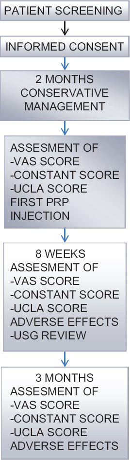
- Original flow diagram for study methodology showing the preinjection and postinjection evaluation of patients using visual analog score, constant, and University of California Los Angeles shoulder score.
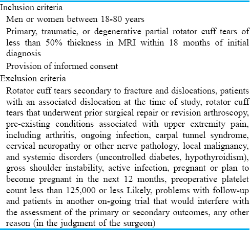
We included active men and women between 18 and 80 years of age with primary, traumatic, or degenerative RC tears within 18 months of initial diagnosis. The provision of informed consent is shown in Table 1. We excluded all patients with RC tears secondary to fracture and dislocations, patients with an associated dislocation at the time of study, RC tears that underwent prior surgical repair or revision arthroscopy, preexisting conditions associated with upper extremity pain, including impingement, arthritis, ongoing infection, carpal tunnel syndrome, cervical neuropathy, other nerve pathologies, local malignancy, and systemic disorders (uncontrolled diabetes, hypothyroidism), gross shoulder instability, active infection, pregnant or plan to become pregnant in the next 12 months, preoperative platelet count <125,000 or less likely, problems with follow-up and patients in another on-going trial that would interfere with the assessment of the primary or secondary outcomes, any other reason (in the judgment of the surgeon)
Platelet-rich plasma preparation and procedure
PRP was prepared using standard techniques. Initially, patient's whole blood was collected with aseptic precautions in acid citrate dextrose tubes. Around 20 ml of patient's whole blood is collected. This whole blood was subjected to centrifugation at 2000 rpm (soft spin). The whole blood will separate into three layers. The supernatant layer of plasma and buffy coat were separated and subjected to centrifugation at 3000 rpm (hard spin). In the final end product, the upper two-third of the tube will be containing platelet poor plasma which is removed, and the lower one-third will be PRP enhanced with superficial buffy coat which will be used for injection. The procedure was performed by a team of radiologist and orthopedic surgeon. The cuff tear size was evaluated using a high-frequency musculoskeletal probe. The procedure initiated with a diagnostic ultrasonogram of the glenohumeral joint. Using a 22-to 25-gauge needle, 2–3 ml of the leukocyte-rich PRP product was injected at the RC tear site, and under ultrasound guidance [Figure 2]. All injections were performed after painting the patient with betadine and sterilizing the USG probe with betadine and surgical spirit, with patients positioned in sitting position.
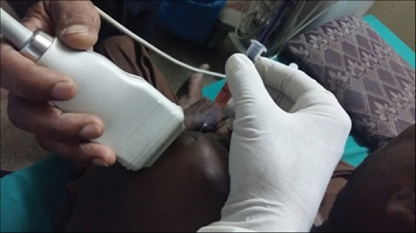
- Original, a 52-year-old male patient with partial supraspinatus tear receiving platelet-rich plasma injection under ultrsound guidance.
This is executed as an OP technique. After injection, patient's arms were supported with a simple sling until pain subsides. Patients were requested to mobilize the shoulder at the earliest. All pain management methods were avoided, including oral and injections of pain medication. Patient follow-up was done by attending surgeon at 8 weeks and 3 months postinjection. The patients completed the VAS for pain, Constant shoulder score, and UCLA shoulder score for quality of shoulder function. They were also asked about adverse events, and this information was verified by their surgeon and by a review of their medical records.
The primary outcome was change in pain severity, measured using a VAS at 8 weeks and 3 months postinjection. Patients had to think their worst pain in their shoulder for the past 24 h on a 10-cm vertical scale, with “0” indicating no pain at all and “10” indicating the maximum pain the patient could imagine using a series of smilies. The VAS is a simple and sensitive measure that can detect minor changes in an individual's perception of pain severity and easy to understood.[11] This scale is widely used and considered to be best in determining fluctuation of pain over time than a questionnaire with a selected number of responses, with good construct validity and high internal consistency. Secondary outcome measures include the Constant and UCLA shoulder score at 8 weeks and 3 months postinjection, as well as the incidence of surgeries and adverse events. Briefly, the constant shoulder score is a questionnaire about the past 4 weeks of shoulder function.[12] It considers shoulder function under 8 headings pain, activity level, arm positioning, forward flexion, lateral elevation, internal rotation, and external rotation. A score difference of >30 is considered as poor, 21–30 fair, 11–20 good, and <11 is excellent. The UCLA shoulder score has questions regarding the past 4 months of shoulder activity under the headings pain, function, active forward flexion, strength of forward flexion, satisfaction of the patient.[13] A score above 27 is graded as excellent/good and below 27 is considered as poor/fair. All adverse events and complications are noted down by the team.
The central committee reviewed all adverse events to see if they were related to the injected product and to be considered an outcome event. After independent assessment, consensus was reached for each decision.
Data Analysis was completed using SPSS for Windows. Descriptive statistics (frequencies for dichotomous data and means and standard deviations (SD) for continuous data) were calculated to describe the demographic characteristics of the patients. Paired t-test was completed at 8 weeks and 3 months for each of the 3 outcome measures to determine whether there was a significant difference in the mean scores between the preinjection and postinjection values. The analysis included t values, significance (P) values, mean differences, and 95% confidence intervals.
RESULTS
The mean age of the study participants was 55 ± 6.4 years and that patient age range was similar across the treatment groups. There were 12 men and 8 women, and sex was also distributed similarly across the treatment groups. Fifteen patients had RC tear in their dominant arm. All patients had either a degenerative RC tear (48%) or a traumatic tear (52%). Out of 20, 17 patients were having supraspinatus tear and two patients with infraspinatus tear and one patient was having subscapularis tear.
An exploratory analysis of VAS pain scores at 8 weeks includes data of only eligible patients. As two patients had already lost follow-up, data of only 20 patients were analyzed. The difference between preinjection and postinjection VAS scores was extremely statistically significant (P < 0.001). The preinjection, mean ± SD VAS pain scores were 5.4 ± 0.92, and after 8 weeks, it is 3.2 ± 0.94, respectively [Figure 3]. After 3 months, the mean ± SD VAS pain scores were 2.55 ± 0.83 with a (P < 0.001).
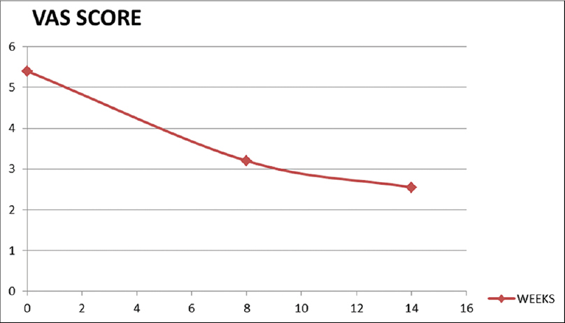
- Original, scattered diagram showing the relationship between preinjection and postinjection (8 weeks and 3 months) VAS score. VAS is plotted along the x-axis and duration of treatment in weeks along the y –axis. The curve shows nonlinear decline in VAS score. VAS: Visual analog score.
The UCLA shoulder score and Constant shoulder score showed statistically significant improvement at 8 weeks and 3 months, respectively (P < 0.001) [Figure 3]. The UCLA shoulder score was increasing from baseline to 8 weeks with initial mean ± SD of 14.05 ± 1.05and at 8th week 25.75 ± 2.75 at (P < 0.001). There was also significant improvement from 8th week to 3 months (29.10 ± 3.34) at P value <0.0001. After 3 months, 11 patients had excellent UCLA score, 6 patients had good, 2 had fair, and one patient had poor score. The Constant shoulder also showed improvement from baseline (42.80 ± 3.53–64.7 ± 4.1 in 8 weeks and77.50 ± 4.45 in 3 months). This was also statistically significant improvement in 8th week and in 3rd month with P < 0.0001 [Figure 4]. An ultrasonogram done at 8 weeks also showed complete healing in 11 patients and partial healing in 5 patients. Nearly 81% of the completely healed RC tears are intratendinous tears [Figure 5a–c].
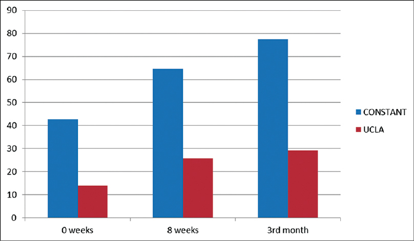
- Original bar diagram plotting improvement of CONSTANT score and University of California Los Angeles score in 8 weeks and 3 months postinjection.
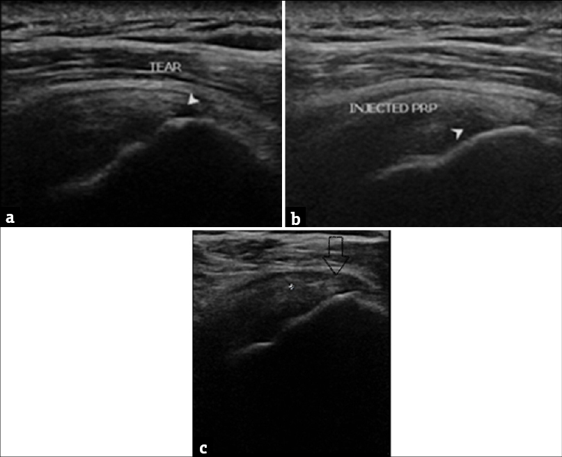
- (a) Original, ultrasound image of a 52-year-old male showing articular sided partial supraspinatus tear following a 2 months of conservative management (b) The same patient after ultrasound-guided platelet-rich plasma injection, arrow mark showing injected platelet-rich plasma (c) The same patient evaluated after period of 8 weeks, ultrasound shows healing in the tear site.
The main adverse events identified by the central committee are: (1) pain and (2) dizziness immediately postinjection. Both of them are not specific for the study event.
DISCUSSION
Our study showed a significant change in the preinjection and postinjection VAS scores at 8 weeks and 3 months while the study was able to demonstrate that the injected product influenced the pain score, the overall decrease in pain from baseline for group demonstrates that ultrasonogram-guided injection can be effective in pain control. Most of the people were satisfied because of the adequate pain control by 3rd month. The secondary objectives which designate the shoulder function exhibit unique progressive pattern. The UCLA shoulder score increases from baseline to 8th week with good patient satisfaction. The 3rd month review also shows a unanimous increase in the score with a definite difference in shoulder abduction. The Constant score shows improvement in 8th week and 3rd month compared to baseline showing improved range of motion in the shoulder. No revision procedures were required till the 3rd month except for one patient for whom arthroscopic RC repair was done because of the unsatisfactory result. The most common adverse effect was postinjection pain which was mainly treated with ice packing. Pain is nonspecific to study event, or it could be technically related. One patient had postinjection dizziness which was not considered to be a study event by Central Committee. There were no other specific adverse effects regarding PRP. These overall findings are consistent with the mounting literature examining the use of PRP in RC injuries.
There are several strengths in the present study. First, the sample of the patients was same, and everybody received PRP injection. Shoulder functions and pain scores were collected at frequent time points postinjection, allowing for an increased likelihood that subtle changes may be noticed. Furthermore, the follow-up was 91% at 8 weeks. The primary outcome measure used for assessing the effect of PRP, was not able to represent each RC muscles individually using specific data.
There are little studies suggesting the effect of ultrasound-guided PRP injection in partial RC RC tears. However, there are studies in augmentation of arthroscopic RC repair using PRP intraoperatively. Saltzman et al. in 2016 concluded that there is improvement in pain and reduction in rehabilitatory period in cases where PRP augmentation was done in patients with RC tear. When PRP is used to augment rotatorcuff repair, it resulted in decreased retear rates, early going back to day-to-day activity and improvement in pain.[14]
Randelli et al. study revealed that when PRP is used for the augmentation of arthroscopically conducted cuff repair, all the fourteen patients had reduction in pain and functional improvement and had no adverse effect as shown by improvement in constant score at 12 weeks following the repair.[6]
Preliminary data suggest that the administration of PRP into injured tissues promotes natural healing mechanisms by release of growth factors and other bioactive substances. This prospective study demonstrates that ultrasound-guided PRP injection resulted in markedly improved clinical, functional, and radiological outcomes.
CONCLUSION
The present study demonstrated that there is conclusive benefit for reducing pain and improving shoulder function in partial RC tears with ultrasound-guided PRP. Therefore, the primary hypothesis is accepted. This study succeeded in demonstrating that those patients receiving PRP injections have decreased pain, healing properties, and superior functional outcomes. However, how PRP works on a partially torn RC remains a challenge and deserves ongoing investigation.
Financial support and sponsorship
Nil.
Conflicts of interest
There are no conflicts of interest.
Available FREE in open access from: http://www.clinicalimagingscience.org/text.asp?2017/7/1/32/212941
REFERENCES
- Partial-thickness rotator cuff tears. Pathogenesis and treatment. Orthop Clin North Am. 1997;28:145-55.
- [Google Scholar]
- Does platelet-rich plasma accelerate recovery after rotator cuff repair? A prospective cohort study. Am J Sports Med. 2011;39:2082-90.
- [Google Scholar]
- Autologous platelet rich plasma for arthroscopic rotator cuff repair. A pilot study. Disabil Rehabil. 2008;30:1584-9.
- [Google Scholar]
- Strategies in biologic augmentation of rotator cuff repair: A review. Clin Orthop Relat Res. 2010;468:1476-84.
- [Google Scholar]
- Platelet rich plasma in arthroscopic rotator cuff repair: A prospective RCT study, 2-year follow-up. J Shoulder Elbow Surg. 2011;20:518-28.
- [Google Scholar]
- Protease-activated receptor-4: A novel mechanism of inflammatory pain modulation. Br J Pharmacol. 2007;150:176-85.
- [Google Scholar]
- The role of platelet-rich plasma in arthroscopic rotator cuff repair: A systematic review with quantitative synthesis. Arthroscopy. 2012;28:1718-27.
- [Google Scholar]
- Protein therapeutics and bone healing. In: Lynch SE, Wisner-Lynch LA, Nevins M, Marx RE, eds. Tissue Engineering: Applications in Oral and Maxillofacial Surgery and Periodontics (2nd ed). Chicago, IL: Quintessence; 2008. p. :3-25.
- [Google Scholar]
- Platelet-rich therapies in the treatment of orthopaedic sports injuries. Sports Med. 2009;35:1-10.
- [Google Scholar]
- A descriptive study of the use of visual analogue scales and verbal rating scales for the assessment of postoperative pain in orthopedic patients. J Pain Symptom Manage. 1999;18:438-46.
- [Google Scholar]
- Age Related Recovery of Shoulder Function After Injury. Cork, Ireland: University College; 1986.
- Does the use of platelet-rich plasma at the time of surgery improve clinical outcomes in arthroscopic rotator cuff repair when compared with control cohorts? A systematic review of meta-analyses. Arthroscopy. 2016;32:906-18.
- [Google Scholar]


