
Translate this page into:
Primary Epithelioid Angiosarcoma of Lung: Radiologic and Clinicopathologic Correlation
Address for correspondence: Dr. Fung Him Ng, Department of Radiology and Organ Imaging, United Christian Hospital, Kowloon, Hong Kong. E-mail: ngphonehim@gmail.com
-
Received: ,
Accepted: ,
This is an open access article distributed under the terms of the Creative Commons Attribution-NonCommercial-ShareAlike 3.0 License, which allows others to remix, tweak, and build upon the work non-commercially, as long as the author is credited and the new creations are licensed under the identical terms.
This article was originally published by Medknow Publications & Media Pvt Ltd and was migrated to Scientific Scholar after the change of Publisher.
Abstract
Primary pulmonary angiosarcoma is extremely rare. It is often characterized by a clinically indolent course and delayed diagnosis. To date, there have been <20 cases reported. By far, little article correlates the clinical presentation, the imaging findings with the pathology. The authors present a case of middle-aged gentleman with primary pulmonary epithelioid angiosacroma which we initially thought as tuberculosis (TB) infection. A 60-year-old gentleman, with a history of 6 months on and off blood stained sputum, was admitted for an episode of massive hemoptysis. Urgent computed tomography (CT) bronchial arteriogram excluded any dilated bronchial artery. Patchy consolidation with multiple small centrilobular ground-glass nodules was noted at left upper lobe. The bronchoscopy was negative for malignancy and infection. Autoimmune workup was negative. Despite negative bronchoscopy, fungal, acid-fast bacilli culture and cytology, and anti-TB treatment were empirically given. However, his hemoptysis was unresolved. He was followed up with high-resolution CT after a month showed an enlarging left upper lobe mass surrounding by a ground glass halo. Left thoracotomy and left upper lobe lobectomy were performed. Epithelioid angiosacroma was found in histology. Radiologic and clinical-pathological findings were correlated in this paper.
Keywords
Hemoptysis
lung cancer
primary epithelioid angiosacroma
pulmonary hemorrhage


INTRODUCTION
Angiosarcoma is a rare malignant neoplasm derived from uncontrolled proliferation of anaplastic vascular endothelial cells that line abnormal blood-filled sacs. Primary angiosarcoma of lung is an extremely rare condition. Only about 20 case reports published in English.[1] It is often characterized by a clinically indolent course and delayed diagnosis. By far, little article correlates the clinical presentation, the imaging findings with the pathology. The authors present a case of middle-aged gentleman with primary pulmonary epithelioid angiosacroma, which we initially thought as tuberculosis (TB) infection.
CASE REPORT
A 60-year-old gentleman, with a history of 6 months on and off blood stained sputum, was admitted for an episode of massive hemoptysis. Urgent computed tomography (CT) bronchial arteriogram was performed to assess the need for bronchial artery embolization. It excluded any dilated bronchial artery. Focal consolidation with multiple small centrilobular nodules and adjacent ground glass changes were found [Figure 1a and b]. Bronchoscopy with biopsy taken was negative for malignancy and infection. Autoimmune workup was negative. Tuberculosis infection (TB) could present with centrilobular nodules and consolidative changes in CT, and relatively common in Asia. The culture sometimes could be false negative in partially treated cases. Combined with the clinical picture, with negative bronchoscopy, fungal, acid-fast bacilli culture and cytology, the case was treated as culture-negative TB. However, his hemoptysis was unresolved. He was followed up with high-resolution CT after a month, showed an enlarged left upper lobe mass surrounding by a ground glass halo [Figure 2a and b] Neoplasm had to be considered. Left thoracotomy and left upper lobe lobectomy were performed. Epithelioid angiosacroma was found in histology. The sections showed an infiltrative hemorrhagic tumor, which eroded the bronchus and was associated with bronchial luminal hemorrhage and diffuse hemorrhage into the adjacent pulmonary parenchyma [Figure 3a]. The tumor comprised sheets of loosely cohesive malignant tumor cells which had eccentric markedly pleomorphic nuclei, prominent nucleoli, vesicular with presence of fine strands of chromatin, and moderate amount of eosinophilic cytoplasm [Figure 3b]. In areas, adenomatoid tumor arrangement, ill-formed ectatic vascular channel formation, and hemorrhagic microcystic arrangement with mitotic figures up to 1.5 per 10 high-power fields. The tumor cells were immunoreactive to vascular markers ERG and CD31 [Figure 3c and d], and are negative to CD34. Some tumor cells showed cytoplasmic staining upon study for epithelial marker CK. Tumor cells were negative to TTF-1. The overall features supported epithelioid angiosarcoma. On review of the intitial CT, there is partial obliteration of the segmental bronchus, these findings would be more suggestive of malignancy than atypical infection such as TB or fungal infection.
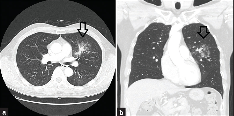
- A 60-year-old gentleman, with a history of 6 months on and off blood stained sputum, was admitted for an episode of massive hemoptysis. (a) Selected axial image of urgent computed tomography thorax (lung window) showed focal consolidation with multiple small centrilobular nodules and adjacent ground glass changes (arrow). (b) Selected coronal image of computed tomography thorax could appreciate the adjacent ground glass changes around the focal consolidation at left upper lobe (arrow).
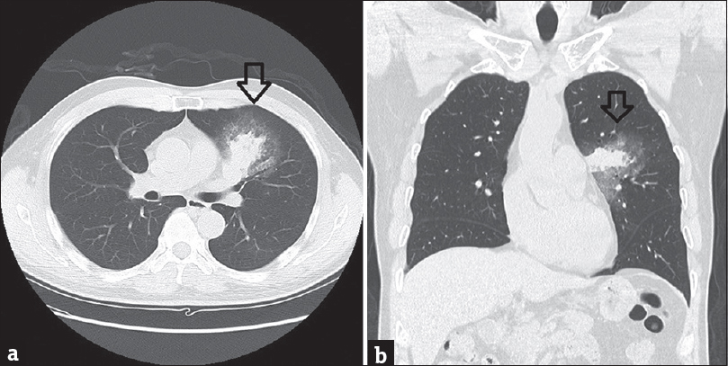
- (a) With repeated hemoptysis, investigation with high resolution computed tomography after a month (selected axial image) showed the left upper lobe mass is significantly enlarged (arrow). (b) Selected coronal image of high resolution computed tomography could appreciate the ground glass halo around the enlarged lung mass (arrow).

- Histopathological examination of lobectomy of left upper lobe. (a) Showed hemorrhage into the bronchiole (arrow). It correlated with the computed tomography images of ground glass changes around the mass. (b) Histopathological examination showed the tumor comprised sheets of loosely cohesive malignant tumor cells which had eccentric markedly pleomorphic nuclei, prominent nucleoli, vesicular with the presence of fine strands of chromatin, and moderate amount of eosinophilic cytoplasm.(high power). (c) Tumor cells stained positive for ERG, a vascular-specific marker (magnification power, ×100). (d) Tumor cells stained positive for CD31, a vascular-specific marker (magnification power, ×40).
DISCUSSION
Primary pulmonary angiosarcoma is difficult to diagnose clinically with nonspecific respiratory symptoms. Diagnosis is often late due to low index of suspicion and usually metastasized.[1234] The most common clinical presentation is hemoptysis associated with pulmonary hemorrhage, which has been described in a handful of cases.[23] It also happened in our presented case.
Etiology remains unknown. Predisposing factors include polyvinyl chloride and thorium dioxide exposure, postmastectomy and postirradiation states, and chronic empyema for pleural angiosarcomas.[4]
Prognosis of pulmonary angiosarcoma has been shown to be poor with almost all patients dead within months of initial presentation. Our presented case was fortunate that the tumor was still localized in the left lung and curative intented left thoracotomy and left upper lobectomy could be performed with negative disease involvement of mediastinal lymph nodes.
Pathologic features
Angiosarcomas are rare malignancies of endothelial origin. These neoplasms are classified into cutaneous, visceral, and soft tissue subtypes. Histologically, angiosarcomas range from well-differentiated tumors with variable endothelial atypia to high-grade spindle cell malignancies. There is a distinctive, unique morphologic subtype of angiosarcoma. The malignant endothelial cells have a predominantly or exclusively epithelioid appearance which defines the epithelioid angoisarcoma.[5] As with all types of angiosarcoma, the epithelioid variant is strongly vimentin positive. Factor VIII-related antigen, CD34 and CD31, which are specific markers for tumors derived from the endothelium. The histologic features of epithelioid angiosarcoma frequently preclude a straightforward diagnosis on routine H- and E-stained microscopic evaluation. Carcinomas will not stain for endothelial markers, including factor VIII, CD31, and CD34.[567]
Radiologic features
Primary pulmonary angiosarcomas may occur either as multifocal lesions or as a solitary nodule.[8] When the involvement is multifocal, chest radiographs show bilateral reticulonodular or alveolar infiltrates, and with or without pleural effusion. This pattern of involvement suggests metastatic cancer and lymphangitic carcinoma. For the solitary form of primary pulmonary angiosarcoma, the size of the lesion may vary from a small nodule to a large mass invading the mediastinum or chest wall.[5] There may be accompanying pleural or pericardial effusions, and the surrounding lung may contain hemorrhage.[12] Erosion of the adjacent bronchial structures may cause hemoptysis.[125] Ground glass changes or halo sign around the tumor corresponding to area of pulmonary hemorrhage was seen in our presented case [Figures 1a, b and 2a, b] Obliteration of the segmental bronchus would be more suggestive for malignancy as TB or fungal infection may also presented as mass-like consolidation with surrounding centrilobular nodules and ground glass changes, which were more common differentials compared with the rarity of this malignancy. This subtle finding was present in our initial CT image [Figure 1a]. Differentiation from the more common bronchogenic carcinoma is practically impossible.
CONCLUSION
Pulmonary angiosarcomas are characterized by an insidious growth and a late stage at presentation when metastases have already occurred. CT would be useful to differentiate malignancy from other causes of hemoptysis, with particular attention to the bronchial wall involvement, but differentiation of pulmonary angiosarcomas from other masses, especially lung cancer, is not possible without biopsy. The exact diagnosis requires histology with immunohistochemical methods in most patients. Among the various immunohistochemical markers used for classification, factor VIII-related antigen, and CD31 are considered specific for tumors derived from endothelium.
Financial support and sponsorship
Nil.
Conflicts of interest
There are no conflicts of interest.
Acknowledgment
Special thanks to Dr. W. H. Lau, Associate Consultant of Department of Pathology, Queen Elizabeth Hospital, Hong Kong, for the digital histological images of the disease.
Available FREE in open access from: http://www.clinicalimagingscience.org/text.asp?2017/7/1/33/213659
REFERENCES
- Primary pulmonary epithelioid angiosarcoma presenting as a solitary pulmonary nodule on image. Pathol Int. 2012;62:424-8.
- [Google Scholar]
- Primary epithelioid angiosarcoma of the lung presenting as pulmonary hemorrhage. Hum Pathol. 1997;28:383-5.
- [Google Scholar]
- Primary epithelioid angiosarcoma of the lung presenting as pulmonary hemorrhage. Asian Cardiovasc Thorac Ann. 2006;14:69-71.
- [Google Scholar]
- Radiographic, CT, and MRI findings in primary pulmonary angiosarcoma. Clin Imaging. 2001;25:337-40.
- [Google Scholar]
- Diagnostic Histopathology of Tumors Vol 1. (3rd ed). Philadelphia, PA: Elsevier Limited; 2007. p. :66-7.
- Epithelioid angiosarcoma of deep soft tissue: A distinctive tumor readily mistaken for an epithelial neoplasm. Am J Surg Pathol. 1991;15:915-24.
- [Google Scholar]
- Epithelioid angiosarcoma: A brief diagnostic review and differential diagnosis. Arch Pathol Lab Med. 2011;135:268-72.
- [Google Scholar]
- Metastatic angiosarcoma of the lung: Spectrum of CT findings. AJR Am J Roentgenol. 2003;180:1671-4.
- [Google Scholar]


