
Translate this page into:
Rare Coexistence of a Cerebellar Hemangioblastoma and Angiomatosis of the Breast without Underlying Phakomatosis
-
Received: ,
Accepted: ,
Abstract
Angiomatosis of the breast is an unusual benign vascular process which may affect middle aged women and simulate carcinoma. We report a unique case of a female patient with cerebellar hemangioblastoma and coexisting breast angiomatosis. We discuss the neuroradiology and breast imaging, illustrating the diagnostic pearls and pitfalls in the setting of this extremely uncommon combination. A 50-year old patient with a history of right-sided cerebellar hemangioblastoma resection 10 years previously presented with a recurrent left sided palpable breast mass. She was referred for triple breast assessment and subsequent ultrasound-guided biopsy. On physical examination, the lesion was hypoechoic, ill-defined and located in the upper outer quadrant as are most breast malignancies. Ultrasound and mammography showed suspicious features. The ipsilateral axilla was normal. Histopathology revealed a diagnosis of breast angiomatosis with no evidence of associated malignancy. Vascular tumors of the breast are very rare, present diagnostic challenges and are prone to local recurrence. Complete excision with clear margins is recommended. Mastectomy is a consideration for diffuse disease that cannot be fully cleared with lumpectomy or Wide local excision. Cerebellar hemangioblastoma and breast angiomatosis is a very unique combination, particularly in the absence of an underlying phacomatosis. Radiological features of angiomatosis mimicking malignancy without pathognomonic imaging signs have been observed. Knowledge of these rare vascular breast tumors is key to making this unusual diagnosis and helps to reduce the number of radical surgical procedures.
Keywords
Angiomatosis of the breast
hemangioblastoma
benign breast lesions
sonography
mammography
biopsy


INTRODUCTION
Vascular tumors of the breast are uncommon neoplasms which can be benign, premalignant, or malignant. Angiomatosis of the breast is a rare condition that can simulate carcinoma and mislead specialist breast imagers regardless of experience. Angiomatosis most commonly appears in isolation, may be large, and, due to shared imaging features, needs to be differentiated from angiosarcoma.[1]
We report a very unique and complex case of a patient with coexisting recurrent breast angiomatosis and cerebellar hemangioblastoma in whom no underlying phakomatosis was identified.
The clinical presentation, physical findings, mammography, ultrasonography, and neuroradiology are presented and discussed, in combination with a review of the literature with regard to this extremely rare combination of pathologies.
CASE REPORT
In 2015, a 47-year-old female was referred to the triple breast assessment clinic with a left-sided breast lump. She had no history of breast pathology. Physical examination revealed a painless, palpable mass. A subsequent ultrasound of the whole left breast was performed which demonstrated a solitary hypoechoic, irregular mass measuring approximately 2 cm located at 2 o’clock in the upper outer quadrant. There was no associated axillary lymphadenopathy.
Bilateral mammography was performed demonstrating an irregular suspicious mass with features suggestive of malignancy, thus assigned BI-RADS 5 [Figure 1].
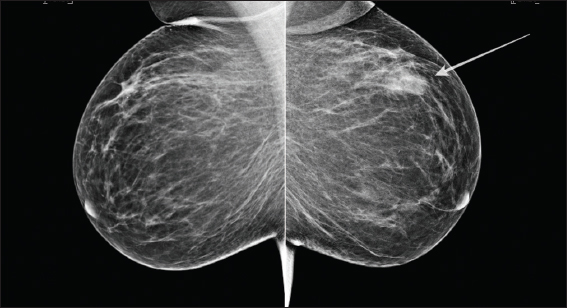
- A 47-year-old female with a history of painless palpable mass within the left breast. Bilateral mammography showing an ill-defined mass in the left upper outer quadrant in keeping with biopsy-proven mammary angiomatosis (white arrow).
After administration of local anesthetic, ultrasound-guided core biopsies using a 14-Gauge needle were performed, and multiple samples sent for histopathologic examination. Histopathology revealed a diagnosis of breast angiomatosis with no evidence of malignancy or atypia.
Given the tendency of these rare tumors to recur, wide local excision with clear margins was performed. The mass was clearly palpable and thus there was no need to perform wire localization. Routine follow-up was advised.
Three years later, the patient re-presented to the triple breast assessment clinic with suspected local recurrence. The mammographic and sonographic features were suspicious, again showing a similar irregular lesion, which was indistinguishable from cancer [Figures 2 and 3].
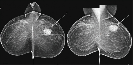
- Three years later, the same patient represented with a history of recurrent left-sided palpable breast mass. Mammography: Local recurrence of the previously diagnosed left-sided angiomatosis demonstrated on mediolateral oblique and craniocaudal views. The mass appears much more dense and is more conspicuous than on prior imaging (white arrows).
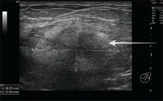
- Three years later, the same patient represented with a history of recurrent left-sided palpable breast mass. Ultrasound of the left upper outer quadrant showing an irregular hypoechoic mass correlating with the mammographic abnormality and palpable lesion (white arrows).
Microscopic examination revealed recurrent angiomatosis with vascular spaces lined by endothelial cells without any features of malignant transformation, nuclear atypia, or low-grade angiosarcoma.
The initial vascular lesion recurred despite the previously noted clear margins, and the complete excision was again recommended.
Thus, it is important to highlight the risk of recurrence with regard to this uncommon entity.
Seven years before her initial breast presentation, she had been referred to the neurology department with nausea, dizziness, and unsteady gait. Neuroradiology demonstrated typical appearances consistent with right-sided cerebellar hemangioblastoma on both computed tomography and postcontrast magnetic resonance imaging (MRI) [Figure 4].
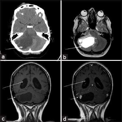
- (a-d) Seven years before the initial symptomatic breast presentation, the patient had been admitted to the hospital with a history of nausea, dizziness, and unsteady gait. Noncontrast computer tomography (a) and T2-weighted magnetic resonance imaging (b) showed a well-defined posterior fossa cystic lesion in keeping with right-sided hemangioblastoma (white arrows). Pre- and postcontrast T1-weighted coronal sequences (c and d) confirm the typical appearances of a hemangioblastoma, i.e., a large cystic component and a small enhancing mural nodule (short white arrows). No enhancement evident in relation to the cystic component. Associated hydrocephalus also evident (long white arrows).
MRI of the brain revealed cerebellar tumor consistent of a large cystic component and a small associated enhancing mural nodule on T1 post-gadolinium sequence. The cystic component remained unchanged on both pre- and postcontrast images. There was associated subsequent hydrocephalus. No other findings or associated symptoms to suggest phakomatosis were evident. After successful neurosurgery, the patient was discharged with no immediate complications.
DISCUSSION
In general, angiomatosis of the breast is extremely rare with few cases reported in the literature.[2] These vascular tumors may demonstrate as palpable masses on physical examination.[3]
The possible age of presentation is between 20 and 60 years.[4] It is important to note that this unusual entity may appear in both sexes, with a female predominance.[4]
Angiomatosis may recur after local excision and thus complete excision with clear margins is recommended.[2] Mastectomy is a consideration for diffuse disease that cannot be fully treated with breast-conserving surgery.[1]
Hemangioblastoma is a benign vascular neoplasm mostly occurring in the cerebellum, which may be related to von Hippel–Lindau disease or appear in isolation.[5] Juvenile pilocytic astrocytoma is the main differential; however, the latter occurs in a younger group of patients.[6,7]
Given the vascular nature of the presented coexisting tumors, both cerebellar hemangioblastoma and the breast angiomatosis, an underlying phakomatosis with regards to von Hippel–Lindau syndrome was a consideration. This has not been confirmed in the subsequent multidisciplinary diagnostic process, and the groups suggested no overt connection between the lesions.
Thus, it remains uncertain if there is a causal relationship between these symptomatic presentations with vascular lesions of the central nervous system and the breast or if it is purely coincidental. The rarity of angiomatosis means research has been limited.[8] To the best of our knowledge, there is no overt association between angiomatosis of the breast and von Hippel–Lindau disease.[9]
However, more cases need to be reported to establish the significance of this combination and to evaluate if there is a predisposing genetic mutation or connection.
CONCLUSIONS
Angiomatosis of the breast is a very unusual vascular tumor, and only limited cases have been reported. Moreover, cerebellar hemangioblastoma and breast angiomatosis is a very exceptional combination and research needs to be performed to establish the exact significance of this coexistence. It remains unknown if there is an underlying genetic abnormality that could contribute to the formation of both vascular tumors in this particular case.
No specific mammographic or sonographic features were visualized to support the diagnostic breast imaging process. Even armed with the broad knowledge of potential vascular breast cancer mimickers, it is not possible to make such a diagnosis based on imaging alone. Thus, histopathological confirmation is required. The mammary angiomatosis can be fully cleared with breast-conserving surgery in most cases, and the overall prognosis is excellent.
Declaration of patient consent
The authors certify that they have obtained all appropriate patient consent forms. In the form the patient(s) has/have given his/her/their consent for his/her/their images and other clinical information to be reported in the journal. The patients understand that their names and initials will not be published and due efforts will be made to conceal their identity, but anonymity cannot be guaranteed.
Financial support and sponsorship
Nil.
Conflicts of interest
There are no conflicts of interest.
REFERENCES
- Focal angiomatosis of the breast with MRI and histologic features. Radiol Case Rep. 2017;12:219-22.
- [Google Scholar]
- Angiomatosis of breast –An extremely rare benign vascular lesion of the breast:A case report. Int Surg J. 2015;2:115-7.
- [Google Scholar]
- Diffuse angiomatosis of the breast –Sonographic appearance. J Clin Ultrasound. 2014;42:498-501.
- [Google Scholar]
- Diffuse dermal angiomatosis of the breast:A series of 22 cases from a single institution. Gland Surg. 2015;4:554-60.
- [Google Scholar]
- Imaging in von Hippel-Lindau Syndrome. Manchester, UK: Medscape; 2015.


