
Translate this page into:
Prevalence and Degree of Breast Arterial Calcifications on Mammography: A Cross-sectional Analysis
Address for correspondence: Dr. Norman Loberant, Department of Radiology, Western Galilee Hospital, Nahariya, Israel. E-mail: nloberant@yahoo.com
-
Received: ,
Accepted: ,
This is an open-access article distributed under the terms of the Creative Commons Attribution License, which permits unrestricted use, distribution, and reproduction in any medium, provided the original author and source are credited.
This article was originally published by Medknow Publications & Media Pvt Ltd and was migrated to Scientific Scholar after the change of Publisher.
Abstract
Objectives:
The purpose of this study is to establish a database including prevalence and degree of breast arterial calcifications (BAC) in our population of women presenting for mammography.
Materials and Methods:
The mammograms of 1786 women over the age of 40 years were examined for the presence and degree of BAC. Statistical analysis was performed to correlate patient's age and ethnic origin with the presence and degree of BAC.
Results:
There was statistically significant and strong correlation between the patient's age and presence of BAC. There was also a less strong yet statistically significant correlation between patient age and degree of BAC. Regression analysis showed the likelihood of BAC at various ages. The prevalence of BAC is only 2% of women under 50 years of age; the prevalence of Grade 2-3 BAC is only 1% in women under 60 years of age.
Conclusion:
There is a predictable increase with age in both prevalence and degree of BAC in women. The presence of high degree BAC in women under 60 years of age or any BAC in women under 50 years of age is unusual.
Keywords
Arterial calcifications
cardiovascular disease
mammography
risk factors

INTRODUCTION

In recent years, evidence has accumulated that breast arterial calcifications (BAC) noted on mammography are indicators of coexisting cardiovascular disease. Cardiovascular diseases are a significant cause of morbidity and mortality in women over 50 years and the feasibility of early diagnosis is important. We examined the prevalence of BAC in a population of women undergoing both screening and diagnostic mammography in order to establish a local database. In addition, we examined the degree of BAC in order to examine the change of this parameter with age. The study was approved by the hospital's institutional review committee.
MATERIALS AND METHODS
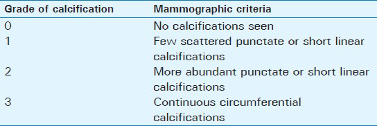
During a 1-year period around 1786 women's (age range 40-93 years) underwent screening or diagnostic examinations in our mammography unit. Two experienced breast radiologists interpreted all mammograms. In addition to the routine screening for intrinsic breast pathology, all cases were evaluated for BAC. We recorded patient age, presence and degree of BAC for each examination. Degree of calcification ranged from 0 to 3; criteria are summarized in Table 1.
The examined population was divided into seven age groups (40-44 years, 45-49 years, 50-54 years, 55-59 years, 60-64 years, 65-69 years, and 70-93 years). For statistical analyses, the midpoints of each age group were used. Presence of BAC in each age group was calculated and the data, in percentages, were arcsine square transformed. The relationship between age and BAC presence was described by Pearson correlation coefficient and a linear regression. Correlation between age and degree of calcification was described with Spearman correlation coefficient.
RESULTS
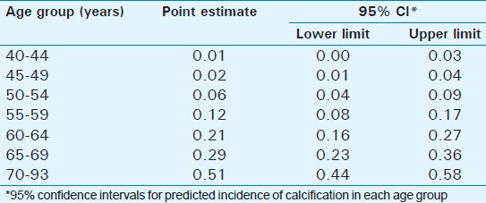
The prevalence of BAC significantly increased with age [Table 2, Figure 1], rising from 1% to 2% in women under the age of 50 years, to over 50% in women over the age of 70 years. Prevalence of BAC in women 50-59 years of age was 14%. Pearson's r was 0.995 (P << 0.001) and the dependence of BAC presence (after transformation) on age was described by the following regression equation: Y = −0.691 + 0.018* (age).
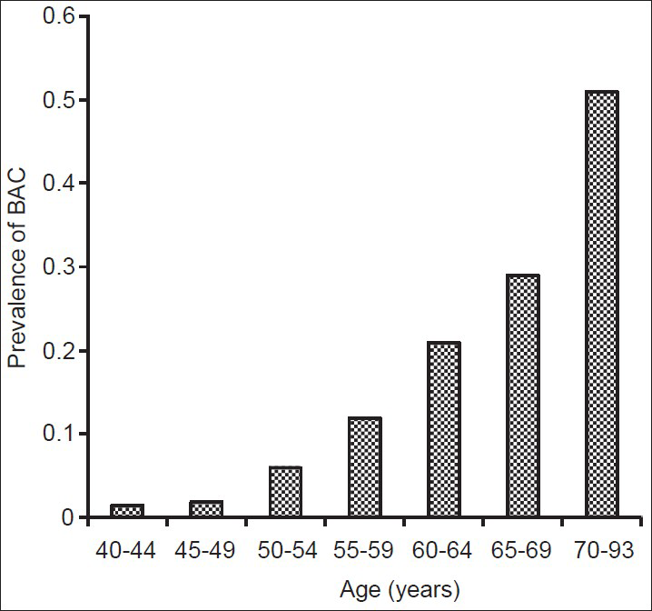
- Prevalence of breast arterial calcification in the different age groups studied.
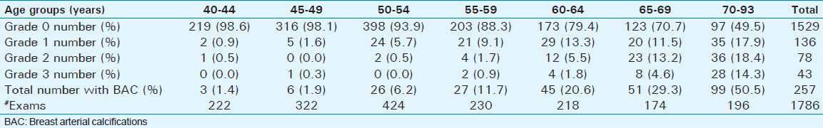
The degree of calcification also rose significantly with increasing age [Table 3, Figure 2] (Spearman's correlation = 0.4, P << 0.001). The positive result indicates a significant, yet weak, correlation.
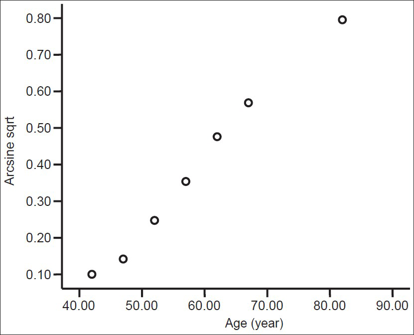
- Presence of calcification in different age groups. Calcification (arcsine sqrt of proportion) versus age.
Of women under 60 years of age with BAC, 80-90% were Grade 1 BAC; in women over 60 years, this dropped to 40-50% of women having Grade 1 BAC, with a concomitant increase in Grades 2 and 3 [Figure 3].
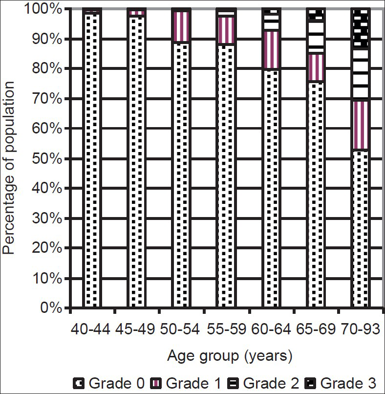
- Graphic representation of distribution of degree of BAC in different age groups.
DISCUSSION
In the United States, about 250,000 women die every year from acute myocardial infarction, whereas 40,000 die from breast cancer. More than 60% of women who die suddenly from coronary heart disease were previously asymptomatic. The prevalence of coronary heart disease is more than 8% in women from ages 55 to 64 years. Asymptomatic individuals may be unaware of harboring risk factors such as hypertension and hyperlipidemia. Some of these risk factors may be discovered with laboratory examinations and physical diagnosis. Imaging examinations can also provide evidence of cardiovascular risk, such as been demonstrated for coronary arterial calcifications on computed tomography (CT),[12] aortic calcifications on plain films,[34] and BAC on mammograms.[56] Extensive investigation in an asymptomatic population is not feasible because of cost. However, since there is significant overlap in the ages of peak vulnerability to breast cancer and to cardiovascular illness, the use of mammography to help stratify the population into higher- and lower-risk subsets could be advantageous.
BAC results from diffuse calcification of the arterial media, as opposed to atherosclerotic calcification of the intima.[7] In their early stage, medial arterial calcifications are punctate in appearance. Coalescence results in linear calcifications; further progression leads to parallel linear calcific opacities. Both atherosclerotic intimal calcifications and calcifications of the arterial media increase with increasing patient age and studies have found a higher incidence of BAC in patients with diabetes, chronic renal failure, and atherosclerotic coronary disease.[89]
In two large studies,[510] the increased risk of cardiovascular events associated with BAC has been calculated as 1.32 for coronary heart disease, 1.8 for myocardial infarction, 1.4 for stroke/transient ischemic attack, 1.52 for heart failure, and 1.5 for thrombosis. These studies were based on the presence or absence of BAC on mammography. However, this subject is still unsettled: A recent study by Maas et al.,[11] found no association between BAC and cardiovascular risk factors. The authors found an association between BAC and age, previous pregnancy and history of lactation. Another study showed no correlation between BAC and coronary heart disease detected on coronary angiography;[12] however, degree of BAC was not considered.
In our study, we planned to record both the presence and degree of BAC in women of various ages. We did not record additional information such as body habitus or concurrent illness, since our intention was to provide a database based only on patient age and mammographic findings. We hypothesize that the use of degree of calcification may aid stratification of the population for planning further investigations to discover cardiovascular disease and its risk factors in asymptomatic women.
Prevalence of BAC has varied in different studies. A study from The Netherlands found 9% prevalence in a screening population aged 50-69 years.[5] A study from California found 2.7% prevalence in women from 50 to 69 years of age and 17.7% in women 70-79 years old.[10]
Our study showed 14.1% prevalence of BAC in the age group 50-69 years, which is significantly higher than previous reports (Chi-square test P < 0.001). The California study was a long-term longitudinal analysis using mammograms obtained over a 30-year period and the authors admit to the possibility of lower sensitivity and possibly lower reporting rates from the reading physicians.[10] In our study, we looked specifically at BAC in order to be as accurate as possible. The BAC prevalence of only 1.6% in women under 50 years confirms the previously published conclusion that further investigation is warranted when BAC are discovered in a woman under 50 years of age.
Few studies have addressed the degree of BAC.[1314] In a preliminary study by Iribarren and Molloi, in 39 women with BAC, quantitation of total BAC was achieved using a densitometric technique in a digital mammography system. Whether quantification is feasible in practice or is clinically relevant awaits further study.[15] Since we had no possibility of quantifying our observations of BAC, we used a qualitative assessment, which combines extent and severity. The increase of BAC severity with age is clear, since 91% of Grade 2 and 3 calcifications occurred in women over 60 years of age, whereas women over 60 years made up only 31% of the examined population. The clinical implication is the observation of higher-grade calcification in a woman less than 60 years is unusual and might merit further investigation.
Limitations of our study include the subjective nature of our mammographic calcification grading system and the lack of additional patient information other than age and mammographic findings. However, the goal of our study was to provide a general database of prevalence and severity of BAC in a population of women presenting for mammography. As such, the relevance of other risk factors is limited in a woman presenting with high grade calcification; whether she has or has no other risk factors does not alter the mammographic finding. Thus, high grade calcifications should not be downplayed because a woman is diabetic.
The appropriate work-up of an asymptomatic woman found to have BAC is not settled. Certainly routine, inexpensive and non-invasive tests are warranted, as they are in all patients of a certain age. The question remains whether more expensive and more invasive tests might be warranted in the case of presence of BAC in a young woman or higher-grade BAC than expected for age. The utility of additional imaging examinations such as carotid sonography, coronary calcium scoring or even CT coronary angiography would have to be established by further study of women with different grades of BAC.
CONCLUSION
According to our results, the infrequency of BAC in a woman less than 50 years of age and of high-grade BAC in a woman less than 60 years of age suggests that findings should be made known to the referring physician for further cardiovascular investigations.
Available FREE in open access from: http://www.clinicalimagingscience.org/text.asp?2013/3/1/36/119013
Source of Support: Nil
Conflict of Interest: None declared.
REFERENCES
- Coronary calcification detected by electron-beam computed tomography and myocardial infarction. The Rotterdam coronary calcification study. Eur Heart J. 2002;23:1596-603.
- [Google Scholar]
- Coronary calcifications in young patients with first, unheralded myocardial infarction: A risk factor matched analysis by electron beam tomography. Heart. 2003;89:625-8.
- [Google Scholar]
- Calcification of the aortic arch: Risk factors and association with coronary heart disease, stroke, and peripheral vascular disease. JAMA. 2000;283:2810-5.
- [Google Scholar]
- Aortic calcification as a predictor of cardiovascular mortality. Lancet. 1986;2:1120-2.
- [Google Scholar]
- Mammograms may convey more than breast cancer risk: Breast arterial calcification and arterio-sclerotic related diseases in women of the DOM cohort. Eur J Cancer Prev. 1996;5:483-7.
- [Google Scholar]
- Breast arterial calcification: Association with coronary artery disease. Work in progress. Radiology. 1995;194:181-3.
- [Google Scholar]
- Breast calcifications due to Mönckeberg medial calcific sclerosis. Radiographics. 1999;19:1401-3.
- [Google Scholar]
- Breast arterial calcifications: Association with diabetes mellitus and cardiovascular mortality. Work in progress. Radiology. 1996;201:75-8.
- [Google Scholar]
- Breast arterial calcifications associated with diabetes and hypertension. J Diabetes Complications. 2004;18:363-6.
- [Google Scholar]
- Breast vascular calcification and risk of coronary heart disease, stroke, and heart failure. J Womens Health (Larchmt). 2004;13:381-9.
- [Google Scholar]
- Arterial calcifications seen on mammograms: Cardiovascular risk factors, pregnancy, and lactation. Radiology. 2006;240:33-8.
- [Google Scholar]
- Breast arterial calcifications on mammograms do not predict coronary heart disease at coronary angiography. Radiology. 2010;254:367-73.
- [Google Scholar]
- Association of breast arterial calcifications detected by mammography and coronary artery calcifications quantified by multislice CT in a population of post-menopausal women. Radiol Med. 2003;106:305-12.
- [Google Scholar]
- Reproducibility of breast arterial calcium mass quantification using digital mammography. Acad Radiol. 2009;16:275-82.
- [Google Scholar]
- Breast arterial calcification: A new marker of cardiovascular risk? Curr Cardiovasc Risk Rep. 2013;7:126-35.
- [Google Scholar]


