Translate this page into:
Prenatal Diagnosis of Lethal Multiple Pterygium Syndrome Using Two-and Three-Dimensional Ultrasonography
Address for correspondence: Prof. Edward Araujo Júnoir, Department of Obstetrics, Federal University of Sāo Paulo (UNIFESP), Rua Carlos Weber, 956 apto. 113 Visage, Vila Leopoldina, Sāo Paulo-SP, Brazil. E-mail: araujojred@terra.com.br
-
Received: ,
Accepted: ,
This is an open-access article distributed under the terms of the Creative Commons Attribution License, which permits unrestricted use, distribution, and reproduction in any medium, provided the original author and source are credited.
This article was originally published by Medknow Publications & Media Pvt Ltd and was migrated to Scientific Scholar after the change of Publisher.
Abstract
Lethal multiple pterygium (LMP) is a series of disorders of fetal formation with a heterogeneous range of manifestations that generally include cystic hygroma, pulmonary hypoplasia, cleft palate, cryptorchidism, joint contractures, fetal akinesia, heart defects, growth restriction, and intestinal malrotation. The prenatal diagnosis of this syndrome is suspected when two-dimensional ultrasound (2DUS) scan shows several malformations.. The three-dimensional ultrasound (3DUS) in rendering mode permits the spatial visualization of these malformations, allowing better understanding of this anomaly by parents. We report a case of a fetus in the second trimester with multiple abnormalities suggestive of LMP that were identified using 2DUS, and emphasize the importance of 3DUS in counseling the parents.
Keywords
Lethal multiple pterygium
prenatal diagnosis
three-dimensional ultrasound
two-dimensional ultrasound
INTRODUCTION

The prevalence of the rare fetal syndrome known as lethal multiple pterygium (LMP) remains unknown. It follows a recessive autosomal inheritance pattern and in some cases, the transmission pattern is linked to chromosome X.[12] LMP is a series of disorders of fetal formation with a heterogeneous range of manifestations that generally include cystic hygroma, pulmonary hypoplasia, cleft palate, cryptorchidism, joint contractures, fetal akinesia, heart defects, growth restriction, and intestinal malrotation.[1–3]
Two-dimensional ultra-sonography (2DUS) used routinely for prenatal examinations offers the possibility of screening for severe malformations that were previously diagnosed only after birth. Likewise, the more recent technique of three-dimensional ultrasonography (3DUS) makes it possible to detect anomalies and enables clearer imaging.[3] We report a case of a fetus in the second trimester with multiple abnormalities suggestive of LMP that were identified using 2DUS and emphasize the importance of 3DUS in counseling the parents, who are faced with a difficult decision.
CASE REPORT
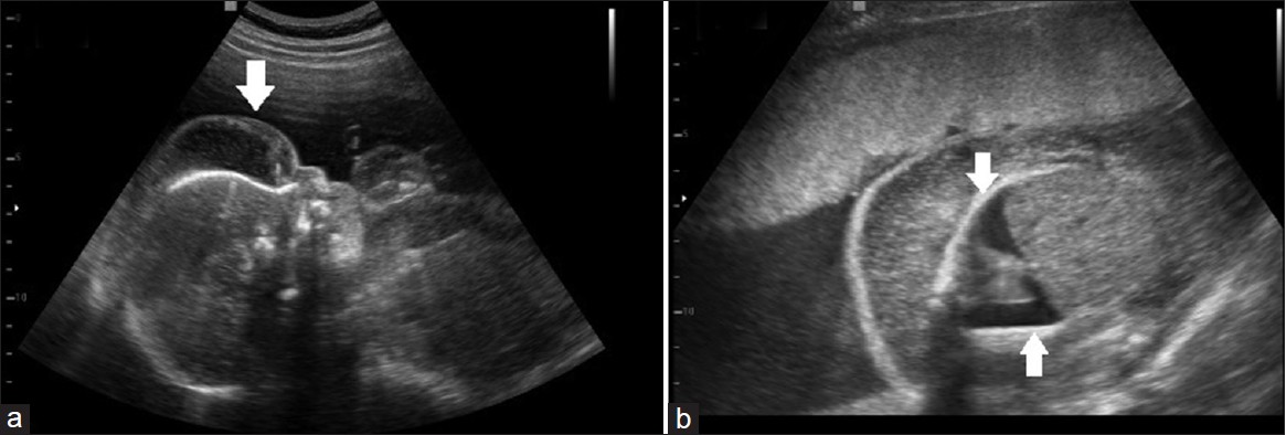
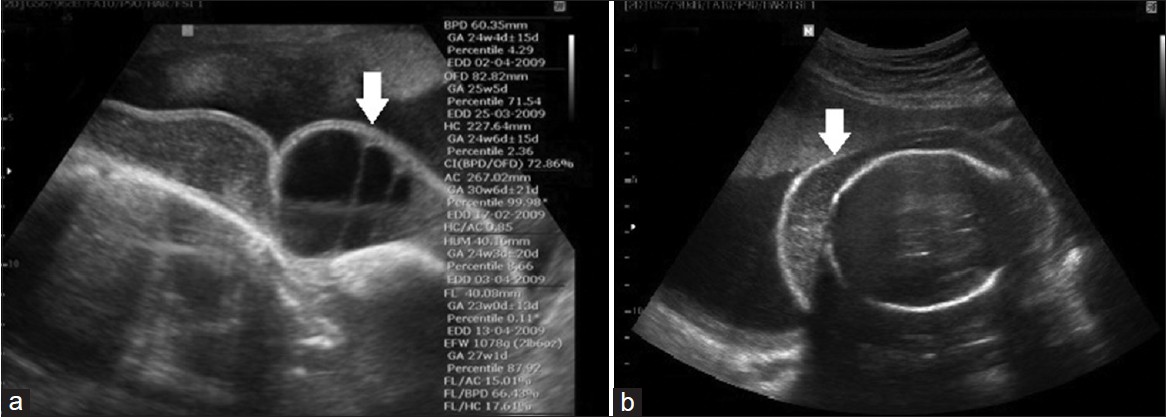
The patient, a 22-year-old primigravida, at her 23rd week of pregnancy was referred to the Department of Gynecology and Obstetrics of the School of Medical Sciences of Santa Casa de Sāo Paulo (FCMSCSP) for a morphological study of the fetus by 2DUS. The examination showed generalized edema, which was most prevalent on the face [Figure 1a], bilateral pleural effusion [Figure 1b], scoliosis, congenital clubfoot, absence of a stomach, hypotrophy and forced flexion of the lower limbs [Figure 2a] and upper limbs [Figure 2b], with fetal akinesia. The hygroma coli was evident in the face, in sagittal [Figure 3a] and axial planes [Figure 3b]. Fetal karyotyping analysis was performed after amniocentesis. The result showed 46XX chromosomes. From the ultrasound findings, a diagnostic hypothesis of LMP was made. In order to view the fetal surface better, a 3DUS, using the MedisonX8 apparatus (Samsung Medison, Seoul, Korea), equipped with a convex volumetric transducer, was performed. In rendering mode, 3DUS made it possible to assess the surface malformations, especially the contracture of the upper limbs [Figure 4]. This was of great importance in enabling the parents to understand the severe malformations, and making it possible for them to receive appropriate counseling. Because Brazilian law does not permit termination of the pregnancy in cases of fetal malformation, the pregnant woman continued to be followed up at the clinic. In subsequent examinations, the presence of progressive pleural effusion, hydrops, and polyhydramnios was observed, finally leading to fetal death in the 27th week. The ultrasound findings and diagnosis of LMP were confirmed from the necropsy.

- (a) 2DUS image of the sagittal plane of the fetal cranium and trunk, shows severe edema on the face (white arrow). (b) 2DUS image of the axial plane of the fetal thorax, reveals severe bilateral pleural effusion with pulmonary hypoplasia (white arrows).

- (a) 2DUS image of the sagittal plane of the fetal lower limbs, shows severe contracture of the lower leg and foot (white arrow). (b) 2DUS image of the sagittal plane of the fetal upper limbs, demonstrates severe contracture of the hands (white arrow).

- (a) 2DUS image of the sagittal plane of the fetal cranium and trunk, shows the hygroma coli (white arrow). (b) 2DUS image of the axial plane of the fetal cranium, reveals the hygroma coli (white arrow).

- 3DUS image in rendering mode, shows severe contracture of the fetal upper limb (white arrow).
DISCUSSION
Increased nuchal translucency and generalized edema that extends across the fetal body during the first trimester may be signs indicative of LMP.[45] However, many other chromosomal syndromes may also present these ultrasound markers.[6] For this reason, in most cases, it is only possible to suspect LMP at a more advanced stage of pregnancy, when the joint contractures become more evident, along with absence of normal fetal movement.[1–3] However, presence of joint contractures and fetal akinesia concomitantly with a normal karyotype is not exclusively a feature of LMP, thus it becomes necessary to make a differential diagnosis with other syndromes such as Bartsocas-Papas, Neu-Laxova, and arthrogryposis multiplex congenita, among others.[47]
LMP is a diverse syndrome, both genetically and clinically. Among the abnormalities that are most observed on 2DUS in the second trimester of pregnancy, persistence of cystic hygroma with fetal hydrops, arthrogryposis with fetal akinesia, pulmonary hypoplasia, cleft palate, heart malformations, and intestinal rotation defects can be highlighted.[127] Furthermore, because the inheritance of this syndrome is mostly recessive, autosomal, and sometimes associated with chromosome X,[12] LMP needs to be diagnosed so that appropriate genetic counseling can be offered to the parents in order to plan for future pregnancies.
This is the first report in the literature regarding 3DUS used for prenatal diagnosis of LMP. Although 3DUS in rendering mode was not essential for the diagnosis in this case, it was shown to be important for enabling the parents to gain adequate understanding of the severe surface malformations, especially the contractures of the upper and lower limbs. In countries that legally permit termination of pregnancy, this may become a diagnostic method that assists greatly in allowing the parents to make this decision, thereby reducing the physical and psychological consequences of maintaining the pregnancy.
CONCLUSION
In summary, we present the first case of prenatal diagnosis of LMP by 3DUS. The 3D image can permit better understanding of the fetal anomalies by parents, allowing them to decide about the termination of the pregnancy.
Available FREE in open access from: http://www.clinicalimagingscience.org/text.asp?2012/2/1/65/103055
Source of Support: Nil
Conflict of Interest: None declared.
REFERENCES
- Prenatal diagnosis and genetic analysis of fetal akinesia deformation sequence and multiple pterygiumsyndrome associated with neuromuscular junction disorders: A review. Taiwan J Obstet Gynecol. 2012;51:12-7.
- [Google Scholar]
- Mutation analysis of CHRNA1, CHRNB1, CHRND, and RAPSN genes in multiple pterygium syndrome/fetal akinesia patients. Am J Hum Genet. 2008;82:222-7.
- [Google Scholar]
- Sonographic features of lethal multiple pterygiumsyndrome at 14 weeks. Prenat Diagn. 2005;25:475-8.
- [Google Scholar]
- First trimester ultrasound diagnosis of lethal multiple pterygium syndrome. Fetal Diagn Ther. 2006;21:466-70.
- [Google Scholar]
- Nuchal translucency and other first-trimester sonographic markers of chromosomal abnormalities. Am J Obstet Gynecol. 2004;191:45-67.
- [Google Scholar]
- Lethal multiple pterygium syndrome.The importance of fetal posture in mid-trimester diagnosis by ultrasound: Discussion and case report. Ultrasound Obstet Gynecol. 1993;3:212-6.
- [Google Scholar]


