
Translate this page into:
Prenatal Diagnosis of Arthrogryposis as a Phenotype of Pena-Shokeir Syndrome using Two- and Three-dimensional Ultrasonography
-
Received: ,
Accepted: ,
This is an open-access article distributed under the terms of the Creative Commons Attribution License, which permits unrestricted use, distribution, and reproduction in any medium, provided the original author and source are credited.
This article was originally published by Medknow Publications & Media Pvt Ltd and was migrated to Scientific Scholar after the change of Publisher.
Abstract
Pena-Shokeir syndrome is a rare autosomal recessive disease, characterized by facial anomalies, arthrogryposis, polyhydramnios, fetal growth restriction, and pulmonary hypoplasia. This report describes the findings of this anomaly with two and three-dimensional ultrasound in a female in her 28th week of pregnancy, who was referred to us because the fetus presented arthrogryposis of unknown cause. These imaging methods allowed adequate evaluation of the fetal malformations and also enabled appropriate counseling of the couple.
Keywords
Arthogriposis
Pena-Shokeir syndrome
prenatal diagnosis
three-dimensional ultrasound
two-dimensional ultrasound

INTRODUCTION

Pena-Shokeir syndrome was described in 1974 and is characterized by multiple joint contractures (arthrogryposis), facial anomalies, polyhydramnios, fetal growth restriction, and pulmonary hypoplasia. Its incidence is estimated at 1 in every 12,000 births. It is an autosomal recessive disease. There are phenotypic variations that show decreased or even absence of movement in the uterine environment, with consequent fetal akinesia, and cause a series of deformities.[1] Around the 14th week of pregnancy, it is possible to diagnosis this disease.[2] We describe the prenatal diagnosis of arthrogryposis that is expressed in Pena-Shokeir syndrome, using two- and three-dimensional ultrasound.
CASE REPORT
A 30-year-old nulliparous female in her second pregnancy was admitted to the Fetal Medicine division of Sāo Paulo Hospital, Paulista School of Medicine, Federal University of Sāo Paulo (UNIFESP), in her 28th week of pregnancy, because of suspected fetal arthrogryposis seen on the second-trimester ultrasound. The two-dimensional ultrasound showed persistent hyperextension of the spine with extension of the head, persistent flexion of the arms and legs, twisting of hands and feet, retrognathia, and signs of pulmonary hypoplasia [Figure 1]. However, three-dimensional ultrasound in rendering mode enabled better diagnosis. Observation of the fetal posture showed persistent flexion of the upper and lower limbs and twisting of the hands and feet, along with greater detailing of the micrognathia [Figure 2]. The fetal weight was below the 10th percentile and the amniotic fluid index showed polyhydramnios. The fetal echocardiography examination identified the presence of a perimembranous interventricular septal defect associated with intermittent fetal bradycardia. The patient did not wish to undergo fetal karyotyping and, therefore, prenatal care was not conducted until the 32nd week of pregnancy, when preterm labor was triggered during a routine consultation, which led to the patient being immediately hospitalized. Corticoid therapy with betamethasone and tocolysis with terbutaline were administered to the patient. Two days after admission, there was progression of labor, and cesarean section was chosen for delivery because the fetus was in a transverse position. The cesarean delivery took place without any problems. The newborn was female, weighed 1050 g and had Apgar scores in the first and fifth minute of 0/0, respectively. After exhaustive resuscitation, the newborn died [Figure 3]. The parturient progressed well and was discharged 2 days after giving birth.
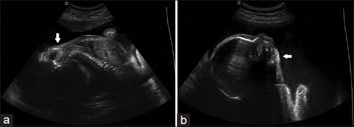
- 30-year-old nulliparous female in her second pregnancy at 28th week of pregnancy, fetus diagnosed with Pena-Shokeir syndrome. Two-dimensional ultrasound (a) longitudinal section through the fetus shows the persistent hyperextension of the spine (arrow). (b) Sagittal section shows the fetal head extension and micrognathia (arrow).
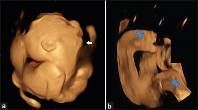
- 30-year-old nulliparous female in her second pregnancy at 28th week of pregnancy, fetus diagnosed with Pena-Shokeir syndrome. Three-dimensional ultrasound images in rendering mode (a) shows hyperextension of the head (white arrow) and micrognathia (red arrow), (b) shows twisting of the feet (blue arrows).
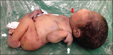
- Baby born at 32nd week of gestation. Postnatal image after unsuccessful attempt to resuscitate the newborn with Pena-Shokeir syndrome. The photograph shows twisted appearance of the hands and feet (white arrows), as well as the micrognathia (red arrow).
DISCUSSION
Arthrogryposis comprises a group of syndromes and deformities that are individually rare, occurring in about 1 in every 3000 births. However, they are not uncommon when grouped together and in more than 50% of the cases, a genetic cause can be identified.[3] The ultrasound diagnosis of arthrogryposis during prenatal care is made initially through the observation of decreased or absent movement of the extremities of the fetus.[4] In the first trimester, early stage of decreased movement and joint contractures can be detected.[5]
Pena and Shokeir described this rare and lethal condition in 1974, which allowed the scientific community to become aware of the gravity of their findings.[1] Limb contracture and the process of muscle atrophy result from lack of adequate movement.[6] Lung development stagnates around the 15th week of pregnancy, in the canalicular phase, in which there is development of pulmonary hypoplasia in the diminished thoracic cavity. This occurs due to the absence of adequate development of the diaphragm and intercostal muscles, since these do not perform early respiratory movements. The decreased fetal swallowing results in polyhydramnios and the underdevelopment of facial muscles explains the craniofacial defects.[6]
Even with a considerable number of differential diagnoses, Pena-Shokeir syndrome shows similarities to trisomy of chromosome 18, such as arthrogryposis and micrognathia. Thus, karyotyping studies are necessary. If the chromosome study is normal and the fetus presents micrognathia, arthrogryposis, and lack of movement, Pena-Shokeir syndrome can then be considered as the diagnostic possibility.[7]
The care that should be provided for subsequent pregnancies for couples who have had a fetus with Pena-Shokeir syndrome include serial assessments using ultrasound, with the aim of achieving early detection of abnormalities. However, since the phenotypic condition is derived from heterogeneous causes, the counseling for couples to provide guidance regarding recurrence is imprecise. It has been estimated that the risk in the next pregnancy is around 0-25%.[8]
Addition of three-dimensional ultrasound in rendering mode makes it possible to evaluate each suspected fetal abnormality in greater detail, thereby satisfactorily complementing the two-dimensional technique; furthermore it permits appropriate parents counseling. Ruano et al.,[9] reported on the benefit of three-dimensional ultrasound in evaluating fixed postural abnormalities of the extremities and of the limbs of the fetus. In our study, this technique was of unquestionable importance, aiding in understanding the disease, particularly as no chromosomal study had been conducted on the fetus.
The other imaging modality to diagnose Pena-Shokeir syndrome is magnetic resonance imaging (MRI). Gupta et al.,[10] reported a case of a fetus at 28-weeks with kyphoscoliosis and reduced movements. MRI using T1-weighted, T2-weighted (half Fourier single-shot turbo spin echo, HASTE) and Tru-FISP sequences was performed to analyze the fetus. Two-dimensional ultrasound and MRI demonstrated distorted spine with scoliosis, persistent flexion of the bilateral wrist, elbow joints, knee joints, and bilateral club foot; however, the retro-micrognathia and pulmonary hypoplasia were also present and were better depicted on MRI images. Although, the MRI is not essential in the diagnosis of Pena-Shokeir syndrome, it should be solicited in cases where there is suspicion of fetal central nervous system anomalies.
CONCLUSION
In summary, two-dimensional ultrasound is necessary for early detection of fetal defects associated with Pena-Shokeir syndrome, especially when combined with three-dimensional ultrasound in rendering mode. Through this technology, the twisting of the limbs and facial abnormalities in the fetus become evident.
Available FREE in open access from: http://www.clinicalimagingscience.org/text.asp?2014/4/1/20/131642
Source of Support: Nil
Conflict of Interest: None declared.
REFERENCES
- Syndrome of camptodactyly, multiple ankyloses, facial anomalies and pulmonary hypoplasia. A lethal condition. J Pediatr. 1974;85:373-5.
- [Google Scholar]
- Ultrasound diagnosis of the Pena Shokeir phenotype at 14 weeks of pregnancy. Prenat Diagn. 1995;15:762-4.
- [Google Scholar]
- Arthrogryposis and fetal hypomobility syndrome. Handb Clin Neurol. 2013;113:1311-9.
- [Google Scholar]
- Fetal akinesia deformation sequence. Pena-Shokeir type I syndrome: New features of an un-uncommon condition. J Obstet Gynecol. 2006;26:818-20.
- [Google Scholar]
- Prenatal diagnosis of distal arthrogryposis type 1. Skeletal Radiol. 1999;28:233-5.
- [Google Scholar]
- Prenatal diagnosis and genetic analysis of fetal akinesia deformation sequence and multiple pterygium syndrome associated with neuromuscular junction disorders: A review. Taiwan J Obstet Gynecol. 2012;51:12-7.
- [Google Scholar]
- Pena-Shokeir phenotype with variable onset in three consecutive pregnancies. Ultrasound Obstet Gynecol. 2001;17:163-5.
- [Google Scholar]
- Prenatal ultrasound of regional akinesia with Pena-Shokier phenotype. Prenat Diagn. 2000;20:422-5.
- [Google Scholar]
- Three-dimensional ultrasonographic appearance of the fetal akinesia deformation sequence. J Ultrasound Med. 2003;22:593-9.
- [Google Scholar]
- Antenatal ultrasound and MRI findings of Pena-Shokeir syndrome. Arch Gynecol Obstet. 2011;283:27-9.
- [Google Scholar]


