
Translate this page into:
Aggressive Angiomyxoma with Perineal Herniation
-
Received: ,
Accepted: ,
This is an open-access article distributed under the terms of the Creative Commons Attribution License, which permits unrestricted use, distribution, and reproduction in any medium, provided the original author and source are credited.
This article was originally published by Medknow Publications & Media Pvt Ltd and was migrated to Scientific Scholar after the change of Publisher.
Abstract
Aggressive angiomyxoma is a rare mesenchymal tumor involving the pelvic-perineal region. It occurs during the third and fourth decade of life and is predominantly seen in females. It presents clinically as a soft tissue mass in variable locations such as vulva, perianal region, buttock, or pelvis. Assessment of extent of the tumor by radiological evaluation is crucial for surgical planning; however, biopsy is essential to establish diagnosis. We present the radiological and pathological features seen in a 43-year-old female diagnosed with abdominal angiomyxoma with an unusual extension to the perineum.
Keywords
Abdominal
angiomyxoma
imaging
tumor

INTRODUCTION

Aggressive angiomyxoma is a benign tumor affecting the pelvis and perineum, predominantly in women. Because of its presentation as a soft mass in various locations such as the vulva, perianal region, buttock, or pelvis, the tumor is often clinically misdiagnosed and initial surgery is usually unsuccessful in extirpating the mass.[1] It was first described in 1983 as a locally infiltrative lesion and was treated with wide local excision.[2] Angiomyxoma is described histologically as a mesenchymal tumor, composed of fibroblasts within a strong myxoid background. Vascular proliferation is also prominent, and virtually no mitoses takes place.[3] Vast majority of angiomyxomas afflict premenopausal females in their third or fourth decades of life with a female to male ratio of 6:1.[45] The term aggressive refers to the locally infiltrative nature of the lesion and its tendency to recur.[6] Although, slow growing, they can attain large sizes (up to 60 cm) but are usually 10-20 cm in their greatest diameter and have a marked tendency to recur locally even decades after resection.[456] With an overall (up to 72%) good prognosis, they have a low metastatic capacity.[56]
A 40-year-old married female presented with a 2-year history of recurrent perineal swelling and hernia in the right buttock with mild distension and heaviness in the lower abdomen. She had normal menstrual history. Patient had been operated 16 months earlier as the same lesion was misdiagnosed as obturator hernia. Eight months after the surgery, the patient again felt a lump in the perineal region. Initially the lesion had been small and had progressively increased to its present size of 15 × 12 cm. Patient had felt increasing fullness and heaviness in her lower abdomen.
Her personal and family history was not significant and systemic examination was within normal limits. Her general examination revealed obesity with a weight of 92 kg with no other significant finding. Local examination revealed slightly distended, soft, non-tender abdomen with no distinct lump/mass palpable, with no organomegaly and no free fluid in the abdomen.
RADIOLOGICAL FEATURES
On ultrasonography of the abdomen, there was a large slightly heterogeneous mass lesion occupying almost the entire abdomen and pelvis [Figure 1]. Contrast enhanced computed tomography (CT) abdomen revealed heterogeneously enhancing, well-defined, soft tissue hypodense mass lesion extending from sub-hepatic level and occupying almost all of the lower abdominal and pelvic cavity extending to the perianal region and beyond into the right buttock [Figures 2 and 3]. The lesion was causing anterior displacement along with compression of uterus and urinary bladder. The mass lesion caused compression of rectum posteriorly along with upward and lateral displacement of bowel loops.
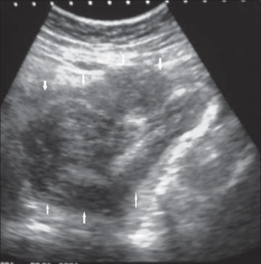
- 43-year-old female with protruding perineal mass diagnosed with angiomyxoma. Ultrasonography abdomen reveals a heterogeneous (predominantly hypoechoic mass involving abdomino-pelvic cavity (arrows).
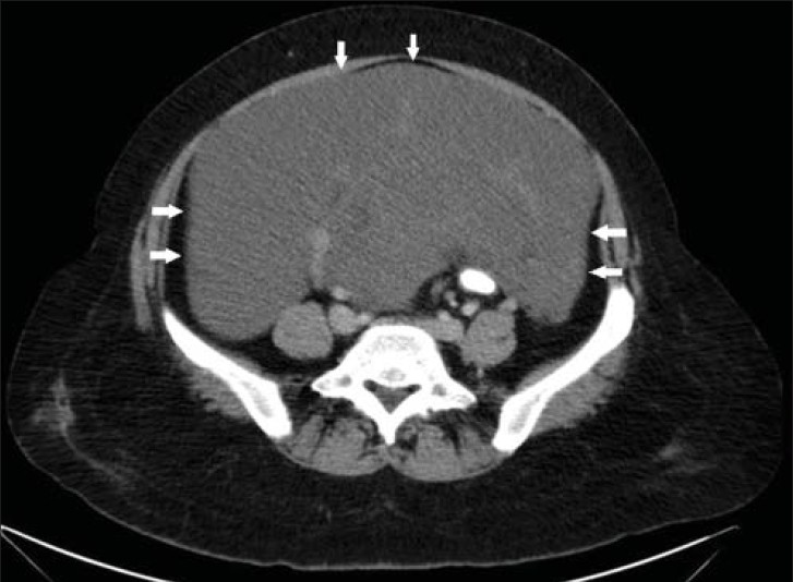
- 43-year-old female with protruding perineal mass diagnosed with angiomyxoma. Contrast-enhanced axial CT scan images of abdomen and pelvis shows a large hypodense mass lesion with well-defined margins (arrow).
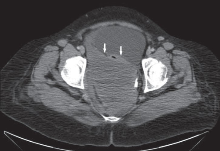
- 43-year-old female with protruding perineal mass diagnosed with angiomyxoma. Contrast-enhanced axial CT scan image of abdomen and pelvis shows a large hypodense mass lesion displacing urinary bladder and uterus anteriorly (white solid arrow).
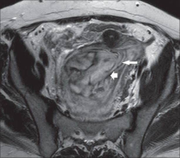
Magnetic resonance imaging (MRI) showed the lesion at the same location and extending in sheet-like layers. The mass revealed predominantly hyperintense signal with hypointense linear foci on T2W [Figures 4–6] and intense contrast enhancement on fat-suppressed TIW images [Figure 7]. Few foci of hyperintensities on TIW images were seen suggestive of fatty changes. No evidence of restricted diffusion was noted.
- 43-year-old female with protruding perineal mass diagnosed with angiomyxoma. T2W axial MRI image shows hyperintense mass with swirled appearance (long arrow) and hypointense linear foci (short arrow).
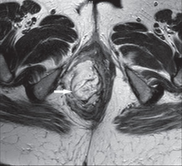
- 43-year-old female with protruding perineal mass diagnosed with angiomyxoma. T2W axial MRI image shows hyperintense mass involving right ischiorectal fossa with swirled appearance (arrow).
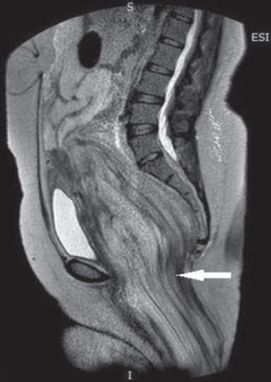
- 43-year-old female with protruding perineal mass diagnosed with angiomyxoma. T2W parasagittal MRI image shows hyperintense mass with swirled appearance herniating into ischiorectal fossa (arrow).
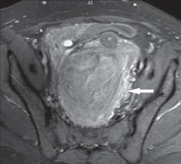
- 43-year-old female with protruding perineal mass diagnosed with angiomyxoma. Contrast enhanced T1W axial MRI image shows intense enhancement of the mass (arrow).
Radiological features were suggestive of a benign mass in the abdomino-pelvic region with perineal herniation.
PATHOLOGICAL FEATURES
A trucut needle biopsy was performed. The biopsy specimens were sent for histological examination. Hematoxylin and eosin staining of the sample revealed presence of scattered spindle cells in an abundant myxoid stroma rich in collagen fibers along with prominent blood vessel of varied caliber [Figure 8]. Histopathological diagnosis was angiomyxoma.
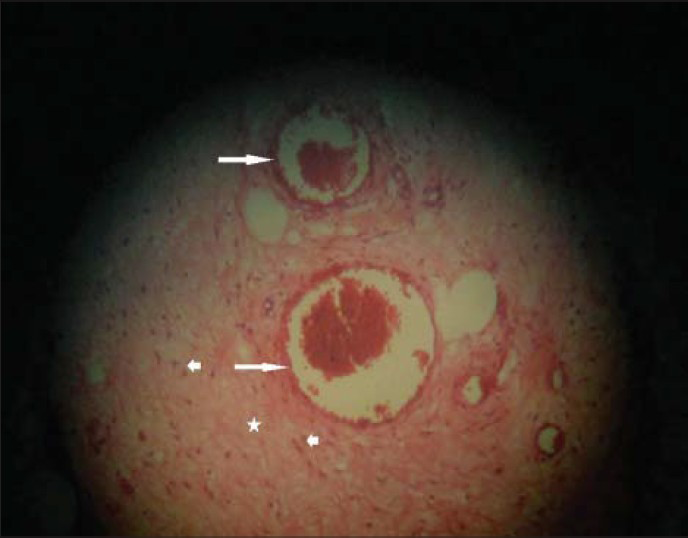
- 43-year-old female with protruding perineal mass diagnosed with angiomyxoma. Histopathological biopsy with hematoxylin and eosin stain (×100) shows medium sized vessels (long arrows) with scattered spindle cells (short arrows) within myxoid stroma (asterix) suggestive of angiomyxoma.
The tumor was operated by abdominal and perineal approach. Intraoperative findings revealed a large tumor occupying almost all of the abdomen lying just below the liver and spleen and originating from the pelvis [Figure 9]. It was a firm, gelatinous, and fleshy mass. Its upper end was free and the mass was occupying the bilateral hypochondriac region. The mass had local infiltration into mesorectal fascia and there was adhesion to the rectum. It was difficult to remove during operation and led to rectal injury during its removal. As a result, patient underwent sigmoid colostomy along with excision of the tumor and perineal hernia repair. A few months later, colostomy reversal surgery was done. Follow-up examinations carried out at 6-months and 12-months post-surgery showed no recurrence of the mass.
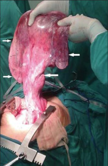
- 43-year-old female with protruding perineal mass diagnosed with angiomyxoma. Intraoperative photograph (mass lifted by the surgeon for demonstration purpose) shows highly vascular fleshy mass (arrows).
DISCUSSION
Aggressive angiomyxoma is a benign mesenchymal tumor of premenopausal females with variable presentation involving vulva, perianal region, buttock, or pelvis. Aggressive angiomyxomas display unusual growth patterns of translevator extension with growth around perineal structures.[1] It is a locally infiltrative lesion usually treated with wide local excision.[2] Histologically, it is described as mesenchymal tumor, composed of fibroblasts within a strong myxoid background and vascular proliferation.[3] Angiomyxomas grow slowly to a large size and have marked tendency to recur locally after resection.[6] It has low metastatic capacity with an overall good prognosis.[56]
Clinical differential diagnosis of a perineal mass in females includes vulvar abscess/neoplasm/cyst, Bartholin abscess, Gartner's duct cyst, vaginal cyst/polyp, vulvar edema, lipoma, canal of Nuck hernia, pelvic floor hernia, and vaginal prolapse.
Histopathological mimics are angiomyofibroblastoma, fibroma, myxofibrosarcoma, myxoid leiomyoma, lymphangioma, neurofibroma, malignant mesenchymoma, malignant fibrous histiocytoma, myxolipoma, sclerosing mesodermal tumor, leiomyosarcoma, and embryonal rhabdomyosarcomas.[7]
Macroscopically angiomyxoma is unencapsulated, poorly circumscribed, and may blend imperceptibly with surrounding soft tissue; size is highly variable ranging from 1 to 60 cm. Tumor is often tan pink to tan gray, bulky and has a rubbery consistency with a glistening, gelatinous cut surface. Areas of congested blood vessels, hemorrhage or fibrosis may be present. On microscopic examination, angiomyxoma shows sparsely cellular tumor composed of eosinophilic stroma studded with numerously haphazardly arranged blood vessels that stand out against the myxoid background. Perivascular rings of condensed collagen may be prominent feature. Soft tissue infiltration is a frequent finding characterized by entrapment of muscle, nerve, and adipose tissue. Tumor cells are cytologically bland and have a spindle, ovoid, or stellate appearance with ill-defined cytoplasmic borders. Nuclear chromatin is evenly dispersed with minimal to no cellular atypia.
Immunohistochemistry does not have any specific marker for angiomyxoma but it does exhibit a characteristic pattern of reactivity. It generally shows diffuse immunopositivity for vimentin and CD-34. Smooth muscle actin highlights myoid bundles and may be positive in individual tumor cells. S-100 reactivity is not a feature of aggressive angiomyxoma but maybe observed in entrapped nerves. Perhaps the most characteristic feature is estrogen receptor and progesterone receptor positivity. Labeling for ki-67 consistently demonstrates a low proliferative index.
Cytogenetic analysis and fluorescent in situ hybridization have confirmed the presence of HMGA2 gene rearrangement in aggressive angiomyxoma and have shown that a subset of them, have the same molecular genetic background as other common mesenchymal tumors.[8]
On imaging, sonography shows a mass that is hypoechoeic or frankly cystic. Angiography shows a generally hypervascular mass. CT shows a hypoattenuating mass. The tumor tends to grow around the structures of the pelvic floor without penetrating the muscles of the vagina and rectum. However, exceptions have been reported. On both CT and MR swirled and layered tissue within the tumor produced a distinctive appearance. This tissue shows slightly lower signal intensity than remainder of the tumor on T1-weighted MR images and enhances with gadolinium. On CT, the swirled appearance is evident only on enhanced scans.[1] High signal intensity on T2-weighted MR images may reflect the myxomatous stroma of these tumors.[1]
The first line of therapy for aggressive angiomyxoma is surgery, although achieving negative resection margins is difficult because of the infiltrative nature of the tumor and the absence of a defined capsule.[9] Several reported attempts using chemotherapy and radiotherapy as part of the treatment for aggressive angiomyxoma have been disappointing, probably due to the low mitotic activity/growth fraction of cells.[10] Primary treatment with GnRH agonists has been carried out successfully as many of angiomyxomas are positive for estrogen and progesterone receptors. Whether treatment is with surgery, hormone therapy, or a combination of both, it is clear that angiomyxoma requires close, long-term follow up to monitor for recurrence and that the individualization of each case is essential for adequate management.[11] Multiple relapses may occur but metastases are unusual. One case of aggressive angiomyxoma in a young female with multiple local recurrences that metastasized to the lung has been reported by Stella et al.[12]
CONCLUSION
Aggressive angiomyxoma is a rare benign mesenchymal tumor of the abdomen, which can infiltrate locally and present unusually as perineal hernia. As it has variable clinical appearance, thorough radiological and pathological examination is essential to establish final diagnosis. Local infiltration is common and great care is needed during surgical excision to avoid any visceral injury. Moreover, there is need for regular follow up at 6-month intervals because of the high rate of recurrence.
Available FREE in open access from: http://www.clinicalimagingscience.org/text.asp?2014/4/1/23/131740
Source of Support: Nil
Conflict of Interest: None declared.
REFERENCES
- Aggressive angiomyxoma: findings on CT and MR imaging. AJR Am J Roentgenol. 1999;172:435-8.
- [Google Scholar]
- Aggressive angiomyxoma of the female pelvis and perineum: Report of nine cases of distinctive type of gynaecologic soft tissue neoplasm. Am J Surg Pathol. 1983;7:463-75.
- [Google Scholar]
- Aggressive angiomyxoma of vulva and vagina: A series of three cases and review of literature. Arch Gynecol Obstet. 2011;283:1145-8.
- [Google Scholar]
- Aggressive angiomyxoma of the vulva: A case report and review of literature. Arch Gynecol Obstet. 2008;277:483-7.
- [Google Scholar]
- Late recurrence of aggressive angiomyxoma of the vulva. J Low Genit Tract Dis. 2013;17:85-7.
- [Google Scholar]
- Aggressive angiomyxoma of the vulva: A précis for primary care providers. Case Rep Obstet Gynecol 2013 2013 183725
- [Google Scholar]
- Aggressive angiomyxoma: A case series and literature review. Eur J Surg Oncol. 2010;36:335-9.
- [Google Scholar]
- Soft tissue tumors: Aggressive angiomyxo. 2007. Atlas Genet Cytogenet Oncol Haematol. Available from: http://atlasgeneticsoncology.org/Tumors/AggresAngiomyxomaID5203.html
- [Google Scholar]
- Aggressive angiomyxoma of the vulva: Case report and literature review. J Int Med Res. 2010;38:1547-52.
- [Google Scholar]
- Aggressive angiomyxoma: A second case of metastasis with patient's death. Hum Pathol. 2003;34:1072-4.
- [Google Scholar]


