Translate this page into:
Plexiform Ameloblastoma of the Mandible
Address for correspondence: Dr. Yadavalli Guruprasad, Department of Oral and Maxillofacial Surgery, AME'S Dental College Hospital and Research Center, Raichur - 584 103, Karnataka, India. E-mail: guru_omfs@yahoo.com
-
Received: ,
Accepted: ,
This is an open-access article distributed under the terms of the Creative Commons Attribution License, which permits unrestricted use, distribution, and reproduction in any medium, provided the original author and source are credited.
This article was originally published by Medknow Publications & Media Pvt Ltd and was migrated to Scientific Scholar after the change of Publisher.
Abstract
Ameloblastoma is a common and aggressive odontogenic epithelial tumor. It has an aggressive behavior and recurrent course, and is rarely metastatic. Ameloblastoma represents 1% of all tumors and cysts that involve the maxillomandibular area and about 10% of the odontogenic tumors. It is primarily seen in adults in the third to fifth decade of life, with equal sex predilection. Radiographically, it appears as an expansile radiolucent, with thinned and perforated cortices, and is known to cause root resorption. As it shares common radiographic features with other lesions such as the giant cell tumor, aneurismal bone cyst, and renal cell carcinoma metastasis, a definitive diagnosis can only be made with histopathology. We present an extensive case of plexiform ameloblastoma of the mandible in a 42-year-old female patient.
Keywords
Mandible
plexiform ameloblastoma
odontogenic tumor
INTRODUCTION

Ameloblastoma is a true neoplasm of the odontogenic epithelium. It is an aggressive neoplasm that arises from the remnants of the dental lamina and dental organ (odontogenic epithelium) and patients usually present late in life after the tumor has achieved considerable size, to cause facial disfigurement.[12] 70% of ameloblastomas develop in the molar-ramus region of the mandible and are occasionally associated with an unerupted third molar teeth.[3] Radiographically an ameloblastoma can be a unilocular or multilocular radiolucent lesion with a honeycomb or soap bubble appearance. It is characterized by slow growth and local infiltration into the adjacent tissues.[34] There are three forms of ameloblastomas, namely peripheral, unicystic, and multicystic tumors. Multicystic ameloblastoma is common and represents 86% of all the cases.[4]
Our patient was a 42-year-old female with a history of swelling in the left mandibular region that had been present for the four-and-a-half years. Physical examination demonstrated a non-tender, left mandibular lesion measuring approximately 15 × 17 × 12 cm with normal overlying skin [Figures 1 and 2]. Panoramic and posterior-anterior (PA) skull radiographs revealed a well-defined, large, expansile, multilocular radiolucent lesion, involving the entire hemimandible with medial displacement of the teeth [Figures 3 and 4]. An axial computed tomography (CT) scan of the mandible showed a well-defined, large, multilocular, expansile radiolucent lesion with erosion, cortical destruction, and thinning of the posterior border of the left ramus of the mandible [Figure 5]. Fine needle aspiration cytology (FNAC) of the lesion was performed, which yielded straw-colored fluid. Subsequently, the patient underwent incisional biopsy of the left mandibular mass confirming the diagnosis of plexiform ameloblastoma. It was predominantly composed of epithelium arranged as a tangled network of anastomosing strands, enclosing cysts of various sizes, suggestive of plexiform ameloblastoma. After confirmation of the diagnosis, the patient underwent left hemimandibulectomy [Figures 6 and 7]. The excised mass was sent for histopathology examination, which further confirmed the diagnosis of plexiform ameloblastoma [Figure 8].
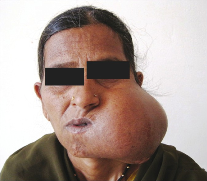
- A frontal view of the patient demonstrates facial asymmetry and a large left mandibular mass.
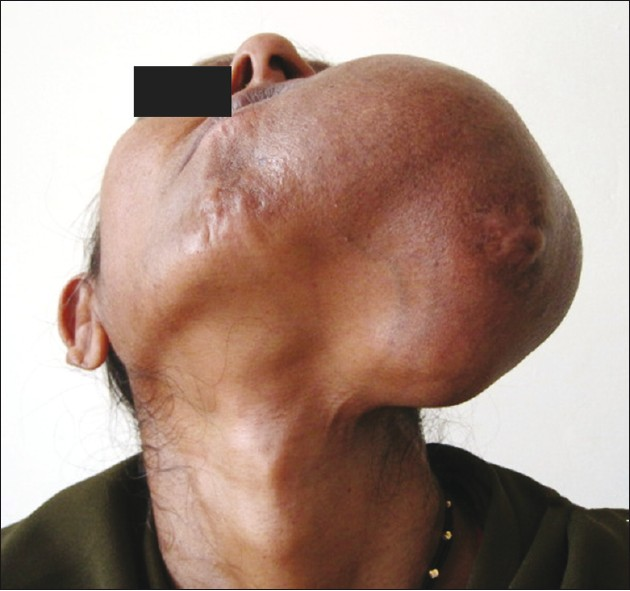
- A submental view of the patient demonstrates a left mandibular mass with its lateral and inferior extension.
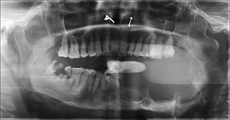
- An orthopantomograph (OPG) demonstrates a large multilocular, expansile lesion causing thinning of the cortical plates involving the whole of the left hemimandible.
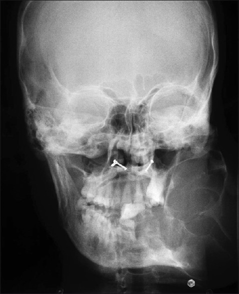
- A posterior–anterior (PA) skull radiograph reveals a multilocular expansile lesion of the left mandible, with medial displacement of the involved teeth.
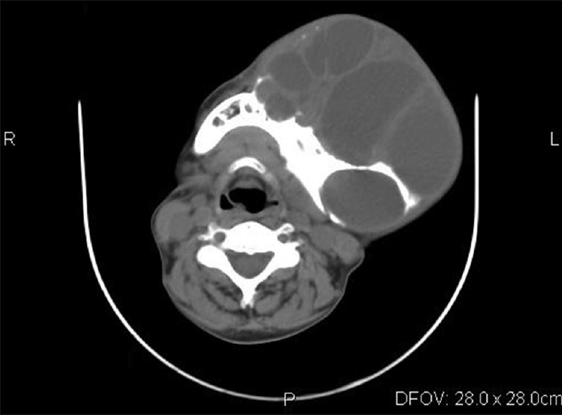
- A non-contrast computed tomography (CT) axial view through the left mandible demonstrates multiple locules, with expansion and thinning of both the cortical plates, and perforation along the posterior border of the left ramus.
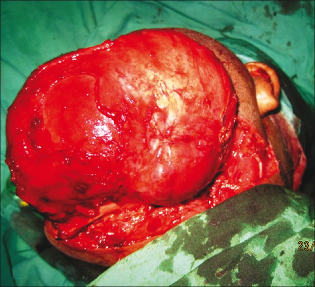
- An intraoperative photograph demonstrates gross appearance of the mass during surgical exploration.
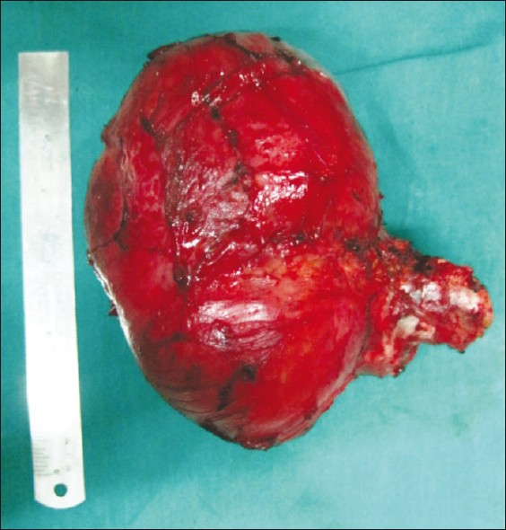
- Photograph of the gross appearance of the mass, measuring 14 × 15 × 10 cm.
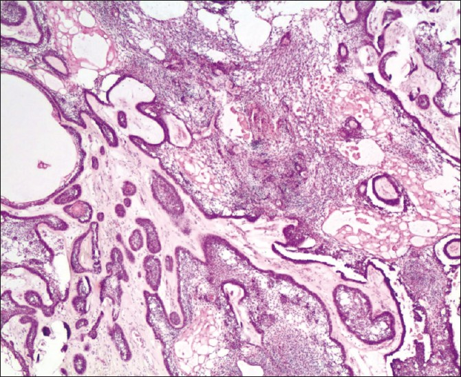
- Photomicrograph demonstrates a plexiform ameloblastoma predominantly composed of the epithelium arranged as a tangled network of anastomosing strands, enclosing cysts of various sizes. (Hemotoxylin and Eosin Stain, 40× magnification).
RADIOLOGICAL FEATURES
The radiographic appearance of an ameloblastoma varies from characteristic soap bubble loculations, to unicystic and multicystic radiolucencies, to subtle appearances such as expanded follicles of erupting teeth.[4] The most common location is the posterior mandible associated with impacted teeth and follicular cysts, causing expansion of the cortical plates with scalloped margins and perforations with resorption of the involved teeth in advanced stages.[45] Radiographically an ameloblastoma may be mistaken for an odontogenic keratocyst, aneurysmal bone cyst, fibrosarcoma, or a giant cell tumor.[5]
HISTOPATHOLOGICAL FEATURES
Ameloblastoma is characterized by the proliferation of epithelial cells arranged on a stroma of conjunctive vascular tissue in locally invading structures that resemble the enamel organ at different stages of differentiation.[5] Diverse histological patterns have been described in the literature and include follicular, plexiform, acanthomatous, papilliferous-keratotic, desmoplastic, granular, vascular and those with dentinoid induction. The tumor found in our patient was an ameloblastoma of the plexiform type.[56] The term plexiform refers to the appearance of anastomosing islands of odontogenic epithelium in contrast to a follicular pattern.
Histopathological features in our case showed anastomosing sheets and cords of odontogenic epithelium. The epithelium displayed a stellate, reticulum-type appearance, arranged as a tangled network of anastomosing strands, enclosing cysts of various sizes.[6]
In conclusion, although ameloblastoma is one of the most common odontogenic tumors, its final diagnosis can only be confirmed with a histopathological examination. Radiographically, ameloblastoma of the mandible can mimic other tumors of the mandible, such as, the odontogenic keratocyst, aneurysmal bone cyst, fibrosarcoma, or a giant cell tumor. A high index of suspicion of ameloblastoma will help triage the patients for further appropriate management.[7]
Source of Support: Nil
Conflict of Interest: None declared.
Available FREE in open access from: http://www.clinicalimagingscience.org/text.asp?2011/1/1/61/91134
REFERENCES
- Mandibular ameloblastoma. A review of the literature and presentation of six cases. Med Oral Patol Oral Cir Bucal. 2005;10:231-8.
- [Google Scholar]
- Clinicopathological analysis of histological variants of ameloblastoma in a suburban Nigerian population. Head Face Med. 2006;2:42-50.
- [Google Scholar]
- Dynamic multislice helical CT of maxillomandibular lesions: Distinction of ameloblastomas from other cystic lesions. Radiat Med. 2001;19:225-30.
- [Google Scholar]
- Clinical and radiologic behaviour of ameloblastoma in 4 cases. J Can Dent Assoc. 2005;71:481-4.
- [Google Scholar]
- Ameloblastoma: A clinical, radiographic and histopathologic analysis of 71 cases. Oral Surg Oral Med Oral Pathol Oral Radiol Endod. 2001;91:649-53.
- [Google Scholar]
- Considerations on a long-term course of a plexiform ameloblastoma with a recurrence in the soft tissue. Rev Med Hosp Gen Mex. 1999;62:48-53.
- [Google Scholar]
- Giant ameloblastoma: Report of an extreme case and description of its treatment. Ear Nose Throat J. 1999;78:568-74.
- [Google Scholar]


