
Translate this page into:
Optimal Use of a Panoramic Radiograph as a Screening Tool for Condylar Resorption in Patients Undergoing Active Orthodontic Treatment: A Case Series

-
Received: ,
Accepted: ,
How to cite this article: Malik S, Singh S, George RT, Kakkar M, Vaid NR. Optimal use of a panoramic radiograph as a screening tool for condylar resorption in patients undergoing active orthodontic treatment: A case series. J Clin Imaging Sci 2020;10:65.
Abstract
Condylar resorption of temporomandibular joint findings in the panoramic radiographs is an indication of bone resorption suggesting possible degenerative joint disease that warrants early screen and subsequent referral to a dedicated specialist. This case series reports three patients that underwent the active orthodontic treatment for the duration of approximately 24–36 months. The patients were asymptomatic at the initial examination. The clinical examination was negative for clicking; the range of motion on opening, lateral excursion, and protrusion was normal. Neither of these patients had a history of rheumatic disease or bruxism. During the later stages of orthodontic treatment, two of the three patients reported mild pain and clicking during mastication, which was also confirmed chairside on clinical evaluation. Patients were referred to the orofacial pain specialist, were they were prescribed specific medication for the symptoms, along with cognitive behavioral therapy, and were further evaluated for splint therapy. Panoramic radiographs taken before the start of the treatment, during the treatment and at the completion of the orthodontic treatments indicate the progression in the resorption of mandibular condyle in all three patients suggesting possible degeneration that warrants further investigation and therapy.
Keywords
Panoramic radiograph
Condylar resorption
Iatrogenic
Active orthodontic treatment

INTRODUCTION
A widely accepted taxonomic classification of temporomandibular disorders (TMD) provided by diagnostic criteria for TMD (DC/TMD) states temporomandibular joint disorders as wide term that includes disk displacement disorders, degenerative joint disease, and subluxation.[1] This classification also includes masticatory muscle disorders, headache attributed to TMD, and associated structures. After chronic low back pain, TMD are second most common musculoskeletal condition among general population USA.[2,3] Epidemiological studies report of 10% TMD’s worldwide with female predominance with age range from 20 to 40 years.[2] TMD are known to be most common reason for non-odontogenic pain in the orofacial region and usually patients present with associated symptoms of otalgia, headaches, or toothaches.[1,4] DC/ TMD is a comprehensive tool to evaluate the TMD’s that are based on physical and psychological criteria.[1,4] The other subjective symptoms that are usually reported are TMJ sounds during jaw function, and deviation or restriction of mandibular movements.[1,4]
The previous studies and investigations have shown higher prevalence of TMD in patients with malocclusion.[5] There are other studies and literatures that state that orthodontic treatments have no effects on the temporomandibular joint. Recent systematic reviews even claim that there are insufficient research data to develop the relationship between orthodontic interventions and TMD.[6-7] Manfredini et al. state that the onset or relief of TMD signs or symptoms during active orthodontic treatment is possibly a casual finding.[5] At the same time, there is enough evidence to support that internal derangements of TMJ can manifest altered craniofacial structures and may represent as facial asymmetry.[8-14] The cephalometric analysis in individuals with TMD as compared to control groups shows significant craniofacial differences.[11,15-20] Panoramic radiographs are another readily available screening tool for observing condylar pathology. Condylar changes indicative of pathology on a panoramic radiograph include condyle flattening, osteophyte formation, and/or vertical ramus asymmetry.[8]
The routine imaging for initial records in an orthodontic setting include panoramic and cephalometric radiographs. There remain several controversies on orthodontic treatment having a cause-effect relationship on TMD, and the exact relationship still remains to be elucidated. This case series reports patients [Table 1] who underwent active orthodontic treatment and exhibited resorption of mandibular condyle on panoramic radiographs suggestive of idiopathic condylar resorption.
| 1st Case | 2nd Case | 3rd Case | |
|---|---|---|---|
| Age | 10 | 12 | 11 |
| Skeletal classification | Class II | Class -I | Class II |
| Angle’s malocclusion | Class II Division 1 | Class II on left and Class I on right | Class II Division I |
| Edgewise application | 0.22 Roth+Herbst | 0.22 Roth | 0.22 Roth+Extractions |
| Finishing arch wire | 19×25 stainless steel | 19×25 stainless steel | 19×25 stainless steel |
| Total duration of the Tx | 36 months | 36 months | 24 months |
The present case series was to assess the clinical and radiographic features of TMJ osteoarthritis in three adolescent patients who were asymptomatic before orthodontic treatment, and to stimulate further trials that can study and establish the possible association between TMJD and orthodontic treatment. Case reports/series have often introduced protocols to orthodontic therapy that have stimulated evidence based research on the topic.[21]
CLINICAL EXAMINATION
Clinical examinations of the patients included assessment of the following: TMJ sounds (clicking or crepitation), range and deviation of mouth opening, tenderness to palpation of the joint and the masticatory muscles, and joint or muscle pain during mouth opening and protrusive or lateral mandibular movements.
Case #1
A 10-year-old female presented with skeletal Class II profile (ANB=5), and Class II Division 1 malocclusion. Patient was in early mixed dentition phase, with increased overjet and deep overbite, patient also presented with midline diastema with lower frenum attachment, bilaterally impacted canines and severe root resorption of the maxillary central incisors. Patient had no history of bruxism or clenching and reported no pain in the TMJ [Figure 1]. Data collection including panoramic imaging before orthodontic treatment, revealed a normal morphology of both right and left mandibular condyles [Figure 2]. Orthodontic treatment with the upper and lower fixed edgewise appliances, (0.22 Roth) was performed subsequent to surgical exposure of the upper first canines. After several steps of rounding and leveling, progress panoramic radiograph was taken and right condylar resorption was noted at this time [Figure 3]. On clinical examination of the TMJ clicking was noted. Patient was referred to orofacial pain specialist and was advised to continue active orthodontic treatment. Herbst appliance was used for 6 months to address the 10 mm overjet. A new panoramic radiograph was taken to evaluate the progression of the orthodontic treatment [Figure 4]. While assessing the new panoramic radiograph, we noticed further progression of condylar resorption on the right side, but patient remained asymptomatic. Finishing and detailing were done in 19 × 25 stainless steel archwires. Final records obtained at the debond visit, which included panoramic radiograph also exhibited further flattening of the right condyle [Figure 5].
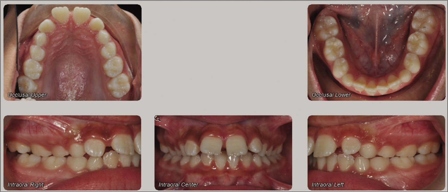
- Initial intra-oral records of 10-year-old female in early mixed dentition. Class II Division 1 malocclusion, with increased overjet and deep overbite, midline diastema, and low frenum attachment.

- A 10-year-old female with bilaterally impacted canines and severe root resorption of the maxillary central incisors. (a) Panoramic radiograph with normal osseous morphology of the TMJ. (b) Tracing of the initial panoramic radiograph.

- Progress panoramic image of the patient taken at age 11 years. (a) Panoramic image shows right-condyle resorption. (b) Tracing of right-condyle showing right-condyle resorption.

- Progress panoramic image of the patient taken at age 12 years. (a) Panoramic image shows flattening of the right-condyle. (b) Tracing of the right condyle showing flattening.

- Panoramic image of the patient taken at age 13 years. (a) Panoramic image shows further flattening of the right-condyle. (b) Tracing of the panoramic image showing further flattening of the right-condyle.
Case #2
A 12-year-old female presented with skeletal Class 1 profile (ANB=1), Angle Class II on the left, and Class I on the right side. The patient was in permanent dentition, with crowding in maxillary and mandibular arches, increased overjet (5 mm) and deep bite. The patient did not report of any history of bruxism or clenching and reported no pain in the TMJ [Figure 6]. Panoramic image before orthodontic treatment revealed a normal morphology of the both right and left mandibular condyles [Figure 7]. Orthodontic treatment with the upper and lower fixed edgewise appliances, (0.22 Roth) appliance was performed, after which panoramic radiograph was taken and bilateral condylar resorption was noted [Figure 8]. On clinical examination of the TMJ clicking was noted, the patient also complained of mild pain on the left side. Patient was referred to orofacial pain specialist and was advised to continue active orthodontic treatment. Finishing and detailing were completed and final records were obtained at the debond visit. Final panoramic radiograph also exhibited further flattening of both right and left condyles, more so on the left condyle [Figure 9].

- Initial intra-oral records of 12-year-old female with Angle Class II on the left and Class I on the right side. Patient was in permanent dentition, with crowding in maxillary and mandibular arches, increased overjet (5 mm) and deep bite.

- Initial panoramic image of 12-year-old female. (a) Panoramic radiograph with normal osseous morphology of the TMJ. (b) Tracing of the right and left condyles.

- Progress panoramic image of the patient taken at age 13 years. (a) Panoramic image shows flattening of both right and left condyles. (b) Tracing of the right and left condyles showing flattening.

- Progress panoramic image of the patient taken at age 14 years. (a) Panoramic image shows further flattening of the right and left condyles. (b) Tracing of the panoramic image showing further flattening of both the right and left condyles.
Case #3
A 11-year-old female presented with skeletal Class II profile (ANB=7), and Class II Division 1 malocclusion. The patient was in mixed dentition, with crowding, increased overjet (7 mm), and deep overbite. The patient did not report of any history of bruxism or clenching and was asymptomatic [Figure 10]. Panoramic image before orthodontic treatment revealed normal morphology of left and right mandibular condyles [Figure 11]. Orthodontic treatment started with hyrax expander, followed by the upper and lower fixed edgewise appliances (0.22 Roth). The patient had extractions of the upper first premolars and lower second premolars after which panoramic radiograph was taken and bilateral condylar resorption was noted [Figure 12]. On clinical examination of the TMJ unilateral clicking on the right TMJ was noted. The patient was referred to orofacial pain specialist and was advised to continue active orthodontic treatment. Finishing and detailing were done in 19 × 25 stainless steel archwires. Final records obtained at the debond visit, which included panoramic radiograph also exhibited further flattening of the right condyle.

- Initial intra-oral records of 11-year-old female with Angle Class II malocclusion. Patient was in mixed dentition, with crowding, increased overjet (7 mm) and deep overbite.

- Initial panoramic radiograph taken beforeprior to orthodontic treatment. (a) Panoramic radiograph revealed normal morphology of the left and right mandibular condyles. (b) Tracing of the right and left condyles.

- Progress panoramic image of the patient at age 12 years. (a) Panoramic image showing further flattening of the left and right condyles. (b) Tracing of the condyles showing flattening of the right and left condyles.
DISCUSSION
The conundrum still prevails between TMD and orthodontic treatments. The previous data analyzed different variables including dental and skeletal occlusion, TMJ symptoms, condylar position, and various orthodontic treatment and its effects on TMJ. Nonetheless, there is not any conclusive evidence suggestive of any correlation between them. The cases presented here may enlighten and educate the dentist about the just as seen in these cases and the current literature it is difficult and almost impossible to foresee which patient might end up with TMJ disorder during or after orthodontic treatment.
Wang et al. showed the occurrence of TMJOA increased sharply in patients in the 11–19 years of age range, which corresponds with the ages when the greatest proportion of adolescents received orthodontic treatment. Some patients in this series were found using regular pre-orthodontic screening radiographic examination.[22] Therefore, it is particularly important for orthodontists to notice the shape and osseous status of articular surface of the condyle when they conduct standard pre-orthodontic radiographic examinations. Unfortunately, there are still many unknown factors related to the cause and progress of TMJOA. Orthodontic therapy and mechanics should be appropriately directed for symptomatic patients, particularly because stress on the joints may result in secondary degeneration of the condyle (secondary OA) and failure of orthodontic treatment itself.
In addition, there are no data which identify a link between active orthodontic intervention and the causation of TMD. However, there are multiple factors that can play a role in the condylar resorption; one of them may be orthodontic forces, which may contribute either to the initiation or progression of the TMD. The apparent random nature of the progression of such a disease process demonstrates the need for a clear, unambiguous informed consent. Furthermore, there is not much evidence available to validate these finding and further studies with regimented protocols and controls are required to better understand the etiology behind these condylar resorptions which are observed during the orthodontic treatments.
SUMMARY
Radiographic abnormalities in the condylar morphology were pathological findings in three ongoing orthodontic treatment cases. These pathological findings along with comprehensive evaluation suggest the development of TMD that need appropriate referral and further investigation. These findings can be presented in symptomatic and subjective symptoms may include joint sound, joint pain, associated headache or abnormal jaw opening, or changes in occlusion. At the same time, asymptomatic patients can present with similar radiographic findings. A complete history review and comprehensive evaluation are required to rule out any other possible diagnosis for autoimmune disorders or idiopathic condylar resorption. Comprehensive evaluation should also include the jaw function and muscle palpation DC/TMD. A close follow-up and diligent monitoring is required during orthodontic treatment. Panoramic radiographs are still used as a screening aid in the detection of osseous changes of the TMJ, so this does not warrant additional imaging to monitor changes that may occur during treatment.
Limitations
Although, we used panoramic radiographs to assess the changes in the articular surface of the condyles, which is essentially a two-dimensional diagnostic imaging tool. Nevertheless, panoramic radiographs still appear to be widely used for screening and preliminary diagnosis of TMJ space deficiencies and bone disorders.[23-25] Honey et al. looked at a sample of skulls and showed that the accuracy of panoramic projection was 0.64 (0.11).[26] Another study indicated that, compared with CBCT, the diagnostic accuracy of panoramic radiography, transpharyngeal, and transcranial projection was 90.64%, 94.10%, and 86.97%, respectively.[27]
Declaration of patient consent
Patient’s consent not required as patients identity is not disclosed or compromised.
Financial support and sponsorship
Nil.
Conflicts of interest
There are no conflicts of interest.
References
- Diagnostic Criteria for Temporomandibular Disorders (DC/TMD) for clinical and research applications: Recommendations of the international RDC/TMD consortium network* and orofacial pain special interest group†. J Oral Facial Pain Headache. 2014;28:6-27.
- [CrossRef] [PubMed] [Google Scholar]
- Definition, epidemiology and etiology of painful temporomandibular disorders In: Fernández-DeLas-Peñas C, Mesa-Jimenez J, eds. Temporomandibular Disorders Manual Therapy Exercise and Needling. Edinburgh: Handspring Publishing; 2018. p. :3-22.
- [Google Scholar]
- Relationship between specific temporomandibular disorders and impaired upper neck performance. Eur J Oral Sci. 2020;128:292-8.
- [CrossRef] [PubMed] [Google Scholar]
- Comparative assessment of condylar position in patients with Temporomandibular Disorder (TMD) and asymptomatic patients using cone-beam computed tomography. Dent Med Probl. 2019;56:81-7.
- [CrossRef] [PubMed] [Google Scholar]
- Orthodontics is temporomandibular disorder-neutral. Angle Orthod. 2016;86:649-54.
- [CrossRef] [PubMed] [Google Scholar]
- Orthodontics for treating temporomandibular joint (TMJ) disorders. Cochrane Database Syst Rev. 2010;7:CD006541.
- [CrossRef] [Google Scholar]
- Orthodontics and temporomandibular disorder: A meta-analysis. Am J Orthod Dentofacial Orthop. 2002;121:438-46.
- [CrossRef] [PubMed] [Google Scholar]
- Predisposing and precipitating factors in temporomandibular disorders. Semin Orthod. 2012;18:1029.
- [CrossRef] [Google Scholar]
- Reliability of a negative clinical temporomandibular joint examination: Prevalence of disk displacement in asymptomatic temporomandibular joints. Oral Surg Oral Med Oral Pathol. 1989;68:5514.
- [CrossRef] [Google Scholar]
- Are temporomandibular disorders associated with facial asymmetry? A systematic review and meta-analysis. Orthod Craniofac Res. ;2020
- [CrossRef] [PubMed] [Google Scholar]
- Comparison of skeletal and dental morphology in asymptomatic volunteers and symptomatic patients with unilateral disk displacement with reduction. Angle Orthod. 2004;74:212-9.
- [Google Scholar]
- Internal derangement of the temporomandibular joint: Radiologic staging with clinical, surgical, and pathologic correlation. Magn Reson Imaging. 1989;7:495-515.
- [CrossRef] [Google Scholar]
- Facial skeleton remodeling due to temporomandibular joint degeneration: An imaging study of 100 patients. AJNR Am J Neuroradiol. 1990;155:373-83.
- [CrossRef] [PubMed] [Google Scholar]
- Observations on individuals with radiographic bilateral condylar remodeling. J Craniomandibular Pract. 1982;1:20-37.
- [CrossRef] [PubMed] [Google Scholar]
- Comparison of skeletal and dental morphology in asymptomatic volunteers and symptomatic patients with bilateral disk displacement without reduction. Angle Orthod. 2004;74:684-90.
- [Google Scholar]
- Comparison of skeletal and dental morphology in asymptomatic volunteers and symptomatic patients with unilateral disk displacement with reduction. Angle Orthod. 2004;74:212-9.
- [Google Scholar]
- Comparison of skeletal and dental morphology in asymptomatic volunteers and symptomatic patients with unilateral disk displacement without reduction. Angle Orthod. 2003;73:121-7.
- [Google Scholar]
- Comparison of skeletal and dental morphology in asymptomatic volunteers and symptomatic patients with normal temporomandibular joints. Angle Orthod. 2003;73:116-20.
- [Google Scholar]
- Comparison of skeletal and dental morphology in asymptomatic volunteers and symptomatic patients with bilateral degenerative joint disease. Angle Orthod. 2003;73:71-8.
- [Google Scholar]
- Comparison of skeletal and dental morphology in asymptomatic volunteers and symptomatic patients with bilateral disk displacement with reduction. Angle Orthod. 2002;72:541-6.
- [Google Scholar]
- Investigation on radiographic signs of osteoarthrosis in temporomandibular joint with cone beam computed tomography in adolescents. Beijing Da Xue Xue Bao Yi Xue Ban. 2013;45:280-5.
- [Google Scholar]
- A survey of panoramic radiographs for evaluation of normal and pathologic findings. Oral Surg Oral Med Oral Pathol. 1980;50:472-8.
- [CrossRef] [Google Scholar]
- Digital panoramic radiography for diagnosis of the temporomandibular joint: CBCT as the gold standard. Braz Oral Res. 2015;29:S1806-83242015000100303.
- [CrossRef] [PubMed] [Google Scholar]
- The interpretation of temporomandibular joint radiographs. S Afr Med J. 1974;48:1905-12.
- [Google Scholar]
- Accuracy of cone-beam computed tomography imaging of the temporomandibular joint: Comparisons with panoramic radiology and linear tomography. Am J Orthod Dentofacial Orthop. 2007;132:429-38.
- [CrossRef] [PubMed] [Google Scholar]
- Diagnostic evaluation of the temporomandibular joint osteoarthritis using cone beam computed tomography compared with conventional radiographic technology. Beijing Da Xue Xue Bao Yi Xue Ban. 2007;39:26-9.
- [Google Scholar]


