
Translate this page into:
Morphometric anlaysis of mandibular coronoid, condyle and sigmoid shape using panoromic view for personal identification in south Indian population
 , Leeba Mathew1, Srikant Natarajan2, Shweta Yellapurkar1,, Shravan Shetty3, Ceena Denny4, Samarika Dahal5

*Corresponding author: Shweta Yellapurkar, Department of Oral Pathology & Microbiology, Manipal College of Dental Sciences, Mangalore, Manipal Academy of Higher Education Manipal, Mangalore, India. shweta.y@manipal.edu
-
Received: ,
Accepted: ,
How to cite this article: Manoj M, Mathew L, Natarajan S, Yellapurkar S, Shetty S, Denny C, et al. Morphometric anlaysis of mandibular coronoid, condyle and sigmoid shape using panoromic view for personal identification in south Indian population. J Clin Imaging Sci 2022;12:25.
Abstract
Objective
The shape of the condyle, coronoid, and the intervening sigmoid notch is unique due to attachments and direction of pull of temporalis muscle, different chewing habits, hormones, etc. So morphometric variation of these mandibular landmarks can be used in individual identification.
Material And Methods
The study was carried out on 500 panoramic view images. Visual tracing assisted assessment of thousand sides (left and right) was done. Sexual dimorphism of left and right mandibular ramus parts (sigmoid notch bordered by coronoid and condylar process). The shape of coronoid process, condyle, and sigmoid notch were interpreted.
Results
The most prevalent shape of sigmoid notch was wide variant (41.2%), condyle was convex type (50%) and coronoid was round (53.4%). Condyle and coronoid showed asymmetry between right and left with p-value 0.001 and 0.019, respectively. This study has illustrated that triangular-shaped coronoid was more in females (p-value 0.012).
Conclusion
Sexual dimorphism is observed in the shape of the mandible due to masticatory activity at the point of insertion of temporalis muscle, biting force, hormones, and various genetic reasons. The results have exemplified that the morphometric variation of condyle, coronoid and sigmoid notch can be used as a tool for personal identification.
Keywords
Morphometry
Sexual dimorphism
Coronoid
Condyle
Sigmoid notch

INTRODUCTION
Forensic odontology is defined as a “branch of dentistry concerned with the correct management, examination, and presentation of dental evidence in criminal or civil legal proceedings in the interest of justice.”[1] Identification is the establishment of a person’s individuality. Fingerprint assessment and DNA profiling are the most common methods used for personal identification. Burnt human remains, or in cases of DNA degradation, it is impossible to use this method.[2]
Application of techniques of biologic anthropology is used to identify an individual from bones. The skeleton changes due to the various factors during the life of the individual. The density of bone and contours can be used to estimate a person’s age, sex, stature, ancestry, and comparative human identification. Pelvic bone and skull show the most apparent sexual dimorphism. Due to the interplay of biological factors, males have a longer skull, on average, more significant muscle development, and more rugged muscle attachment than females. Measurement of morphometric parameters and morphological variations of bone can identify a person. At birth, our bones are primarily soft cartilage. As they grow, hard bones replace the cartilage at different centers of growth. We can use this appearance to estimate the age of the individual up to 20 years as the bone growth is complete thereafter. After growth completion, degenerative bone changes, suture closure stages, etc. are used to estimate the individual’s age.[3] Many skull characteristics such as nasal opening, zygomatic bone, maxillary bone, and teeth are used to determine the race from the skeletal remaining.[4]
The mandible is the most durable and sexually dimorphic bone of the skull, and it is also known to resist post-mortem changes. Mandible and tympanic ossicles are the only mobile bones of the skull.[5] Muscle attachment and function may dynamically affect the shape and orientation of the contours of the mandible. Attachment and action of temporalis and masseter muscle alter the shape of condyle and coronoid. Genetic determinants, hormonal variation in condyle, and coronoid itself can lead to different “shapes” of the sigmoid notch.[6]
In the present study, we accessed the morphological parameters of the shape of condyle, coronoid, and the intervening sigmoid notch as evolutionary markers to establish its efficiency in individual identification.
MATERIALS AND METHODS
Five hundred radiographs of panoramic view were obtained from the Department of Oral Medicine and Radiology. The radiographs were obtained using Planmeca Promax 2D S3digital panoramic X-ray, Helsinki Finland. Taken at 68kv, 10 mA exposed for 15.8 seconds having exposure of 131mGy*cm2. Approval from the institutional ethics committee was obtained before the study (reference number:19052). Written consent for use of radiographs was obtained from the subjects before the evaluation. The radiographs were anonymized to prevent disclosure of patient identity before evaluation. A qualified radiologist having an experience of more than 10 years assessed the radiographs. Visual tracing of the outline of the coronoid and condyle was done to identify the morphology of coronoid, condyle, and sigmoid notch. The patients were distributed in the age range of 21–58 years and were categorized into four groups based on age, 20–30 years, 31–40 years, 41–50 years, and >50 years, respectively. Criteria given by Smita Tapas et al. were used to evaluate the shape of the mandibular condyle, coronoid, and intervening sigmoid notch.[6] The shapes were interpreted as a triangular, round, beak, or flat coronoid process [Figure 1A], round, angled, convex, or flat condylar process [Figure 1B], and sloping, wide, or round sigmoid notch [Figure 1C].
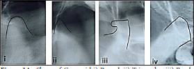
- Shape of Coronoid i) Round, ii) Triangular, iii) Break, iv) Flat

- Shape of Condyle i) Wide, ii) Round, iii) Sloping

- Shape of Sigmoid notch i) Round, ii) Convex, iii) Angled, iv) Flat
The panoramic view radiographs were assessed systematically for alterations like developmental, traumatic, or other diseases if present were excluded from the study. Thousand sides (500 left and 500 right) were assessed, and comparisons of the shape of the condyle, coronoid, and intervening sigmoid notch were made between age, and gender. The proportions of the number of each shape of the condyle, coronoid, and sigmoid notch were described using number and percentage and sexual dimorphism was evaluated using chi-square tests. Analysis was done using IBM SPSS 20.0 (IBM, Chicago) and a significance level of 0.05 (95% confidence interval) was taken for statistical analysis.
RESULTS
One thousand sides (left and right) of 500 Panoramic view images constituted 217 (434 sides) males and 283 (566 sides) females. Tests of proportion evaluated the shapes of condyle, coronoid and sigmoid notch. The comparison between both sides was done using the McNemars chi-square test, and between the gender and different age groups using chi-square tests of association. On analysis of the symmetry of the shapes, the sigmoid notch was relatively symmetric. The majority of the cases had a wide pattern (41.2%). Only 23% of the cases were not concordant with 16% having the combination of sloping and wide type combination on the left and right sides. The condylar pattern was also symmetric, with half the cases having a convex shape (50.0%). Only 27.2% of the patients were not concordant, with 11.8% having the combination of round and convex type combination (p-value = 0.001). Coronoid patterns showed more than half the cases having a round pattern (53.4%). Only 26.2% of the cases were not concordant, with 16.8% having triangular and round type combinations (p-value = 0.019) [Table 1]. On comparison of the shape of the sigmoid notch between females and males in 500 patients using the chi-square test, we found that there was a marginally higher proportion of sloping patterns in females with a higher proportion of round and wide patterns in males both in right and left sigmoid notch (p = 0.095 and p = 0.078 in right and left side, respectively). The shape of the condyle showed a more round and flat shape in males. In the coronoid process, the right coronoid showed a significant difference between the gender. There were 9.2% cases that were flat in males as compared to only 3.5% in females. Similarly, there was a higher proportion of triangular shape of coronoid in females accounting for 32.2% and only 22.6% in males (p-value of 0.012). A similar trend was not evident in the left coronoid, suggesting asymmetry in the shape of the coronoid [Table 2].
| Agreement of pattern | Combination of the patterns noted | Chi-square value (p-value) |
|||||||||
|---|---|---|---|---|---|---|---|---|---|---|---|
| Sloping | Wide | Round | Sloping-wide | Sloping-round | Wide-round | ||||||
| Sigmoid notch | 135 (27.0%) |
206 (41.2%) |
44 (8.8%) |
80 (16.0%) |
7 (1.4%) |
28 (5.6%) |
6.536 (0.0880) |
||||
| Round | Angled | Convex | Flat | Round-angled | Round-convex | Round-flat | Angledconvex | Angled-flat | Convex-flat | ||
| Condyle | 78 (15.6%) |
31 (6.2%) |
250 (50.0%) |
5 (1.0%) |
15 (3.0%) |
59 (11.8%) |
8 (1.6%) |
43 (8.6%) |
2 (0.4%) |
9 (1.8%) |
21.875 (0.001) |
| Triangular | Round | Beak | Flat | Triangular-round | Triangular-beak | Triangular-flat | Round-beak | Round-flat | Beak-flat | ||
| Coronoid | 96 (19.2%) |
267 (53.4%) |
1 (0.2%) |
5 (1.0%) |
84 (16.8%) |
6 (1.2%) |
5 (1.0%) |
5 (1.0%) |
29 (5.8%) |
2 (0.4%) |
15.223 (0.019) |
| Categories | N | Sex | Chi-square | p-value | ||
|---|---|---|---|---|---|---|
| Female [N (%)] | Male [N (%)] | |||||
| Shape of right sigmoid notch | Sloping | 184 | 113 (39.9) | 71 (32.7) | 4.708 | 0.095 |
| Wide | 249 | 139 (49.1) | 110 (50.7) | |||
| Round | 67 | 31 (11) | 36 (16.6) | |||
| Shape of left sigmoid notch | Sloping | 173 | 109 (38.5) | 64 (29.5) | 5.106 | 0.078 |
| Wide | 271 | 147 (51.9) | 124 (57.1) | |||
| Round | 56 | 27 (9.5) | 29 (13.4) | |||
| Shape of right condyle | Round | 135 | 70 (24.7) | 65 (30) | 4.158 | 0.245 |
| Angled | 64 | 34 (12) | 30 (13.8) | |||
| Convex | 291 | 175 (61.8) | 116 (53.5) | |||
| Flat | 10 | 4 (1.4) | 6 (2.8) | |||
| Shape of left condyle | Round | 103 | 53 (18.7) | 50 (23) | 2.256 | 0.521 |
| Angled | 58 | 31 (11) | 27 (12.4) | |||
| Convex | 320 | 189 (66.8) | 131 (60.4) | |||
| Flat | 19 | 10 (3.5) | 9 (4.1) | |||
| Shape of right coronoid | Triangular | 140 | 91 (32.2) | 49 (22.6) | 10.916 | 0.012 |
| Round | 319 | 176 (62.2) | 143 (65.9) | |||
| Beak | 11 | 6 (2.1) | 5 (2.3) | |||
| Flat | 30 | 10 (3.5) | 20 (9.2) | |||
| Shape of left coronoid | Triangular | 147 | 91 (32.2) | 56 (25.8) | 3.824 | 0.281 |
| Round | 333 | 182 (64.3) | 151 (69.6) | |||
| Beak | 4 | 1 (0.4) | 3 (1.4) | |||
| Flat | 16 | 9 (3.2) | 7 (3.2) | |||
The majority of the individuals had a wide pattern followed by a sloping pattern of sigmoid notch in all age groups. (p-value of 0.59 on the right and 0.343 on the left side). Evaluation of the left condyle shape showed a significant difference in the age groups with a p-value of 0.036. The right condyle, however, was found to be not significant with a p-value of 0.182. The left condyle significantly showed a round pattern in the age group of more than 50 years as well as less than 30 years. This indicated that condyle is more rounded in the young and old age groups whereas they are more angled or convex in the middle age groups. When we compare the shape of the right and left coronoids, there was no significant difference. The majority of the coronoid processes were rounded followed by triangular shapes. (p-value of 0.284 on the right and 0.193 on the left side) [Table 3].
| Categories | N | Age | Chi-square | p-value | ||||
|---|---|---|---|---|---|---|---|---|
| <30 years [N (%)] | 31-40 years [N (%)] | 41-50 years [N (%)] | >50 years [N (%)] | |||||
| Shape of right sigmoid notch | Sloping | 184 | 93 (37.1) | 36 (43.4) | 25 (32.1) | 30 (34.1) | 4.643 | 0.59 |
| Wide | 249 | 123 (49) | 40 (48.2) | 43 (55.1) | 43 (48.9) | |||
| Round | 67 | 35 (13.9) | 7 (8.4) | 10 (12.8) | 15 (17) | |||
| Shape of left sigmoid notch | Sloping | 173 | 93 (37.1) | 31 (37.3) | 22 (28.2) | 27 (30.7) | 6.764 | 0.343 |
| Wide | 271 | 128 (51) | 48 (57.8) | 46 (59) | 49 (55.7) | |||
| Round | 56 | 30 (12) | 4 (4.8) | 10 (12.8) | 12 (13.6) | |||
| Shape of right condyle | Round | 135 | 77 (30.7) | 18 (21.7) | 17 (21.8) | 23 (26.1) | 12.593 | 0.182 |
| Angled | 64 | 21 (8.4) | 12 (14.5) | 15 (19.2) | 16 (18.2) | |||
| Convex | 291 | 148 (59) | 52 (62.7) | 44 (56.4) | 47 (53.4) | |||
| Flat | 10 | 5 (2) | 1 (1.2) | 2 (2.6) | 2 (2.3) | |||
| Shape of left condyle | Round | 103 | 62 (24.7) | 12 (14.5) | 9 (11.5) | 20 (22.7) | 17.939 | 0.036 |
| Angled | 58 | 23 (9.2) | 12 (14.5) | 16 (20.5) | 7 (8) | |||
| Convex | 320 | 155 (61.8) | 56 (67.5) | 49 (62.8) | 60 (68.2) | |||
| Flat | 19 | 11 (4.4) | 3 (3.6) | 4 (5.1) | 1 (1.1) | |||
| Shape of right coronoid | Triangular | 140 | 72 (28.7) | 30 (36.1) | 20 (25.6) | 18 (20.5) | 10.874 | 0.284 |
| Round | 319 | 154 (61.4) | 49 (59) | 53 (67.9) | 63 (71.6) | |||
| Beak | 11 | 5 (2) | 1 (1.2) | 3 (3.8) | 2 (2.3) | |||
| Flat | 30 | 20 (8) | 3 (3.6) | 2 (2.6) | 5 (5.7) | |||
| Shape of left coronoid | Triangular | 147 | 78 (31.1) | 27 (32.5) | 20 (25.6) | 22 (25) | 12.365 | 0.193 |
| Round | 333 | 159 (63.3) | 52 (62.7) | 56 (71.8) | 66 (75) | |||
| Beak | 4 | 2 (0.8) | 2 (2.4) | 0 (0) | 0 (0) | |||
| Flat | 16 | 12 (4.8) | 2 (2.4) | 2 (2.6) | 0 (0) | |||
DISCUSSION
Forensic analysis by comparing antemortem and post-mortem radiographs is one of the most preferred methods for personal identification. Panoramic images are the most frequently advised radiographs for diagnosing and monitoring various treatment procedures in dentistry. When retained, they can serve as antemortem records in the personal identification process. Developmental discrepancies, hereditary determinants, and functional variation that arise during the growth process are the major cause of morphological variation of anatomic structure. In this study, the articular and functional parts of the mandible (condyle) coronoid and intervening sigmoid notch were evaluated in panoramic radiographs.[6]
Rounding of the coronoid process was the most common type followed by triangular shape. The right coronoid form showed a significant difference between the gender. Males showed more prevalence of flat coronoid process whereas triangular pattern was more common in females. Our findings were opposite to the findings in the North Indian population as research by Tapas et al., and in the Maharashtrians, as shown by D Jadav S and Kadam et al.[7-9] Hook/beak shape followed by the triangular shape of coronoid was most prevalent in the West Indian population. This difference in both sexes might be due to different chewing habits, hormonal variations, and attachment variations.[10] Studies have shown that males have higher muscle forces that accelerate the mandible faster during the chewing cycle, leading to a greater velocity of movement.[11] This higher force may elongate the coronoid due to the action of the temporalis muscle.[12] The genetic composition is the most important contributing factor to sexual dimorphism.[13] Testosterone also affects the remodeling of bone.[14] The change in form of the coronoid correlates with a larger functional load initially which gets remodeled as a rounded followed by the triangular shape, flat shape and least being a beak. There are multiple theories proposed to explain the elongated coronoid process. They include temporalis hyperactivity, dysfunction of the temporomandibular joint caused by chronic disc displacement, dental causes like the guidance of occlusion and variation in condylar inclination, and other factors like hormonal stimulus, nutrition, and genetic inheritance.[12]
The convex shape was the most common form of condyle, seconded by the round shape in our study. In the study conducted by Sahithi et al.,[4] males predominantly had angular condyles and females had rounded condyles in the South Indian population. Mandibular growth in vertical as well as horizontal directions is influenced by the condylar cartilage. Changes can be brought about by pathologies such as ankyloses, condylar hyperplasia, infections, tumors, and fractures.[6] Even after growth cessation, the mandibular and temporal parts of the TMJ are both thought to maintain their capacity for remodeling.[15] Bone can change its shape by regulating bone formation and resorption, often responding to forces. One of the purposes of this shaping is to limit any risk of fracture. So as age advances, condyle gets remodeled as rounded.[16] Condylar morphology is shown to be influenced by the loss of dentition. Dentition status, as well as chewing habits of dentate individuals, significantly determine the morphology of condyle.[17]
Many researchers have shown sexual dimorphism of ramus of the mandible. A further high degree of accuracy in predicting sex using ramus height, condylar height, and intercondylar distance has been noted.[18-24]
The shape of the sigmoid notch was predominantly wide followed by sloping and round forms. This is similar to results published by Sahithi et al.,[4] Ashwini et al.,[25] and Saraswathi et al.[12]
Merits and demerits
Although panoramic view has limitations in terms of magnification errors and distortion it has many advantages that cannot be ignored. Few of them being economical, nominal radiation dosage, rapid turnaround time, and wide coverage area. Along with the other facts that contrast, brightness amplification, and image expansion give an accurate and repeatable means of measuring the selected spots.[26]
CONCLUSION
Genetic and epigenetic factors give an individual a distinctive phenotype. Due to differences in insertion and action of muscles of mastication (especially temporalis muscle), occlusal load, hormones, and various genetic reasons the shape of the coronoid, condyle, and intervening sigmoid notch varies by gender and on the right and left side. The results have exemplified that the morphometric variation of condyle, coronoid and sigmoid notch can be used as a tool for personal identification.
Declaration of patient consent
Patient consent is not required as there are no patients in this study.
Financial support and sponsorship
Nil.
Conflicts of interest
There are no conflicts of interest.
References
- Forensic dentistry: Oral and maxillofacial Pathology, first South Asian edition. Vol 2016. New Delhi: Elsevier; p. :818-25.
- [Google Scholar]
- Morphological variations of the coronoid process, condyle and sigmoid notch as an adjunct in personal identification. J Med Radiol Pathol Surg. 2017;4:1-5.
- [CrossRef] [Google Scholar]
- A study of morphological variations of the human ear for its applications in personal identification. Egypt J Forensic Sci. 2019;9:1-11.
- [CrossRef] [Google Scholar]
- Reveal the concealed - morphological variations of the coronoid process, condyle and sigmoid notch in personal identification. Egypt J Forensic Sci. 2016;6:108-13.
- [CrossRef] [Google Scholar]
- A study on coronoid process of the dry adult human mandibles. J Anat Soc India. 2016;65:9-14.
- [CrossRef] [Google Scholar]
- A panoramic study of the morphology of mandibular condyle in a sample of population from Basrah city. Int J Morphol. 2020;38:1707-12.
- [CrossRef] [Google Scholar]
- Morphological variations of coronoid process in dry adult human mandibles. IJBAMR. ;2014(3):401-5.
- [Google Scholar]
- Variations in the shapes of the coronoid process of adult human mandible in Marathwada and Western Maharashtra region. MIMJ. 2017;4:17-9.
- [CrossRef] [Google Scholar]
- Variation in the shape of coronoid process in dry mandible of maharashtra population. Int J Anat Res. 2015;3:895-8.
- [Google Scholar]
- Morphology of coronoid process and sigmoid notch in orthopantomograms in South Indian population. World J Dent. 2013;4:1-3.
- [CrossRef] [Google Scholar]
- Influence of age on adaptability of human mastication. J Neurophysiol. 2004;92:773-9.
- [CrossRef] [PubMed] [Google Scholar]
- The Coronoid -an area less explored among temperomandibular joints. Int J Curr Res. 2016;8:42664-70.
- [Google Scholar]
- Anatomical study of various shapes of mandibular coronoid process in relation to gender & age. IOSR-JDMS. 2014;13:09-14.
- [CrossRef] [Google Scholar]
- Skeletal sexual dimorphism: Relative contribution of sex steroids, GH-IGF1 and mechanical loading. J Endocrinol. 2010;207:127-34.
- [CrossRef] [PubMed] [Google Scholar]
- Analysis of condylar morphological variations using digital panoramic radiographs-a retrospective study. Indian J Public Health Res Dev. 2019;10:3450-3.
- [CrossRef] [Google Scholar]
- How curved are your bones? London: Dr. Hajar Razi, Katja Schulze; 2020 Researchers find smart bones curve to protect against fracture (Internet). 2020 (Modified March 13 2021 Cited November 13 2021,11:06am)
- [Google Scholar]
- Evaluation of normal morphology of mandibular condyle: A radiographic survey. J Clin Imaging Sci. 2020;10:1-16.
- [CrossRef] [PubMed] [PubMed Central] [Google Scholar]
- Importance of sexual dimorphism of the maxillary sinus and mandibular inter coronoid distance of vijayawada city population in andhra pradesh: An original research. J Forensic Sci Med. 2021;7:91-5.
- [CrossRef] [Google Scholar]
- Morphology of condyle- a radiographic study. J Chitwan Med Coll. 2022;12:17-20.
- [CrossRef] [Google Scholar]
- Evaluation of sexual dimorphism with mandibular parameters by digital panoramic radiography. Open Dent J. 2020;14:172-7.
- [CrossRef] [PubMed] [Google Scholar]
- Gender determination by morphometric analysis of mandibular ramus in Sriganganagar population: A digital panoramic study. Indian J Dent Res. 2020;31:444-8.
- [CrossRef] [PubMed] [Google Scholar]
- Mandibular ramus sexual dimorphism using panoramic radiography. Avicenna J Dent Res. 2020;12:97-102.
- [CrossRef] [Google Scholar]
- Length of the ramus of the mandible as an indicator of chronological age and sex: A study in a group of Egyptians. Forensic Sci Int: Reports. 2020;2
- [CrossRef] [Google Scholar]
- Morphometric analysis of the ramus of the mandible for sex determination. Int J Dentistry Oral Sci. 2021;08:1821-82.
- [Google Scholar]
- Morphological variations of condylar process and sigmoid notch using orthopantomograms in western part of Maharashtra population. Int J Appl Dent Sci. 2018;4:160-3.
- [CrossRef] [Google Scholar]
- Orthopantomography and use of mandibular indices for the evaluation of gender distribution in Navi Mumbai population - a retrospective, single blind study. J Pharm Res Int. 2021;3:87-94.
- [CrossRef] [Google Scholar]


