More results...

Generic selectors
Exact matches only
Search in title
Search in content
Post Type Selectors
Filter by Categories
Abdominal Radiology, Original Research
Abdominal Radiology, Pictorial Essay
Anthropology and Dental Radiology, Original Research
Breast Imaging, Case Report
Breast Imaging, Original Research
Breast Imaging, Pictorial Essay
Breast Imaging, Review Article
Cardiopulmonary Imaging, Case Report
Cardiopulmonary Imaging, Case Series
Cardiopulmonary Imaging, Education
Cardiopulmonary Imaging, Original Research
Cardiopulmonary Imaging, Pictorial Essay
Cardiopulmonary Imaging, Review Article
Case Report
Case Report, Breast Imaging
Case Report, Cardiopulmonary Imaging
Case Report, Diagnostic Radiology
Case Report, Gastrointestinal Imaging
Case Report, Genitourinary and Gynecologic Imaging
Case Report, Neuroradiology/Head and Neck Imaging
Case Report, Vascular and Interventional Radiology
Case Series
Case Series, Abdominal Radiology
Case Series, Diagnostic Radiology
Case Series, Musculoskeletal Imaging
Case Series, Vascular and Interventional Radiology
DENTAL ARTICLE
Dental Radiology, Case Report
Dental Radiology, Case Series
Dental Radiology, Original Research
Diagnostic Radiology, Case Report
Diagnostic Radiology, Case Series
Diagnostic Radiology, Original Research
Diagnostic Radiology, Pictorial Essay
Diagnostic Radiology, Review Article
Editorial
Education
Education, Diagnostic Radiology
Education, Imaging science
Education, Original Research
Emergency, Original Research
Erratum
Gastrointestinal Imaging, Case Report
Gastrointestinal Imaging, Case Series
Gastrointestinal Imaging, Original Research
Gastrointestinal Imaging, Pictorial Essay
General and Emergency Radiology, Original Research
General and Emergency Radiology, Review Article
Genitourinary and Gynecologic Imaging, Case Report
Genitourinary and Gynecologic Imaging, Original Research
Imaging Science, Original Research
Interventional Radiology, Original Research
Letter to Editor
Musculoskeletal Imaging, Case Report
Musculoskeletal Imaging, Original Article
Musculoskeletal Imaging, Original Research
Musculoskeletal Imaging, Pictorial Essay
Musculoskeletal Imaging, Review Article
Neuroradiology , Review Article
Neuroradiology Head and Neck Imaging, Pictorial Essay
Neuroradiology, Case Report
Neuroradiology, Pictorial Essay
Neuroradiology/Head and Neck Imaging, Case Report
Neuroradiology/Head and Neck Imaging, Case Series
Neuroradiology/Head and Neck Imaging, Original Research
Neuroradiology/Head and Neck Imaging, Review Article
Neuroradiology/Spine Imaging, Original Research
Notice of Retraction
Nuclear Medicine, Case Report
Nuclear Medicine, Original Research
Nuclear Medicine, Pictorial Essay
Original Article
Original Article, Neuroradiology
Original Research
Original Research Article
Original Research, Cardiopulmonary Imaging
Original Research, Dental Radiology
Original Research, Descriptive Study
Original Research, Diagnostic Radiology
Original Research, Gastrointestinal Imaging
Original Research, Genitourinary and Gynecologic Imaging
Original Research, Musculoskeletal Imaging
Original Research, Neuroradiology/Head and Neck Imaging
Original Research, Nuclear Medicine
Original Research, Pediatric Imaging
Original Research, Vascular and Interventional Radiology
Pediatric Imaging, Case Report
Pediatric Imaging, Pictorial Essay
PICTORIAL ESSAY
Pictorial essay, Musculoskeletal Imaging
Pictorial essay, Neuroradiology/Head and Neck Imaging
PICTORIAL REVIEW
Radiologic-Pathologic Correlation
RADIOLOGICAL-PATHOLOGICAL CORRELATION
Radiology Business, Original Research
Research Article
Review Article
Review Article, Diagnostic Radiology
Review Article, Gastrointestinal imaging
Review Article, Musculoskeletal
Review Article, Musculoskeletal Imaging
Review Article, Neuroradiology/Head and Neck Imaging
Review Article, Vascular and Interventional Radiology
Technical Innovation
Technical Innovation, Cardiopulmonary Imaging
Technical Innovation, Gastrointestinal Imaging
Ultrasound, Case Report
Ultrasound, Original Research
Ultrasound, Review Article
Vascular and International Radiology, Case Report
Vascular and Interventional Radiology, Case Report
Vascular and Interventional Radiology, Case Series
Vascular and Interventional Radiology, Original Research
Vascular and Interventional Radiology, Pictorial Essay
Vascular and Interventional Radiology, Review Article
Vascular and Interventional Radiology, Short Communication
Vascular and Interventional, Original Research
View/Download PDF

Lymphangioma Circumscriptum in an Adult: An Unusual Oral Presentation

Department of Oral Medicine and Radiology, S.R.M. Kattankulathur Dental College, Potheri, Chennai, Tamil Nadu, India
Department of Oral Medicine and Radiology, Sree Mookambika Institute of Dental Science, Kulasekharam, Kanyakumari, Tamil Nadu, India
Department of Oral Medicine and Radiology, Saveetha Dental College, Chennai, Tamil Nadu, India

Address for correspondence: Dr. C. Ganesh, No. 28, Ponni Amman Koil Street, Alandur, Chennai - 600 016, Tamil Nadu, India. E-mail: drganesh921@gmail.com

Licence

This is an open-access article distributed under the terms of the Creative Commons Attribution License, which permits unrestricted use, distribution, and reproduction in any medium, provided the original author and source are credited.

Disclaimer:
This article was originally published by Medknow Publications & Media Pvt Ltd and was migrated to Scientific Scholar after the change of Publisher.

Abstract

Lymphangioma is a benign hamartomatous tumor of lymphatic vessels. This lymphatic malformation is characterized by an abnormal proliferation of lymphatic vessels. Extra-oral lymphangiomas occur more frequently in the neck region predominantly in the posterior triangle, while intra-oral lymphangiomas are commonly seen in the tongue mainly on the dorsum surface. Various imaging modalities such as ultrasound and color Doppler are very useful in viewing the extent of the lesion. In most of the cases, surgical excision is the treatment of choice. The prognosis is good for most patients, but recurrence has also been reported in some cases, presumably because the lesion is interwoven between muscle fibers, preventing complete removal. This case report discusses the clinical features, color Doppler imaging, histopathology, and treatment of lymphangioma.

Keywords

Color Doppler
lymph vessels
lymphangioma

INTRODUCTION

Lymphangioma is a benign hamartomatous tumor of lymphatic vessels.[1] Described for the first time by Redenbacher in 1828, currently the lymphangiomas are classified as malformations and not as neoplasms.[2] Lymphangioma circumscriptum is usually seen in the extremities and genitals.[3] Lymphangioma is observed after birth or manifests before 2 years of age; consequently, diagnosis for these lesions is rarely performed in adult patients.[4] This case report of lymphangioma circumscriptum, had a very rare site of occurrence in the tongue and the lesion developed in an older patient.

A 29-year-old female patient reported to the Department of Oral medicine and Radiology with a complaint of growth on the right side of the tongue that had developed 17 years earlier. According to the patient, hyperplastic growth was observed on the dorsum surface of the right side of tongue at the age of 12 years but was left untreated. The lesion was initially small and gradually increased to the present size in the next 5-6 years and involved the entire dorsal surface of the tongue on the right side and had remained stable since then. The patient was moderately built and vital signs were within normal limits and there were no visible sins of distress at the time of examination.

On intra-oral inspection, a single, localized, sessile lesion was seen on the right dorsum of the tongue [Figure 1]. The growth measured about 4.5 cm anteroposteriorly, 3.1 cm mesiolaterally and extended 1 mm from the tip of tongue anteriorly and 2 cm in front of sulcus terminalis posteriorly, medially from the midline of the tongue and laterally extended to the lateral borders of the tongue [Figure 2]. Mucosa over the tongue was well-defined reddish in color with a small area of purplish hue seen on the lateral borders of the tongue and had a pebbly surface [Figure 1]. On palpation, it was soft to firm in consistency, mildly tender, pebbly surface, edges were well-defined, with normal surrounding structure. Diascopy, a technique for examining blanching ability of erythematous skin lesions was performed by applying pressure on the lesion with a finger or glass slide and observing change in color of the skin. This test is used to determine if a lesion is vascular or nonvascular or hemorrhagic. Hemorrhagic lesions and nonvascular lesions do not blanch, but vascular lesions blanch. The diascopy test performed on the lesion showed blanching, which suggested it to be a vascular lesion [Figure 3].
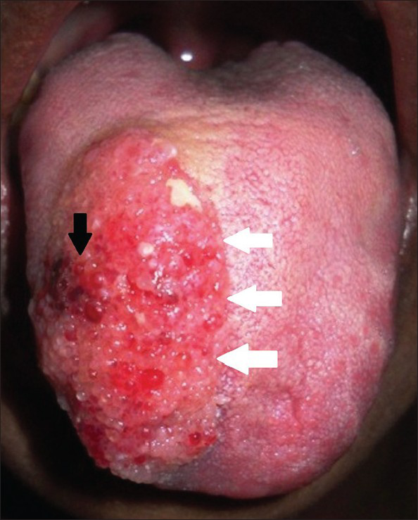
29-year-old female with swelling on the right side of her tongue diagnosed as lymphangioma circumscriptum. Intra-oral picture frontal view shows pebbly, vesicle-like feature with so-called “frog-egg” or “tapioca-pudding” appearance (black arrow) and medially extends till midline of tongue (white arrows).
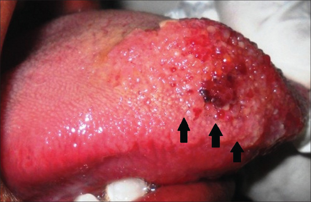
Figure 1
29-year-old female with swelling on the right side of her tongue diagnosed as lymphangioma circumscriptum. Intra-oral picture frontal view shows pebbly, vesicle-like feature with so-called “frog-egg” or “tapioca-pudding” appearance (black arrow) and medially extends till midline of tongue (white arrows).
29-year-old female with swelling on the right side of her tongue diagnosed as lymphangioma circumscriptum. Intra-oral picture lateral view shows that the lesion laterally extends till the lateral borders of the tongue (black arrows).
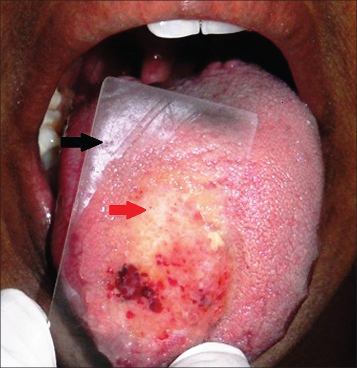
Figure 2
29-year-old female with swelling on the right side of her tongue diagnosed as lymphangioma circumscriptum. Intra-oral picture lateral view shows that the lesion laterally extends till the lateral borders of the tongue (black arrows).
29-year-old female with swelling on the right side of her tongue diagnosed as lymphangioma circumscriptum. Diascopy test performed by pressing a glass slide (black arrow) on the lesion results in blanching (red arrow) which suggests that lesion could be a vascular lesion.
Figure 3
29-year-old female with swelling on the right side of her tongue diagnosed as lymphangioma circumscriptum. Diascopy test performed by pressing a glass slide (black arrow) on the lesion results in blanching (red arrow) which suggests that lesion could be a vascular lesion.

RADIOLOGIC FEATURES

Soft-tissue ultrasonograph of the tongue demonstrated a well-defined heterogeneous soft-tissue mass of 2.3 cm × 3.3 cm × 2.4 cm on the right half of the tongue [Figure 4]. Dilated arteries and veins were seen in the right submental region on color Doppler studies suggesting that it could be a vascular malformation [Figure 5].
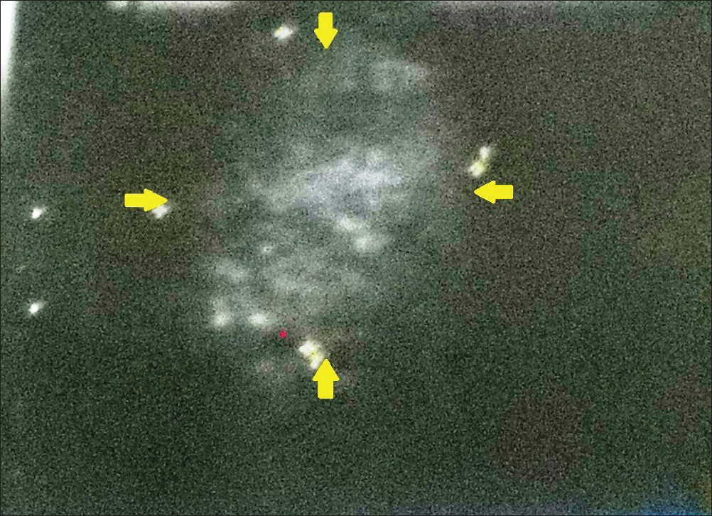
29-year-old female with swelling on the right side of her tongue diagnosed as lymphangioma circumscriptum. Soft-tissue ultrasonograph of the tongue demonstrates a well-defined heterogeneous soft tissue mass of 2.3 cm × 3.3 cm × 2.4 cm (yellow arrows) seen on the right half of the tongue suggestive of a soft tissue tumor.
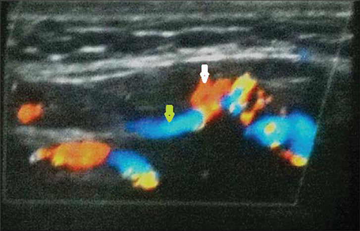
Figure 4
29-year-old female with swelling on the right side of her tongue diagnosed as lymphangioma circumscriptum. Soft-tissue ultrasonograph of the tongue demonstrates a well-defined heterogeneous soft tissue mass of 2.3 cm × 3.3 cm × 2.4 cm (yellow arrows) seen on the right half of the tongue suggestive of a soft tissue tumor.
29-year-old female with swelling on the right side of her tongue diagnosed as lymphangioma circumscriptum. Color Doppler imaging in longitudinal section through the right side of the tongue and sub mental region shows heterogeneous mass along with dilated arteries (white arrow) and veins (yellow arrow) suggestive of a vascular malformation.
Figure 5
29-year-old female with swelling on the right side of her tongue diagnosed as lymphangioma circumscriptum. Color Doppler imaging in longitudinal section through the right side of the tongue and sub mental region shows heterogeneous mass along with dilated arteries (white arrow) and veins (yellow arrow) suggestive of a vascular malformation.

PATHOLOGIC FEATURES

Incisional biopsy under local anesthesia was performed on the dorsal surface of the tongue and the tissue specimen was sent for histopathological examination. Microscopic section revealed tongue mucosa with marked irregular acanthosis and papillomatosis with thin elongated rete pegs. Adjacent subepithelial tissue showed cavernous vascular spaces lined by flattened epithelium. Deep submucosa showed diffuse chronic inflammation. Deep muscle layer appeared normal suggestive of “lymphangioma circumscriptus” [Figure 6]. Patient was referred to the Department of oral and maxillofacial surgery for further treatment.
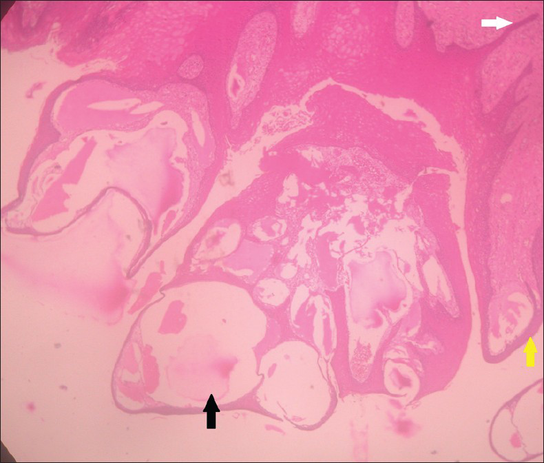
29-year-old female with swelling on the right side of her tongue diagnosed as lymphangioma circumscriptum. Hematoxylin and eosin stained soft-tissue section under ×40 magnification shows elongated rete ridges (white arrow), papillamatosis (yellow arrow), and cavernous spaces filled with lymph represented as eosinophilic stain (black arrow) suggestive of lymphangioma circumscriptum.
Figure 6
29-year-old female with swelling on the right side of her tongue diagnosed as lymphangioma circumscriptum. Hematoxylin and eosin stained soft-tissue section under ×40 magnification shows elongated rete ridges (white arrow), papillamatosis (yellow arrow), and cavernous spaces filled with lymph represented as eosinophilic stain (black arrow) suggestive of lymphangioma circumscriptum.

DISCUSSION

Lymphangiomas are hamartomatous, congenital malformations of the lymphatic system.[5] They are believed to arise from lymph sac sequestration and enlarge due to inadequate drainage, from lack of communication with the central lymphatic channels or excessive secretion of lining cells.[6]

Lymphangioma is observed on birth or manifests before 2 years of age. In the case reported here, the lesion developed in a patient older than the common age group. The patient was 29-years old at the time of diagnosis and had reported that the problem had persisted for 17 years.[4]

Lymphangioma have a marked predilection for the head and neck region, which accounts for about 75% of all cases.[1] Lymphangiomas have been classified into:

  • Lymphangioma simplex (capillary lymphangioma) consists of small, capillary-sized vessels

  • Cavernous lymphangioma is composed of large, dilated lymphatic vessels

  • Cystic lymphangioma (cystic hygroma) exhibits large macroscopic cystic spaces.[7]

This is also classified according to the clinical presentation into macrocystic (cavities larger than about 2 cm3), microcystic (cavities smaller than about 2 cm3) and mixed, combining these two types.[8]

Oral lymphangiomas occur at various sites, but are most frequently found on the anterior two-thirds of the tongue, where they often result in macroglossia.[1] The lesion can also present in the palate, buccal mucosa, gingival, and lips.[7] Uncommon occurences involve the soft palate and retromolar region.[9] Clinically, the lesions present superficially as a pebbly, vesicle-like feature with so-called “frog-egg” or “tapioca-pudding” appearance. Deeper tumors present as soft, ill-defined masses.[1]

The diagnosis of lymphangiomas is not difficult, being mainly clinical. In lesions with atypical clinical features, a definitive diagnosis should be made through biopsy and histopathological examination, along with imaging studies which are very useful in confirming the diagnosis.[2] Imaging modalities, especially color Doppler can identify the vascular malformation in cases of lymphangioma circumscriptum. Vascular malformations do not contain hyperplastic cells but consist of progressively enlarging aberrant and ectatic vessels composed of a particular vascular architecture such as veins, lymphatic vessels, venules, capillaries, arteries, or mixed vessel types. Vascular malformations are appropriately named by their predominant vessel type (e.g., venous malformations, arteriovenous malformations).[10] In this case it was due to lymphatic vessels and hence it is termed as lymphatic malformation.

The histopathology of lymphangioma circumscriptum is characterized by solitary and grouped dilated cystic spaces in the papillary derma. The cystic spaces are lined by endothelial cells often containing red blood cells, as well as lymphatic fluid. In the deep derma and subcutaneous fat, dilated lymphatic vessels are also seen, some of it containing thickened muscular walls.[2] In this case, histological study showed irregular acanthosis and papillomatosis with thin elongated rete pegs. Adjacent subepithelial tissue showed cavernous vascular spaces lined by flattened epithelium. Deep submucosa shows diffuse chronic inflammation. Deep muscle layer appear normal suggestive of “lymphangioma circumscriptus.”

The objectives of treatment of lymphangiomatous macroglossia are preservation of taste, restoration of tongue size for articulation, correction of mandibular and dental deformities and cosmesis, which in turn are treated using different treatment modalities such as surgical excision, radiation therapy, cryotherapy, electrocautery, sclerotherapy, steroid administration, embolization and ligation, laser surgery and radiofrequency tissue ablation technique. Among these treatment modalities, surgical excision continues to be integral to management in many cases.[5]

CONCLUSION

Although rarely encountered in the oral cavity, lymphangiomas represent a condition that must be recognized. Their early recognition allows proper initiation of treatment and prevents the occurrence of the complications. Color Doppler imaging definitely helps to identify the increased vascularity seen in lymphangioma circumscriptum and also the extent of the lesion, which is essential in optimizing surgical and other treatment modalities.

ACKNOWLEDGMENT

The authors would like to acknowledge Dr. N. Gnansundaram, Former Professor and Head, Department of Oral Medicine and Principal, Tamil Nadu Government Dental college, India for his valuable suggestions and to the Department of Oral Medicine and Radiology, Saveetha Dental College and Hospital, Chennai, India.

Source of Support: Nil

Conflict of Interest: None declared.

REFERENCES

  1. , , , , . Soft tissue tumors. In: , ed. Oral and Maxillofacial Pathology (2nd ed). New Delhi, Philadelphia: Saunders Publishers; . p. :475-7.
    [Google Scholar]
  2. , , , , . Lymphangioma of the oral cavity. Rom J Morphol Embryol. 2006;47:373-7.
    [Google Scholar]
  3. , , , . Lymphangioma circumscriptum of the tongue: Successful treatment using intralesional bleomycin. J Laryngol Otol. 2009;123:1390-2.
    [Google Scholar]
  4. , . Parotid area lymphangioma in an adult: Case report. J Oral Maxillofac Surg. 2004;62:1320-3.
    [Google Scholar]
  5. , , , , . Lymphangioma of tongue: A rare entity. Indian J Dent Adv. 2011;3:635-7.
    [Google Scholar]
  6. , , , , . Lymphangioma of the tongue. Natl J Maxillofac Surg. 2011;2:86-8.
    [Google Scholar]
  7. , , , , , , . Oral lymphangioma: A case report. J Indian Soc Pedod Prev Dent. 2005;23:185-9.
    [Google Scholar]
  8. , , , , , , . Utility of radiofrequency ablation for haemorrhagic lingual lymphangioma. Int J Pediatr Otorhinolaryngol. 2008;72:953-8.
    [Google Scholar]
  9. , , , , , , . Oral lymphangioma: Case report. Rev Sul-Bras Odontol. 2011;8:352-6.
    [Google Scholar]
  10. , , , . Diagnosis and management of hemangiomas and vascular malformations of the head and neck. Oral Dis. 2010;16:405-18.
    [Google Scholar]
Show Sections