
Translate this page into:
HRCT Correlation with Round Window Identification during Cochlear Implantation in Children
-
Received: ,
Accepted: ,
This is an open-access article distributed under the terms of the Creative Commons Attribution License, which permits unrestricted use, distribution, and reproduction in any medium, provided the original author and source are credited.
This article was originally published by Medknow Publications & Media Pvt Ltd and was migrated to Scientific Scholar after the change of Publisher.
Abstract
Objective:
To determine the accuracy of High Resolution Computer Tomography (HRCT) temporal bone measurements in predicting the actual visualization of round window niche as viewed through posterior tympanotomy (i.e. facial recess).
Materials and Methods:
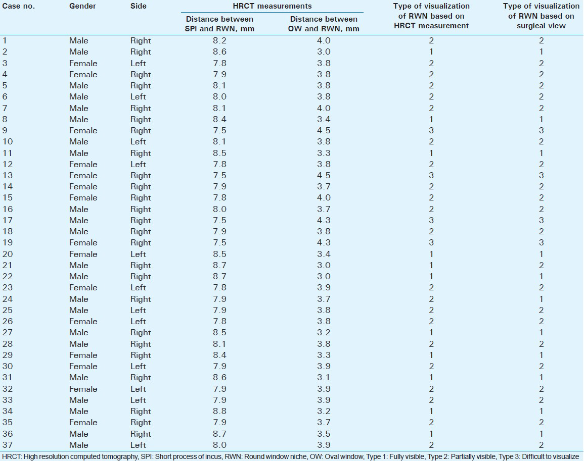
This is a prospective study of 37 cochlear implant candidates, aged between 1and 6 years, who were referred for HRCT temporal bone during the period December 2013 to July 2014. Cochlear implantation was done in 37 children (25 in the right ear and 12 in the left ear). The distance between the short process of incus and the round window niche and the distance between the oval window and the round window niche were measured preoperatively on sub-millimeter (0.7 mm) HRCT images. We classified the visibility of round window niche based on the surgical view (i.e. through posterior tympanotomy) during surgery into three types: 1) Type 1- fully visible, 2) Type 2- partially visible, and 3) Type 3- difficult to visualize. The preoperative HRCT measurements were used to predict the type of visualization of round window niche before surgery and correlated with the findings during surgery.
Results:
The mean and standard deviation for the distance between the short process of incus and the round window niche and for the distance between the oval window and the round window niche for Types 1, 2, and 3 were 8.5 ± 0.2 mm and 3.2 ± 0.2 mm, 8.0 ± 0.4 mm and 3.8 ± 0.2 mm, 7.5 ± 0.2 mm and 4.4 ± 0.2 mm respectively, and showed statistically significant difference (P < 0.01) between them. The preoperative HRCT measurements had a sensitivity and specificity of 92.3% and 96.2%, respectively, in determining the actual visualization of round window niche.
Conclusion:
This study shows preoperative HRCT temporal bone measurements are useful in predicting the actual visualization of round window niche as viewed through posterior tympanotomy.
Keywords
Facial recess
round window niche
short process of incus

INTRODUCTION

Cochlear implantation is performed in patients with profound sensori neural hearing loss, including children with hearing impairment.[1] Posterior tympanotomy is the surgical approach used in cochlear implant surgery for accessing the middle ear and exposing the round window niche.[2] The clear visualization of the round window niche and round window membrane through the facial recess is a prime requisite for the Ear, Nose, and Throat (ENT) surgeon when inserting the electrode into the cochlea. Due to the variations in the position of the round window, the visualization of the round window niche can become difficult for a surgeon. The variations in the position of the round window niche are caused by rotation of the cochlea in the horizontal and vertical axes.
The study was undertaken to assess whether the preoperative High Resolution Computed Tomography (HRCT) measurements are useful in predicting the actual visualization of the round window niche seen during posterior tympanotomy for cochlear implant surgery.
MATERIALS AND METHODS
Institutional ethical committee approval was obtained for this study. Informed consent was obtained from the parents. HRCT images of cochlear implant candidates, aged between 1 and 6 years, who were referred for HRCT temporal bone between December 2013 and July 2014, were studied prospectively. All the children had bilateral sensory neural hearing loss. Cochlear implantation was done in 37 ears. Among these, in four cases, there was difficulty in visualization of the round window niche through posterior tympanotomy approach. The cochlear implant surgery was performed by an ENT surgeon who had experience of more than 8 years in cochlear implant surgeries.
HRCT measurements were performed by two readers. Reader A (PhD research scholar), who is an expert in reading temporal bone scans, performed all the measurements. All measurements were taken by Reader A at two different sessions. The measurements were read again by another Reader B (radiologist) who has experience of more than 10 years in interpretation of temporal bone scans.
Data acquisition and measurement protocol
CT imaging was performed using Brilliance CT 16-slice (Brilliance CT; Philips Medical systems, Cleveland, OH, USA). The images for temporal bone were obtained with a slice thickness of 0.7 mm. The CT scan data were acquired at 120 kVp, 250 mA, and imaging matrix of 512 × 512. The axial images were obtained parallel to orbito-meatal base line.
Preoperative HRCT measurements
The distance between the short process of incus and the round window niche and the distance between the oval window and the round window niche measurements were taken preoperatively on Philips workstation with magnification and electronic caliper capabilities. The measurements were noted in millimeters. These measurements were done on sub-millimeter (0.7 mm) images.
The distance between the short process of incus and the round window niche (cranio-caudal) is defined as the distance between the tip of the short process of incus and the edge of tegmen of the round window niche at the midpoint of anterior and posterior borders. The oblique axial reconstructed image [Figure 1a] showing the incus and malleus head was chosen. Oblique coronal reformation was done by drawing a plane passing through the body of incus and through the center of the round window niche on the oblique axial image. The short process of incus was identified on the reformatted images [Figure 1b] and localized using two lines drawn manually. These lines are extrapolated on the oblique coronal image showing the round window niche. Then the distance [Figure 1c] between the tip of the short process of incus and the edge of tegmen of the round window niche at the midpoint of anterior and posterior borders was measured.

- 4-year-old male with history of sensory neural hearing loss. (a) Oblique axial HRCT reconstructed image of the right ear shows incus and malleus head (white arrow) and also shows the plane passing through the body of incus (white line). (b) Oblique coronal HRCT reconstructed image (double arrow) shows the tip of short process of incus in fossa incudis. (c) Oblique coronal HRCT reformatted image shows the distance between the short process of incus and the edge of tegmen of round window niche at the midpoint of anterior and posterior borders.
The edge of tegmen of the round window niche was used as it was consistently reproducible. Though the round window membrane is an important landmark for the surgeons, it is difficult to visualize it on HRCT images. So, the edge of tegmen of the round window niche was chosen for the measurements.
The distance between the oval window and the round window niche (cranio-caudal) is defined as the distance between the midpoint of the oval window and the edge of tegmen of the round window niche measured at the midpoint of anterior and posterior borders.
The oblique axial image showing the incus and malleus head was chosen. Oblique coronal image was obtained by drawing a plane passing through the body of incus and the midpoint of round window niche on oblique axial image. Using these steps, the location of round window niche becomes reproducible. Then the distance [Figure 2] between the midpoint of the oval window and the edge of tegmen of the round window niche at the midpoint of anterior and posterior borders was measured.

- 1-year-old female with history of sensory neural hearing loss. Oblique coronal HRCT reformatted image (white arrow) of the left ear shows the distance between oval window and round window niche.
Classification of visibility of round window niche
We classified the visibility of round window niche based on surgical view (i.e. through posterior tympanotomy) during surgery into three types:
-
Type 1: Fully visible [Figure 3]
-
Type 2: Partially visible [Figure 4]
-
Type 3: Difficult to visualize [Figure 5].

- 4-year-old male with history of sensory neural hearing loss. Intraoperative photograph of cochlear implant surgery of the right ear shows Type 1 fully visible round window niche (white arrow).

- 4-year-old male with history of sensory neural hearing loss. Intraoperative photograph of cochlear implant surgery of the right ear shows Type 2 partially visible round window niche (white arrow).

- 6-year-old male with history of sensory neural hearing loss. Intraoperative photograph of cochlear implant surgery of the right ear shows Type 3 difficulty to visualize round window (white arrow).
In cases where the round window niche is difficult to visualize, the surgeon needs to drill deeper into the facial recess and adjust the microscope depending on the type of rotation, resulting in an increase in surgery time. The preoperative measurements were used to predict the type of visualization of round window niche before the surgery. However, the surgeon was blind to the measurements and the type of visualization of round window niche.
Statistical analysis
The data were analyzed with Statistical Package for Social Sciences (SPSS Inc., Chicago, IL, USA) version 16.0. To compare the HRCT measurements between the three different types, one-way analysis of variance and Tukey's post hoc test were used. The Receiver Operating Characteristic (ROC) curve was used to find the sensitivity and specificity of the HRCT measurements in predicting the visualization of round window niche. The intra- and inter-observer variability was calculated for preoperative HRCT measurements.
RESULTS
Of the 37 cochlear implant patients, 30% (n = 11) were classified as Type 1, 60% (n = 22) as Type 2, and 10% (n = 4) as Type 3. Table 1 shows the means of preoperative HRCT measurements for the three types.

There was a statistically significant difference (P < 0.01) in HRCT measurements among the three types [Table 1].
The mean distance between the short process of incus and the round window niche for Types 1, 2, and 3 was 8.5 ± 0.2 mm, 8.0 ± 0.4 mm, and 7.5 ± 0.2 mm, respectively [Figure 6].

- Histogram compares mean distance between short process of incus and round window niche for the three types. The mean distance decreases in Type 2 and Type 3 when compared to Type 1.
The mean distance between the oval window and the round window niche for Types 1, 2, and 3 was 3.2 ± 0.2 mm, 3.8 ± 0.2 mm, and 4.4 ± 0.2 mm, respectively [Figure 7].

- Histogram compares mean distance between oval window and round window niche for the three types. The mean distance increases in Type 2 and Type 3 compared to Type 1.
Intra-observer variability
There was no statistically significant difference between the measurements performed by Reader A at different time periods, according to paired Student's t-test.
Inter-observer variability
Intra-class correlation coefficients produce measures of consistency of values in which readers differ. Intra-class correlation coefficient of less than 0.4 indicates lesser reproducibility and greater than 0.75 suggests excellent reproducibility. For all the measurements in this study, the intra-class correlation coefficient was 0.99, suggesting good reproducibility.
Of the 37 cases, 11 cases showed fully visible round window niche (Type 1; based on surgery); of them, 10 cases correlated with the HRCT measurements corresponding to Type 1, while 1 case (24th) was identified as partially visible in HRCT study.
Twenty-two cases showed partially visible round window niche (Type 2; based on surgery); of them, 21 cases correlated with the HRCT measurements corresponding to Type 2, while 1 case (21st) was identified as fully visible in HRCT.
In four cases, the round window was difficult to visualize (Type 3) in the surgical view; all the four cases correlated with HRCT measurements that identified the cases as difficult to visualize [Table 2].
The sensitivity and specificity of HRCT measurements in predicting the visualization of round window niche were 92.3% and 96.2%, respectively. The ROC curve [Figure 8] has an area of 0.9, which shows that HRCT measurements have good accuracy in predicting the actual visualization round window niche in the surgical view.
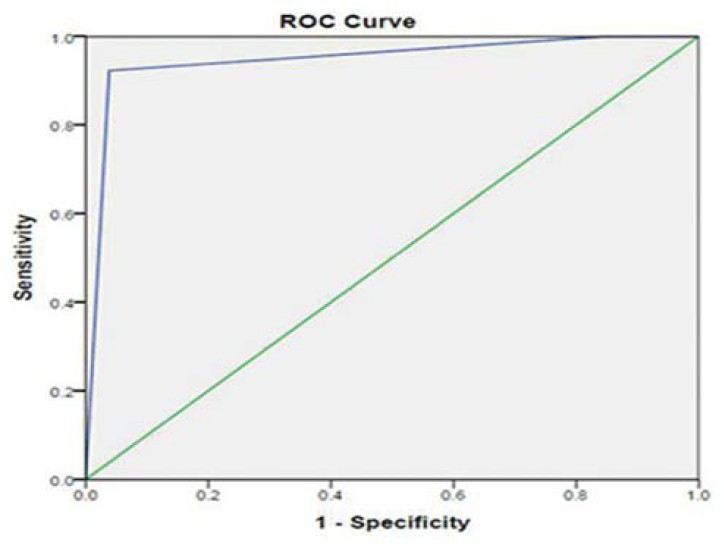
- Receiver operator characteristic (ROC) curve has an area of 0.9, which shows that HRCT measurements have good accuracy in predicting the actual visualization of round niche during posterior tympanotomy. Green line (diagonal) represents the curve for a perfectly useless test. Blue line represents the curve for HRCT measurements.
DISCUSSION
Earlier it was thought that the preoperative CT measurements were not able to predict the complications faced during cochlear implant surgery. Bettman et al.,[3] reported in their study that preoperative CT measurements, such as facial recess width and the angle between facial recess and basal turn of the cochlear, were not useful in predicting the problems encountered during surgery and that the advances in CT, such as Multislice CT, could improve diagnostic accuracy.
In the present study, we determined if the HRCT measurements taken between references points in the middle ear are useful in predicting the actual visualization of the round window niche when viewed through facial recess. The short process of incus is a definitive land mark for surgeons in identifying the round window niche. Hence in our study we measured the distance between the short process of incus and round window niche to identify the variations in position of round window niche. We found in our study that as the distance between the tip of short process of incus (fossa incudis) and the round window niche decreases, the visibility of round window niche through posterior tympanotomy becomes difficult. The probable reason for this could be as the tip of short process of incus (fossa incudis) is placed posteriorly, if the round window niche is displaced further posterior and superior (which is an anatomic variation) the distance between the short process of incus to round window niche will decrease.
We also found in our study that as the distance between the oval window and the round window niche increases, the visibility of round window niche seen during posterior tympanotomy decreases. The probable reason for this could be that the facial recess which acts as a window through which the round window is viewed has a width of 3.8–4.0 mm, as reported by Su et al.,[4] and Young et al.[5] As the distance between the oval window and the round window niche increases (>4 mm), the visibility of the round window niche decreases. This may be a limitation of the facial recess approach.
Lloyd et al., stated that the difficult cochlear implant cases had shown an obtuse angle of the basal turn of the cochlea.[6] McRackan et al., reported that there are significant differences in the anatomical landmarks useful for cochlear implantation between pediatric patients and adults.[7]
Of the 37 cases in this study, 11 cases were classified as fully visible, 22 as partly visible, and 4 cases were classified as difficult to visualize based on the surgical view.
In fully visible Type 1, out of 11 cases, HRCT measurements were able to predict the full visualization of the round window niche in 10 cases, and 1 case (24th) was found to be fully visible in the surgical view and was predicted to be partially visible based on the HRCT measurements.
In partially visible Type 2, out of 22 cases, HRCT measurements were able to predict the partial visualization of the round window niche in 21 cases, and 1 case (21st) was found to be partially visible in the surgical view and was predicted to be fully visible based on HRCT measurements.
In difficult to visualize Type 3, out of four cases, HRCT measurements were able to predict difficulty in visualization of round window niche in all the four cases.
Our study found preoperative HRCT measurements of the distance between anatomical landmarks of the middle ear seen on the scan was a useful method for determining the actual visualization of round window niche during surgery, as viewed through posterior tympanotomy.
Limitations
There were some pitfalls in our study. We have not analyzed the association of gender and right versus left ear with either the CT measurements or surgeon's visibility of the round window niche. As the lines in making measurements are drawn manually, care must be taken in taking measurements and predicting the visualization based on these measurements. The small number of cases is another limitation of this study. A study of larger number of cases is required to confirm these findings.
CONCLUSION
From this study, we conclude that the distance between the short process of incus and the round window niche and the distance between the oval window and the round window niche can help in determining the extent of visibility of the round window seen during posterior tympanotomy. These measurements can be used as a guide by the surgeons in locating the round window niche and planning surgery.
Available FREE in open access from: http://www.clinicalimagingscience.org/text.asp?2014/4/1/70/148264
Source of Support: Shri. N.P.V. Ramasamy Udayar Chancellor Research Fellowship, Sri Ramachandra University
Conflict of Interest: None declared.
REFERENCES
- The posterior route to middle ear: Posterior tympanotomy. Laryngoscope. 1967;77:306-16.
- [Google Scholar]
- Cochlear orientation and dimensions of the facial recess in cochlear implantation. ORL J Otorhinolaryngol Relat Spec. 2003;65:353-8.
- [Google Scholar]
- Anatomical measurements of the cochlear aqueduct, round window membrane, round window niche, and facial recess. Laryngoscope. 1982;92:483-6.
- [Google Scholar]
- Developmental changes in cochlear orientation implications for Cochlear implantation. Otol Neurotol. 2010;31:902-7.
- [Google Scholar]
- Comparison of cochlear implant relevant anatomy in children versus adults. Otol Neurotol. 2012;33:328-34.
- [Google Scholar]


