
Translate this page into:
Hemimandibular Hypertrophy - Hybrid Variants: Report of Two Cases
Address for correspondence: Dr. Ravi Prakash Sasankoti Mohan, C/o Dr. R. P. Singh, Dhanwantri Nursing Home, Sarai Khalsa, Behind Head Post Office, Moradabad - 244 001, Uttar Pradesh, India. E-mail: sasan_ravi@rediffmail.com
-
Received: ,
Accepted: ,
This is an open-access article distributed under the terms of the Creative Commons Attribution License, which permits unrestricted use, distribution, and reproduction in any medium, provided the original author and source are credited.
This article was originally published by Medknow Publications & Media Pvt Ltd and was migrated to Scientific Scholar after the change of Publisher.
Abstract
Hemimandibular hypertrophy and its variants result from unilateral excessive growth of the mandible and involve both the body and ramus of mandible. This causes facial asymmetry and in turn accompanying psychological problems. In this report we discuss use of imaging in diagnosis of these lesions and investigate the different variants.
Keywords
Facial asymmetry
hemimandibular elongation
hemimandibular hypertrophy
hybrid phenotypes

INTRODUCTION

Hemimandibular hypertrophy along with its variants was first described by Obwegeser and Makek as a developmental deformity of unknown etiology affecting the mandible unilaterally.[1] The etiologic factors are not yet completely understood. Various studies have assumed lowered genetic control over the formation and development of bilateral structures of the face or environmental influences and accidents during development to be the cause.[23] Diagnosis of these lesions often poses a challenge to junior dentists. Imaging plays a valuable role in the diagnosis of these conditions. In this article we present variants of hemimandibular hypertrophy with radiographic features and differential diagnosis.
CASE REPORTS
Case 1
A 34-year-old male patient reported to the out-patient department with a complaint of asymmetric appearance of his face. He had been involved in a road accident 15 years ago that had caused severe trauma to his chin. There were no other contributory findings in his dental, medical, and family history. On clinical examination, marked facial asymmetry was observed in the lower region of the face with deviation of the chin to the left. The mandibular body and ramus appeared longer and wider on the right side as compared to the left side [Figure 1a]. Intra-oral examination revealed transverse canting of occlusal plane on the right side. Posterio-anterior view of the skull demonstrated elongation of the condyle on the right side. Height of the ramus and body of the mandible were also larger along with transverse canting of the occlusal plane on the right side [Figure 1b]. Three-dimensional computed tomography (CT) image showed elongation and enlargement of condyle, ramus, and body of the mandible on the right side [Figure 1c]. Scintigram demonstrated hot spots indicative of increased uptake of radioisotope in the condylar region on the right side [Figure 1d]. Thus on the basis of clinical and radiographic features, diagnosis of hemimandibular hypertrophy-elongation hybrid variant of the right side was confirmed. Patient was referred to the oral and maxillofacial surgery department, where orthognathic surgery was done and both function and esthetics of the patient's facial structure were restored [Figure 1e].
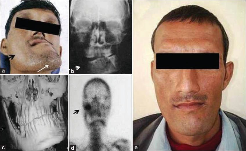
- Case 1. 34-year-old male patient presenting with facial asymmetry due to hemimandibular hypertrophy hybrid variant. (a) Photograph of the face shows elongation of mandible on the right. (black arrow). Deviation of chin to the left. (white arrow). (b) X-ray posterio-anterior view of skull shows mandibular enlargement and transverse canting of occlusal plane on the right side (white arrow). (c) Three-dimensional computed tomography shows right sided hemimandibular hypertrophy along with bilateral crossbite. (d) Scintigram demonstrates hot spot (arrow) in the condylar region, indicative of increased activity in the mandible.on the right side. (e) Clinical photograph of the face post-treatment.
Case 2
An 18-year-old female patient reported to the out-patient department with a complaint of gradually developing asymmetry of her face. She first noticed asymmetry of her face after a trauma she suffered about 5 years earlier. Since then the condition had gradually deteriorated and attained the present status. No information relevant to the condition was elicited from past dental, medical, and family history. On extra-oral examination, marked facial asymmetry was observed in the lower face region with deviation of chin toward the left side. Enlargement of the right side of mandible was clearly seen [Figure 2a]. On intra-oral examination, mandibular central incisor midline was found not to coincide with facial midline. Open bite was present on the right posterior tooth region [Figure 2b]. Panoramic radiograph revealed elongation of condyle, condylar neck, ramus and body of mandible on the right side. Horizontal dimension of the mandibular body was also larger as compared to that on the left side. The distance between the mandibular roots and the inferior alveolar nerve on the right side was more than that on the left side [Figure 2c]. Further, CT demonstrated elongation and medial rotation of right lower border of the mandible [Figure 2d and e]. On the basis of all these findings, diagnosis of hemimandibular hypertrophy-elongation hybrid type was confirmed.
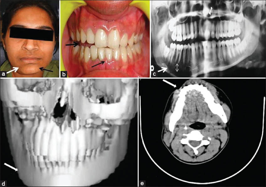
- Case 2. 18-year-old girl with asymmetry of the face due to hemimandibularr hypertrophy hybrid.variant. (a) Photograph of the face shows enlargement of mandible on the right side. (white arrow) with deviation of chin to the left. (black arrow). (b) Intra-oral view reveals open bite. (thick arrow). Mandibular central incisor midline does not coincide with facial midline. (thin arrow). (c) Panoramic radiograph demonstrates elongation (arrow) and increased distance between the mandibular molar roots and inferior alveolar nerve canal on the right side (double-headed arrow). (d) Three-dimensional computed tomography (CT) shows elongation and enlargement of the mandible on the right side (arrow). (e) CT axial view reveals asymmetry due to increased growth of the mandible on the right side (arrow).
DISCUSSION
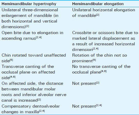
Condylar hypertrophy can result in a range of clinical manifestations leading to facial asymmetries. Obwegeser and Makek classified condylar hyperplasia into two distinct types namely hemimandibular hypertrophy and hemimandibular elongation.[13] Table 1 lists the differences between hemimandibular hypertrophy and hemimandibular elongation. Later, certain cases were seen with overlapping features and were categorized as a hybrid variant that showed a combination of hemimandibular hypertrophy and hemimandibular elongation. Hemimandibular elongation presents with a marked lateral displacement of the chin to the unaffected side and depressed commissure of lips on the affected side. Due to the asymmetric shift of underlying skeletal component to one side, occlusion gets de-arranged and clinically the mandibular dental centerline does not coincide with the midfacial line. Crossbite is observed on the unaffected side.[23] Since there is minimal or almost no discrepancy in the vertical component to the abnormal mandibular growth, open bites or transverse canting of the maxillary occlusal plane are not evident. Radiographically, there is elongation of the body of the mandible on the affected side.[2]
Hemimandibular hypertrophy is a three-dimensional developmental enlargement of one side of the mandible including the condyle, condylar neck, ramus and body along with medial rotation.[2] The anomaly terminates exactly at the symphysis of the affected side and for this reason it is called hemimandibular hypertrophy.[3] It is distinct from hemimandibular elongation as the latter shows increase only in the horizontal component. The etiology of hemimandibular hypertrophy is still under discussion. In the literature, genetic factors, circulatory problems, hormonal disturbances, traumatic lesions and arthrosis have been proposed as the etiologic factors of the disease.[3] There is an increase in the height of the affected side, giving the face a rotated appearance. If the anomaly occurs before adolescent growth spurt then the maxilla follows the downward growth of the mandible and the teeth of the affected side are at a lower level, resulting in tilting of the occlusal plane in the transverse dimension. If the maxilla is unable to follow the mandibular growth, an open bite becomes evident on the affected side. All these findings were present in Case 1. Panoramic radiograph shows that both the ascending ramus and condyle are elongated vertically along with thickening of the condylar neck. The inferior border is bowed downward to a lower level compared to the unaffected side. Due to elongation of the mandibular body, the distance between the molar roots and the mandibular canal is increased.[245] Histological examination of the mandibular head reveals signs of growth. There is over activity in the articular cartilage. The thickness of the proliferative zone increases, the fibro-cartilaginous zone becomes hypertrophic, endochondreal bone formation occurs while the articular zone remains remarkably intact.[678] Technetium-99 scinti-scanning is an essential diagnostic tool. In this procedure, radioactive isotope Technetium-99 methylene diphosphonate is used. It can be used to determine the side that is affected, to demarcate between abnormal condylar growth and generalized mandibular growth, and finally to assess whether hyperplasia is still active or has become stable.[57] Hybrid forms are the combination of unilateral hemimandibular hypertrophy and hemimandibular elongation and lead to marked facial asymmetry.[7] Such cases present with both vertical elongation and lateral displacement, causing large degree of facial asymmetry (seen in Case 2).[5678]
Due to overlapping features, there are certain conditions such as hemifacial hypertrophy, hemifacial atrophy of opposite side, hyperactive condyle, monostotic fibrous dysplasia and segmental odontomaxillary dysplasia that need to be considered in differential diagnosis.[2] In cases of hemifacial hypertrophy, entire half of the face is involved and the affected side shows enlarged teeth with rapid eruption of dentition. Other features include premature loss of primary teeth along with enlarged tongue and alveolar bone. Secondly, it is often associated with mental deficiency, skin abnormalities, compensatory scoliosis, Wilms tumor and Beckwith-Weidemann syndrome. Monostotic fibrous dysplasia and segmental odontomaxillary dysplasia can be differentiated due to their unique radiographic features.[24] Hemifacial hypoplasia can be differentiated by the presence of degenerative soft-tissue changes.[2] Acquired mandibular asymmetries involve pain, symptomatic changes, alterations in facial appearance and function with time. The volume of facial muscles remains unchanged. Other features include temporomandibular joint (TMJ) crepitation, limited mandibular movements, severe crossbite and irregular condyle anatomy. Whereas, developmental changes do not involve pain, symptoms usually remain unchanged over time, no functional changes take place in the TMJ, there may be limited protrusion without limiting mandibular rotation movements, a pronounced dental compensation in the asymmetric mandible may be present and the condyle remains pronounced and smooth, even in the presence of volumetric changes.[9]
Mandibular hypertrophy can be seen in Proteus syndrome that is characterized by striking facial abnormalities, one of two subforms show unilateral condylar overgrowth causing progressive craniofacial asymmetry.[10] Unilateral overgrowth of mandible can be clearly seen in female patients with Klinefelter's syndrome (47, XXY). Finally, there are craniofacial malformations that do not affect the jaws directly, but lead to indirect, presumably compensatory, alterations of otherwise relatively normal condylar growth. For example, in cases of craniosynostoses such as Crouzon, Pfeiffer, and Apert syndromes, unusual transverse mandibular growth may be regarded as an attempt at adapting to the impaired expansion of the cranial vault.[10] Treatment for hemimandibular hypertrophy varies according to the age of the patient. In a growing child, prime goal is to obtain a proper functional occlusion, which is achieved by occlusal adjustments and maxillary expansion. However, in an adult patient apart from orthodontic procedures, an additional muscle memory deprogramming with diagnostic splints is indicated to set the centric relation position.[4]
CONCLUSION
Imaging is essential for diagnosis of hemimandibular hypertrophy and its variants. Patients suffering from hemimandibular hypertrophy often seek seclusion due to abnormal shape of the face and thus early diagnosis with computed tomography can aide in identifying the underlying malformations. Appropriate treatment following correct diagnosis can help them regain their lost confidence.
Available FREE in open access from: http://www.clinicalimagingscience.org/text.asp?2013/3/1/34/116199
Source of Support: Nil
Conflict of Interest: None declared.
REFERENCES
- Hemimandibular hyperplasia: Hemimandibular elongation. J Maxillofac Surg. 1986;14:183-208.
- [Google Scholar]
- Diagnostic imaging of temporomandibular joint. In: White SC, Pharoah MJ, eds. Oral Radiology Principles and Interpretations (5th ed). St. Louis, Missouri: Mosby; 2008. p. :550-4.
- [Google Scholar]
- Mandibular condyle hyperplasia. Therapeutic review. Rev Stomatol Chir Maxillofac. 1996;97:145-60.
- [Google Scholar]
- Hemimandibular hyperplasia treated by early high condylectomy: A case report. Int J Adult Orthodon Orthognath Surg. 2001;16:227-34.
- [Google Scholar]
- Hyperplastic conditions of the mandibular condyles. Korean J Oral Maxillofac Radiol. 2003;33:207-9.
- [Google Scholar]
- Three-dimensional facial growth studied by optical surface scanning. J Orthod. 2000;27:31-8.
- [Google Scholar]
- Skeletal anchorage system for open-bite correction. Am J Orthod Dentofacial Orthop. 1999;115:166-74.
- [Google Scholar]


