
Translate this page into:
Giant Pindborg Tumor (Calcifying Epithelial Odontogenic Tumor): An Unusual Case Report with Radiologic-Pathologic Correlation
Address for correspondence: Dr. Satya Ranjan Misra, Department of Oral Medicine and Radiology, Institute of Dental Sciences and SUM Hospital, Bhubaneswar - 751 003, Odisha, India. E-mail: drsatyaranjanmds@gmail.com
-
Received: ,
Accepted: ,
This is an open-access article distributed under the terms of the Creative Commons Attribution License, which permits unrestricted use, distribution, and reproduction in any medium, provided the original author and source are credited.
This article was originally published by Medknow Publications & Media Pvt Ltd and was migrated to Scientific Scholar after the change of Publisher.
Abstract
Odontogenic tumors develop in the jaws from odontogenic tissues such as enamel organ, Hertwig epithelial root sheath, dental lamina, and so on. A variety of tumors unique to the maxilla and mandible are therefore seen. Calcifying epithelial odontogenic tumor (CEOT) is a rare, aggressive, benign odontogenic tumor of epithelial origin accounting for only about 1% of all odontogenic tumors. It is eponymously called “Pindborg tumor”, as it was first described by Pindborg in 1955. The origin of this locally invasive tumor remains unknown. It is thought to arise from stratum intermedium. It commonly affects the posterior mandible manifesting as a slow-growing asymptomatic swelling often associated with an impacted tooth. We report a case of CEOT, for which, owing to its huge size we have proposed the term “giant” Pindborg tumor (CEOT). This is probably the largest case of this tumor reported so far in the English literature. The present case also has the classic yet rare “driven snow” appearance of the tumor on radiographs.
Keywords
Calcifying epithelial odontogenic tumor
CEOT
driven snow appearance
odontogenic tumors
Pindborg tumor

INTRODUCTION

Calcifying epithelial odontogenic tumor (CEOT) was first described by J. J. Pindborg in 1955 and was eponymously called Pindborg tumor by Shafer et al.,[1] in 1963. Although it is established that CEOT arises from the epithelial components of the enamel organ, but whether the stratum intermedium, the dental lamina remnants or the reduced enamel epithelium are involved in the etiology still remains mysterious. It is an uncommon tumor as it accounts for only about 1% of all odontogenic neoplasms including odontomes.[2] While it is slow-growing, it can be locally aggressive with a tendency to recur.
CASE REPORT
A 38-year-old female patient reported to the dental hospital with an 8-year history of swelling on the right side of the face. The patient had initially noticed a small swelling on the right side of the face which progressively grew to attain its present size, due to which mastication and speech were compromised.
On clinical examination, a huge, well-circumscribed swelling was seen on the right side of the face, ovoid in shape, extending from the right ramus crossing the midline up to the left parasymphysis region of the mandible, measuring 12 × 10 cm in greatest diameter, bony hard in consistency, and nontender on palpation [Figures 1, 2]. Mouth opening was restricted; the interincisal distance being 20 mm. Intraorally, a large swelling was seen extending from tooth 33 up to the right retromolar region with bicortical expansion, obliterating the right buccal sulcus, with lobulations present on the lingual aspect [Figure 3]. Both the maxillary and mandibular teeth were displaced and were mobile on the affected side.

38-year-old female patient with a huge swelling on the right side of the face diagnosed with calcifying epithelial odontogenic tumor. Front view shows the swelling extending from the right ramus (arrow black outline) crossing the midline up to the left parasymphysis region (solid black arrow) of the mandible.
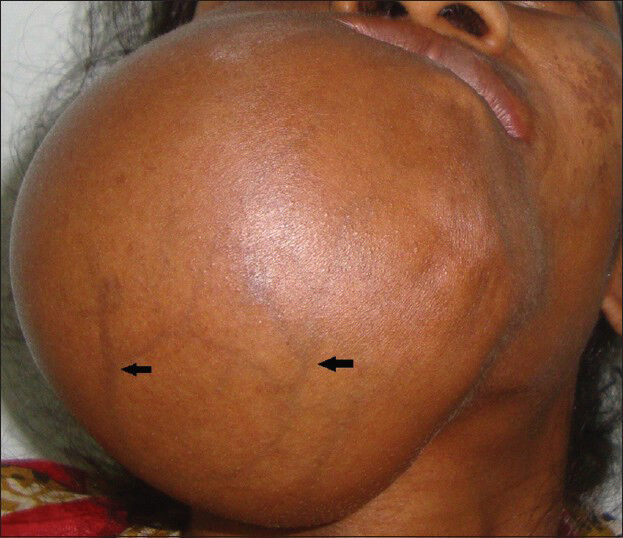
38-year-old female patient with a huge swelling on the right side of the face diagnosed with calcifying epithelial odontogenic tumor. Inferior profile view shows numerous tortuous veins (black arrows) owing to the stretching of the skin overlying the huge swelling.

38-year-old female patient with a huge swelling on the right side of the face diagnosed with calcifying epithelial odontogenic tumor. Intraoral view reveals a swelling with bicortical expansion, obliterating the buccal sulcus, (arrow black outline) lobulations present lingually (solid black arrows) with displacement of mandibular and maxillary teeth (white arrows) on the right side.
Owing to the slow-growing nature, well-circumscribed appearance, bony hard consistency and its presence in the tooth bearing region, a provisional clinical diagnosis of benign bony odontogenic neoplasm, was made. Ameloblastoma, CEOT, and ossifying fibroma were considered in clinical differential diagnosis.
RADIOLOGIC FEATURES
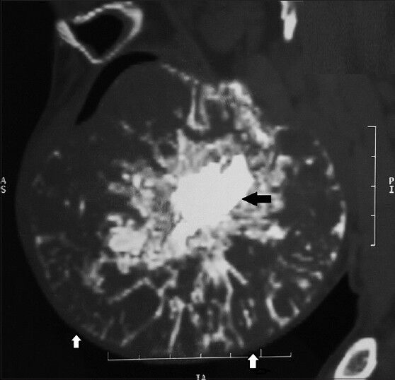
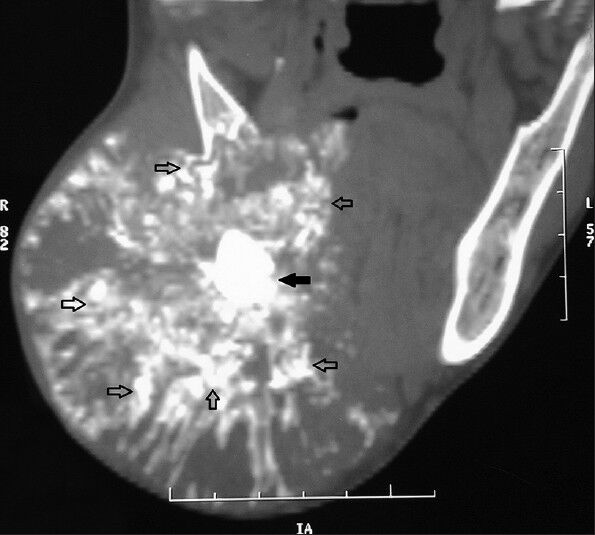
Occlusal and panoramic radiographs revealed a single well-defined circular mixed radiopaque-radiolucent lesion with bicortical expansion, on the right side of the mandible with displacement of the mandibular and maxillary teeth. There was the presence of a radiopaque mass in the center of the lesion, which could possibly be an impacted tooth or odontome with radiopaque streaks, which were suggestive of “driven snow” appearance [Figure 4]. Computed tomography (CT) images in axial, coronal, and sagittal planes [Figures 5–7] showed a large well-defined lytic soft tissue lesion measuring 12 × 10 cm in size having a predominantly calcified component in a snowflake pattern seen in the right mandible. Multiple ossicles and ossific densities with 700-1600 Hounsfield units were seen throughout the lesion. The entire tumor mass was well-circumscribed with expansion of the cortices without perforation [Figures 8 and 9]. The radiologic appearances were suggestive of a calcified tumor, but a definitive diagnosis could only be a histopathologic confirmation.

38-year-old female patient with a huge swelling on the right side of the face diagnosed with calcifying epithelial odontogenic tumor. Panoramic radiograph shows a single well-defined circular mixed radiopaque-radiolucent lesion on the right side of the mandible with displacement of the mandibular and maxillary teeth. Presence of a radiopaque mass in the center of the lesion (black arrow) with radiopaque streaks has the appearance of “driven snow”.
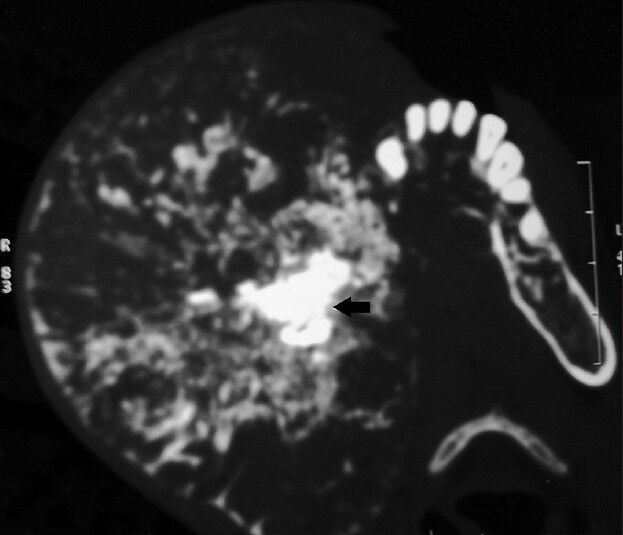
38-year-old female patient with a huge swelling on the right side of the face diagnosed with calcifying epithelial odontogenic tumor. Axial view computed tomography scan shows a huge well-defined heterodense tumor mass on the right side of the mandible with the presence of a hyperdense irregular mass at the center of the lesion (black arrow).
38-year-old female patient with a huge swelling on the right side of the face diagnosed with calcifying epithelial odontogenic tumor. Sagittal view computed tomography scan reveals a well-defined heterodense tumor mass with an irregular central hyperdense mass (black arrow) and expanded inferior border (white arrow) of the mandible without any perforation.
38-year-old female patient with a huge swelling on the right side of the face diagnosed with calcifying epithelial odontogenic tumor. Coronal view computed tomography scan shows a well-defined heterodense tumor mass on the right mandible having a central irregular hyperdense mass (solid black arrow) with multiple irregular calcifications with a “snow flake” pattern (black outline arrows).
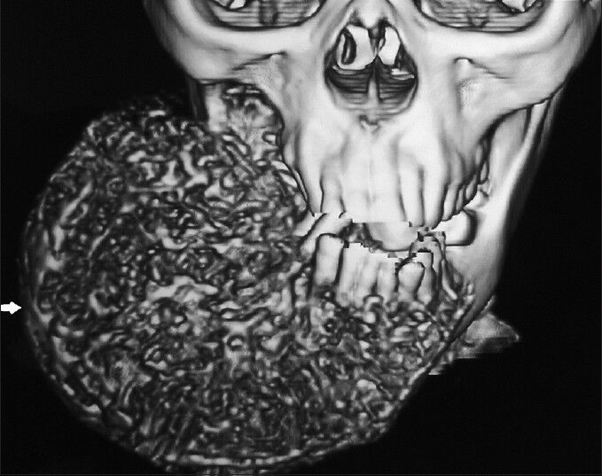
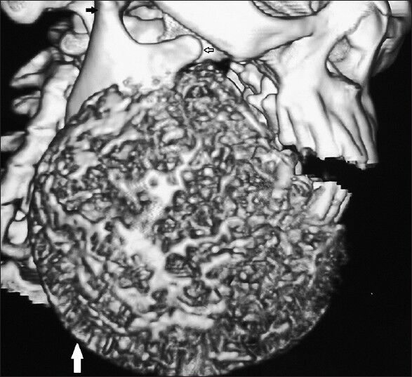
38-year-old female patient with a huge swelling on the right side of the face diagnosed with calcifying epithelial odontogenic tumor. A multidimensional computed tomography reconstruction using a hard tissue algorithm exhibits the extent of the well-circumscribed giant tumor (white arrow).
38-year-old female patient with a huge swelling on the right side of the face diagnosed with calcifying epithelial odontogenic tumor. A three-dimensional reconstructed profile image using a bone window exhibits the predominantly calcified well-defined tumor mass on the right side (white arrow) which has clearly spared the condyle (solid black arrow) and coronoid process (arrow black outline) on the affected side.
PATHOLOGIC FEATURES
An incisional biopsy was done and histopathological evaluation revealed polygonal epithelial components in sheets and islands dispersed throughout the connective tissue matrix along with numerous circular ring-like calcifications [Figure 10]. The polygonal epithelial cells exhibited distinct intercellular bridges, which are the characteristic feature of squamous cells. The nuclei were centrally located and often had prominent nucleoli. Other features of nuclei such as pleomorphism and hyperchromatism were also evident. Occasional binucleated and trinucleated cells were seen [Figure 11]. Rounded, pale eosinophilic masses along with calcifications in the form of Liesegang rings, correlating with radiographic findings of radiopaque streaks were found within sheets of tumor cells.

38-year-old female patient with a huge swelling on the right side of the face diagnosed with calcifying epithelial odontogenic tumor. Hematoxylin and eosin stained biopsy section (×20) shows epithelial cells in sheets and islands dispersed throughout the connective tissue matrix along with numerous circular ring like calcifiactions (black arrows).
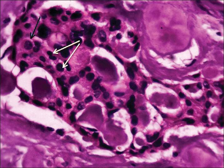
38-year-old female patient with a huge swelling on the right side of the face diagnosed with calcifying epithelial odontogenic tumor. Hematoxylin and eosin-stained biopsy section shows polygonal squamous epithelial cells exhibiting distinct intercellular bridges (black arrow) along with cellular and nuclear polymorphism (white arrows), and areas of irregular calcification and eosinophilic material.
Correlating the clinical, radiological, and histological features, a final diagnosis of CEOT was given. Hemimandibulectomy followed by microvascular reconstruction using a free fibula graft to restore the huge surgical defect was done.
DISCUSSION
CEOT was described by J. J. Pindborg in 1955 but it also been referred to as “adenoid adamtoblastoma” by Thoma, “unusual ameloblastoma” by Tuy and “cystic odontoma” by Stoppack. Even today, it is commonly known as Pindborg tumor. CEOTs occur over a broad age range; however, they are slightly more common between 3rd and 6th decades of life.[1] There is no gender predilection. About half of the cases of CEOT are associated with an impacted tooth, usually with a mandibular molar tooth.[2] Clinically, two types of CEOT are encountered, the central (intraosseous) type and the peripheral (extraosseous) type.[23] The intraosseous variant is seen in 95% cases and two-thirds of these occur in the premolar-molar region of the mandible. The rare peripheral type is seen in an anterior gingiva. The CEOT has also been reported as hybrid tumor in combination with adenomatoid odontogenic tumor (AOT).[3]
CEOT usually manifests as an asymptomatic, slow-growing swelling as seen in the present case except in this case, it had attained a bizarre size. The tumor grows by infiltration, producing cortical expansion, tooth movement, and root resorption.[4] However, maxillary tumors may cause pain, nasal obstruction, epistaxis, headache, and proptosis.[5] Although CEOT is a benign neoplasm, its biologic behaviour is variable, ranging from very slight to moderately invasive.[6] In the present case due to the huge size of the tumor and the posterior mandibular location, ameloblastoma was the first tumor considered in the diff erential diagnosis. To the best of our knowledge, never before as such a large CEOT been reported in the literature.
The radiographic appearance of CEOT is variable and depends on the stage of development. It ranges from unilocular pericoronal radiolucency, pericoronal radiolucency with irregular calcific flecks, mixed radiopaque-radiolucent lesion, driven-snow appearance to an entirely radiopaque mass.[4] In the present case, a radiopaque driven-snow appearance was seen, which though classic is rarely seen. A radiopaque irregular mass was seen in the center of the tumor, which could have been a calcified mass like a complex odontome or the crown of an impacted tooth. When associated with an unerupted/impacted tooth the radiopacities may be seen clustering near the tooth crown with the apex perforating the cortex, which is confused with other lesions associated with impacted tooth such as dentigerous cyst, AOT, and the calcifying odontogenic cyst.[45] Multiplanar CT images and computer-aided three-dimensional reconstructions are helpful in delineating the extent of the lesion which is essential for surgical planning.[7]
Histologically, CEOT comprises of solid epithelial layers or islands with variable thickness and prominent borders. There are sheets of polyhedral epithelial cells with eosinophilic cytoplasm, showing nuclear polymorphism, but mitoses are rare. Acellular hyalinised stromal bridge, interspersed with foci of an amyloid-like substance may be seen along with a variable amount of round, conglomerate, or concentric laminar calcifications known as Liesegang rings.[123] Other variants, such as a clear cell variant of CEOT, which is reportedly more aggressive, CEOT with cementum and bone-like materials, and a noncalcifying CEOT with Langerhans cells have also been described.[89]
CEOT has to be treated surgically but depending upon the lesion, simple curettage, enucleation, marginal resection with tumor-free normal margins, or hemimandibulectomy/hemimaxillectomy is performed. Such cases need to have a long-term follow-up to rule out recurrence, as recurrence occurs in 15% of the cases.[10]
CONCLUSION
CEOTs are uncommon odontogenic tumors, which do not have a pathognomonic clinical or radiologic appearance thereby posing a diagnostic challenge. Understanding the imaging characteristics of the lesion on CT is useful in diagnosis, while histopathologic findings confirm the diagnosis. Being a locally aggressive tumor, resection with adequate normal margins is mandatory when treating large lesions to prevent its recurrence. Proper reconstruction of the bony defect and subsequent follow-ups are required to decrease the morbidity associated with large tumors.
Available FREE in open access from: http://www.clinicalimagingscience.org/text.asp?2013/3/2/11/124056
Source of Support: Nil
Conflict of Interest: None declared.
REFERENCES
- Cysts and tumors of odontogenic origin. In: Rajendran R, Sivapathasundharam B, eds. Shafer's Textbook of Oral Pathology (6th ed). New Delhi: Elsevier; 2009. p. :278-81.
- [Google Scholar]
- Calcifying epithelial odontogenic tumors. In: Reichart PA, Philipsen HP, eds. Odontogenic Tumors and Allied Lesions. London: Quintessence Publishing; 2004. p. :93-104.
- [Google Scholar]
- Odontogenic cysts and tumors. In: Neville BW, Damm DD, Allen CM, Bouquot JE, eds. Oral and maxillofacial pathology (3rd ed). Philadelphia: W.B. Saunder; 2008. p. :623-5.
- [Google Scholar]
- Radiological and clinical features of calcifying epithelial odontogenic tumour. Dentomaxillofac Radiol. 2001;30:22-8.
- [Google Scholar]
- Calcifying epithelial odontogenic tumour: A case report. J Maxillofac Oral Surg. 2010;9:302-6.
- [Google Scholar]
- Calcifying epithelial odontogenic tumor associated with the left mandibular first premolar: A case report and literature review. J Korean Assoc Oral Maxillofac Surg. 2012;38:166-70.
- [Google Scholar]
- CT imaging findings of a calcifying epithelial odontogenic tumour. Br J Radiol. 2012;85:e014-6.
- [Google Scholar]
- Clear cell calcifying epithelial odontogenic (Pindborg) tumor involving the maxillary sinus: A case report and review of literature. J Oral Maxillofac Pathol. 2012;16:454-9.
- [Google Scholar]
- Calcifying epithelial odontogenic tumor (Pindborg Tumor) Head Neck Pathol. 2011;5:137-9.
- [Google Scholar]


