
Translate this page into:
Conservative Management of Type III Dens in Dente Using Cone Beam Computed Tomography
-
Received: ,
Accepted: ,
This is an open-access article distributed under the terms of the Creative Commons Attribution License, which permits unrestricted use, distribution, and reproduction in any medium, provided the original author and source are credited.
This article was originally published by Medknow Publications & Media Pvt Ltd and was migrated to Scientific Scholar after the change of Publisher.
Abstract
Dens in dente, also known as dens invaginatus, dilated composite odontoma, or deep foramen caecum, is a developmental malformation that usually affects maxillary incisor teeth, particularly lateral incisors. It may occur in teeth anywhere within the jaws, other locations are comparatively rare. It can occur within both the crown and the root, although crown invaginations are more common. The use of cone beam computed tomography (CBCT) is very helpful in endodontic diagnosis of complex anatomic variations. In this case we demonstrate the use of CBCT in the evaluation and endodontic management of a Type III dens in dente (Oehler's Type III).
Keywords
Cone beam computed tomography
dens in dente
and endodontic management

INTRODUCTION

“Dens in Dente” is a developmental variation that is thought to arise as a result of an invagination in the surface of the tooth crown before calcification has occurred.[1] On the radiograph, the affected teeth show an infolding of the enamel and dentine which may extend deep into the pulp cavity and sometimes even reach the root apex. Tooth crown and root may exhibit variations in size and form.
This anomaly was first reported by Ploquet in 1794. In 1856, a dentist named ‘Socrates’ first described dens in dente in human teeth. Numerous authors have reported this anomaly beginning with Baume in 1874 and Busch in 1897.[2] This developmental malformation is known by numerous names including Dens Invaginatus, Dilated Composite Odontome, Invaginated Odontome, Dilated Gestant Odontome, Tooth Inclusion, and Dentoid In Dente. The reported incidence for dens invaginatus ranges from 0.04 - 10%.[34] Teeth commonly involved are the maxillary lateral incisors and usually bilateral occurrence is observed (43%).[45]
Dens in dente are classified into three types based on the extension level of the invagination.[6] Type I represents an enamel invagination confined to the tooth crown; Type II is an enamel-lined form that invades the root as a blind sac and may communicate with the pulp; and Type III is a severe form where the invagination extends beyond the cementoenamel junction and exhibits a second foramen into the lateral periodontal ligament or the periapical tissue.
Root canal treatment of teeth with complex root canal anatomy such as dens in dente can be problematic because infected pulpal tissues may be in inaccessible areas of the canal system.
The cleaning and debridement of such root canal systems are therefore challenging. Conventional diagnostic aids like radiographs play an important role in assessment of complex root canal morphologies. These modalities, however, do not provide detailed information of the complexity as a result of their inherent limitations. This calls for the use of more advanced imaging modalities such as CBCT, which can help the clinician in making a more accurate diagnosis. We present here a case of Type III dens in dente in a maxillary lateral incisor in which non-surgical endodontic treatment was performed and post-obturation CBCT was taken to assess the possibility of further treatment.
CASE REPORT
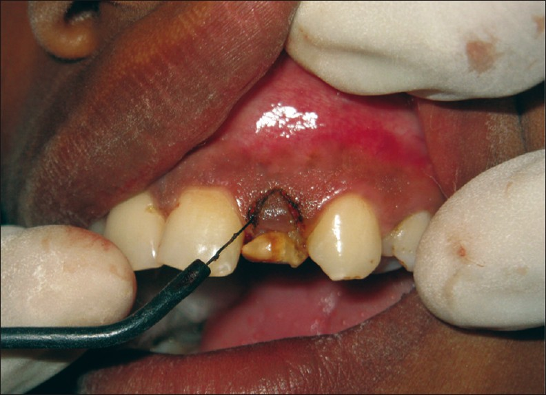
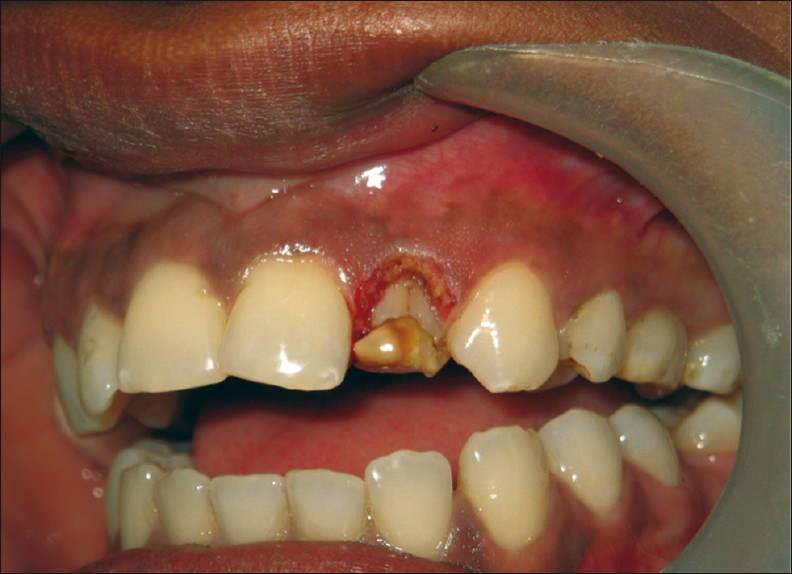
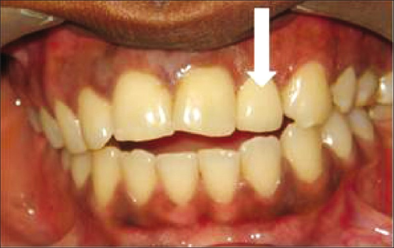
A healthy 18-year-old female patient reported to our dental clinic with a complaint of pain and swelling in her upper left lateral incisor [Figure 1]. Patient history indicated repeated palatal swelling and pain in the same tooth for the past 2 years. On clinical examination, the upper left lateral incisor was found discoloured with folding of the tooth crown palatally. The tooth did not respond to electric pulp vitality test using an Analytic Pulp Tester (Analytic Technology Corp., Richmond, VA, USA.), although all the adjacent teeth responded within normal limits. Radiographic examination revealed the presence of an invagination along the entire length of the root [Figure 2].
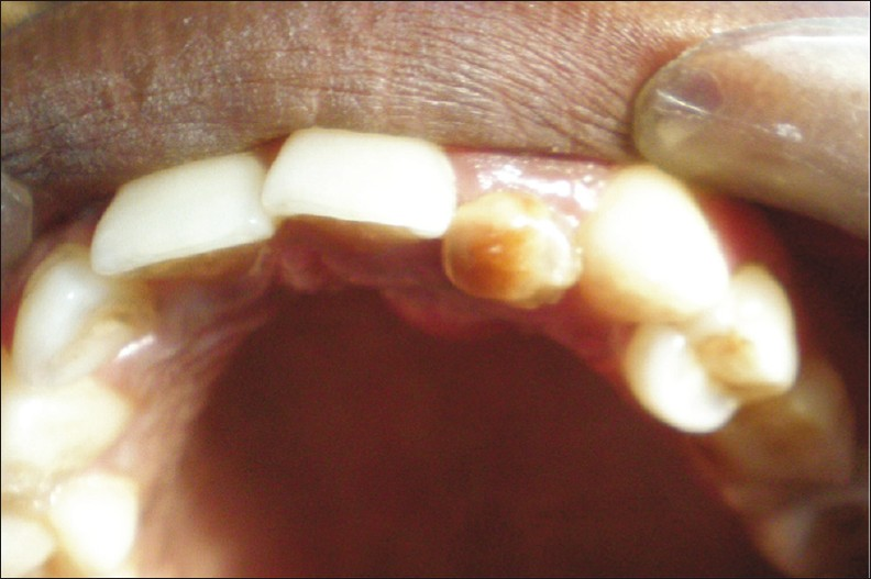
- Photograph shows discoloration and malformation of the left lateral incisor.
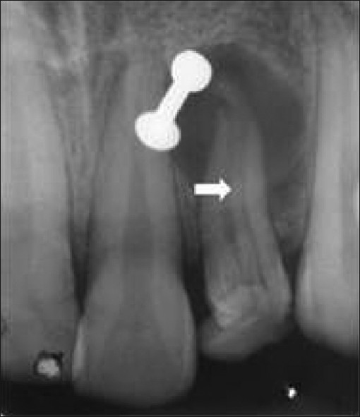
- Radiograph shows presence of an invagination along the entire length of the root (white arrow). The white object in the image is the patient's nose ring which was difficult to remove when taking the radiograph.
Based on the clinical and radiographic finding, presence of Type III dens in dente was confirmed and a definite treatment plan was formulated. The treatment planned included non-surgical endodontic therapy, crown lengthening, raising a palatal flap to seal the groove, reinforcement of crown, permanent restoration, and surgical apical curettage, if necessary.
Treatment started with conventional endodontic procedure. Without local anesthesia, rubber dam (Hygenic dental dam, Coltene whaldent Switzerland) was applied on the tooth, and endodontic access was performed with a tapered safe-ended endoacess diamond point (Maillefer-Dentisply, Baillagues, Switzerland). Using a DG16 endodontic explorer (Hu-Friedy, Germany) and a size 15 K file (Mani Inc, Tochigi, Japan) two orifices were identified. The two canals were negotiated, and working length was established using radiography [Figure 3]. The two canals were prepared to working length by step back technique to a size 55 K file (Master apical file, Mani Inc, Tochigi, Japan) and to a size 80 K file (Final file, Mani Inc, Tochigi, Japan); the canal was then flared by using Gates Glidden drills sizes 1-4 (Maillefer-Dentisply, Baillagues, Switzerland). During the instrumentation, the canals were irrigated with 5.25% sodium hypochlorite (KMC Pharmacy, Manipal, India). Following the final irrigation with 15% ethylenediaminetetraacetic acid (Sigma Aldrich, Germany) and then 5.25% sodium hypochlorite (KMC Pharmacy, manipal, India), the canals were dressed with calcium hydroxide paste (UltraCalâ, Ultradent) and the access was sealed with temporary filling material (Caviton; GC Corporation). After 3 weeks, obturation was done using cold lateral condensation of Gutta-percha cones (Maillefer-Dentisply, Baillagues, Switzerland)) using zinc oxide eugenols cement (Kalzinal, Dentsply UK) as sealer.
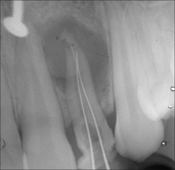
- Working length radiograph confirms the presence of two separate root canals.
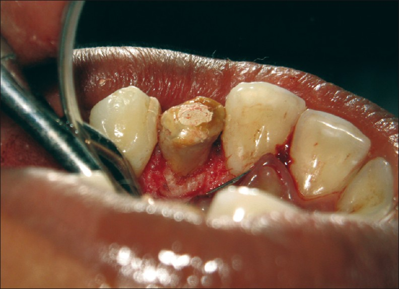
Post-obturation radiograph showed a dentine bridge along the entire length of the root canal [Figure 4]. Post endodontic rehabilitation was started with crown lengthening, which was done using electrocautery [Figure 5]. Crown lengthening revealed the presence of invagination as a developmental groove on the labial aspect [Figure 6].
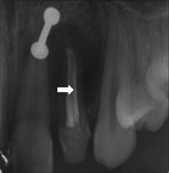
- Post-obturation radiograph shows a dentine bridge along the entire length of the root canal (white arrow).
- The image shows the lengthening of the crown done with electrocautery.
- The photograph shows the presence of invaginations.
The palatal flap was raised to expose the root surface. Palatal surface of the root also had similar type of invagination. Infolding of the tooth crown was evident from the palatal aspect. Palatal and labial grooves were sealed with Light cure glass ionomer cement (GC Corporation, Tokyo, Japan) and sutures (5-0 TevedkR) were placed [Figure 7]. A provisional crown was placed in order to maintain the gingival contour.
- The image shows the palatal and labial groove sealed with light cure glass ionomer cement.
CBCT (KODAK 900 cone beam CT digital X-ray) scan of the maxilla was taken and a three dimensional reconstruction of the involved tooth was done [Figure 8]. The 3D reconstruction showed the developmental groove along the entire length of the root, both labially and palatally [Figure 9].
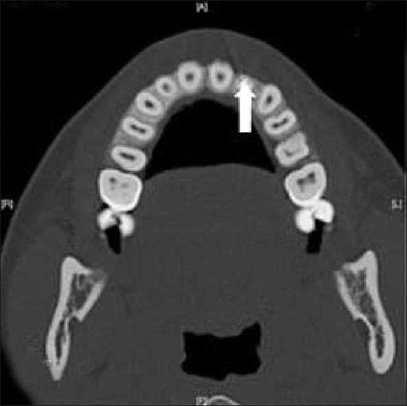
- Computed tomographic (CT) view of the maxilla reveals two obturated canals (white arrow).
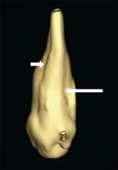
- Three dimensional reconstruction of the involved tooth shows developmental groove along the entire length of the root, both labially and palatally (white arrows).
The CBCT showed a bony lesion, with eroded palatal and labial cortical plates [Figure 10]. In the next appointment, the temporary restoration was placed inside the access cavity and some amount of obturation material was removed. The tooth was reinforced by allowing composite (3M Espee) to flow into the access cavity. A provisional crown was fabricated and placed over the tooth. In the following appointment, a permanent porcelain fused metal crown was cemented [Figure 11]. The patient returned six months later and was found to be asymptomatic. Clinical examination was unremarkable; there was a reduction in the size of the periradicular radiolucency.

- CT image shows a bony lesion with eroded palatal and labial cortical plates.
- The photograph shows the tooth (white arrow) with a permanent ceramic crown.
DISCUSSION
Several theories have been put forward for the etiology of dens invaginatus.[7] These include folding in of the enamel organ during the development of the tooth germ because of pressure from adjacent developing tooth germs (Atkinson 1943, Segura et al. 2002), rapid in-growth of a portion of the internal enamel epithelium into the developing adjacent dental papilla (Rushton 1937), failure of a small area of the internal enamel epithelium to grow whilst the adjacent epithelium continues to grow and develop normally (Kronfeld 1934), absence of certain inter-cellular signal molecules causing dental anomalies (Dassule et al. 2000), and infection during tooth development (Fischer 1936).
In this case, the cause of the periradicular periodontitis was most probably bacterial ingress into the invagination. The dens invaginatus tooth described in this case was a Type III invagination (Ohlers 1957). Invagination allows for the entry of irritants into an area, which is separated from the pulp by only a thin layer of enamel and dentine that may be hypomineralized and presents a predisposition for caries.[8] This results in pulp necrosis within a few years of eruption, abscess formation, cysts, displacement of teeth, and even leads to internal resorption.[3]
Periapical radiographs are limited in revealing the type, extension, and complex morphology of dens invaginatus, as well as, the actual bone loss when compared to tomographic techniques. More advanced imaging techniques, such as CBCT, may aid the diagnosis as well as the management plan and follow-up of teeth with this developmental defect. Cone-beam computed tomography (CBCT) is a useful tool for the management of complex endodontic problems, because it enables one to acquire three-dimensional information on the morphology of root canals, teeth, and periapical areas with radiation doses lower than the conventional CT.[9] With CBCT, a three-dimensional volume data is acquired in the course of a single sweep of the scanner, using a simple, direct relationship between the sensor and the source. The reconstructed images from the CBCT data are particularly useful in assessing the true nature of the invagination, in particular, the relationship of the invagination with the root canal.[7]
In this case, post-obturation CBCT helped to assess the course of invagination along the length of the root and destruction of both labial and palatal cortical plate. It also helped to assess the course of treatment avoiding unnecessary invasive treatment that could have been not possible because of abnormal internal morphology and inaccessible areas.
Treatment of teeth affected by Type III invagination with pulp necrosis and periapical lesion is usually challenging to the clinician. Different treatment options may be applied depending on the morphological complexity of the affected tooth. These include non-surgical endodontic treatment of the root canal and the invagination, a combined endodontic and surgical treatment, intentional replantation, or extraction.[9] In this case, non-surgical endodontic treatment was carried out in an attempt to debride the canal space as much as possible. This was based on the widely accepted concept that non-surgical endodontic treatment should be the first line of treatment and surgical treatment considered as an option only when the non-surgical treatment failed.
CONCLUSION
Historically, endodontic treatment of teeth with severe dens invaginatus was deemed impractical. Treatment options were limited to extraction. The dramatic improvements in endodontic armamentarium and diagnostic aids like CBCT have made possible the conservative treatment of such anomalies. The clinician should be aware of this anomaly because of the risk of apical inflammatory disease. Prophylactic restoration of the palatal pits of these teeth is important to avoid possible biologic injury and related inflammation.
Available FREE in open access from: http://www.clinicalimagingscience.org/text.asp?2012/2/1/51/100372
Source of Support: Nil
Conflict of Interest: None declared.
REFERENCES
- Developmental distrubences of oral and para-oral structures. In: Rajendran R, ed. Shafer's Text book of Oral pathology (6th ed). Haryana, India: Elsevier; 2009. p. :57.
- [Google Scholar]
- Dens invaginatus: Aetiology, classification, prevalence, diagnosis and treatment considerations. Int Endod J. 1997;30:79-90.
- [Google Scholar]
- Dens invaginatus type –III: Report of a case and ten year radiographic follow-up. Int Endod J. 2002;35:873-9.
- [Google Scholar]
- Dens invaginatus. Part 1: Classification, prevalence and etiology. Int Endod J. 2008;41:1123-36.
- [Google Scholar]
- Non-surgical management of infected type III dens invaginatus with vital surrounding pulp using contemporary endodontic techniques. Aust Endod J. 2008;34:4-11.
- [Google Scholar]
- Dens invaginatus (dilated composite odontome). I. variations of the invagination process and associated anterior crown forms. Oral Surg Oral Med Oral Pathol. 1957;10:1204-18.
- [Google Scholar]
- The use of cone beam computed tomography in the conservative management of dens invaginatus: A case report. Int Endod J. 2010;43:707-13.
- [Google Scholar]
- Surgical treatment of a periradicular lesion on an invaginated maxillary lateral incisor. Int Endod J. 1997;30:145-9.
- [Google Scholar]
- Clinical management of dens invaginatus in a maxillary lateral incisor with the aid of conebeam computed tomography – a case report. Dent Traumatol. 2011;27:478-83.
- [Google Scholar]


