Translate this page into:
Carcinoma en Cuirasse from Recurrent Breast Cancer seen on FDG-PET/CT
Address for correspondence: Dr. Aung Zaw Win, San Francisco VA Medical Center, 4150 Clement Street, San Francisco, California - 94121, USA. E-mail: aungzwin@gmail.com
-
Received: ,
Accepted: ,
This is an open access article distributed under the terms of the Creative Commons Attribution-NonCommercial-ShareAlike 3.0 License, which allows others to remix, tweak, and build upon the work non-commercially, as long as the author is credited and the new creations are licensed under the identical terms.
This article was originally published by Medknow Publications & Media Pvt Ltd and was migrated to Scientific Scholar after the change of Publisher.
Abstract
Our patient was a 36-year-old female diagnosed with Grade II ER+/PR−/Her-2 − ductal carcinoma in situ (DCIS) in the left breast. She underwent left lumpectomy and received treatment with tamoxifen and radiotherapy. Three years later, she presented with multiple diffused skin nodules on the chest and upper left arm. 18F-fluorodeoxyglucose-positron emission tomography/computed tomography (FDG-PET/CT) exam showed widespread metastasis in the chest, upper left arm, left axillary lymph nodes, and left suprascapular muscle. FDG-PET/CT imaging of breast carcinoma en cuirasse is very rare. FDG-PET/CT is useful in detecting recurrent breast cancer.
Keywords
Breast cancer
carcinoma en cuirasse
FDG-PET/CT
metastasis

INTRODUCTION
The major sites of breast cancer metastasis in decreasing prevalence are the lungs, bone, liver, adrenal, and brain.[1] Cutaneous metastasis from internal malignancies is rare with a reported incidence between 0.7% and 9% and it may be a sign of cancer recurrence.[2] Skin metastases from breast cancer have many forms: Telangiectatic carcinomas, erysipeloid carcinomas, “en cuirasse” carcinomas, alopecia neoplastica, and a zosteriform type.[3] Carcinoma en cuirasse is very rare and occurs in only 3% of cutaneous metastasis cases.[3] Carcinoma en cuirasse is caused by lymphatic spread. Carcinoma en cuirasse is described as small, multiple erythematous nodules which can enlarge and coalesce to form plaques.[3]18F-fluorodeoxyglucose-positron emission tomography/computed tomography (FDG-PET/CT) can scan the whole body for metastases in one exam, whereas serial CTs are required to accomplish the same task.
CASE REPORT
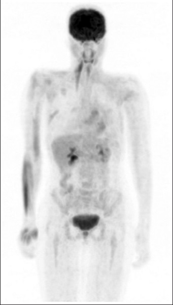
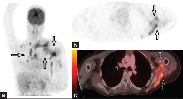
The patient was a 36-year-old female who presented with pain under the left arm. The ultrasound revealed a 2.5 cm irregular mass in the upper outer quadrant of the left breast and a subsequent mammogram showed an asymmetric density in the same region. Left lumpectomy removed a 4.9 cm mass and pathology confirmed it as ER+/PR−/Her-2– intermediate-grade ductal carcinoma in situ (DCIS) with positive margins. Left breast re-excision was done and there was no evidence of residual disease. Sentinel lymph node biopsy was negative and FDG-PET/CT showed only postsurgical changes [Figure 1]. She was then treated with tamoxifen and radiotherapy. About 3 years later, the patient presented with multiple skin nodules on the chest and upper left arm. FDG-PET/CT revealed extensive metastatic disease involving left axillary lymph nodes, left suprascapular muscle, chest, and left upper arm [Figure 2a–c]. Biopsies were done on the chest, left suprascapular muscle, and left axillary lymph nodes and they showed poorly differentiated ER+/PR−/Her-2− adenocarcinoma. She was experiencing progressive pain and weakness in the left upper extremity.

- 36-year-old female presented with pain under the left arm and was diagnosed with intermediate-grade DCIS. FDG-PET/CT Maximum Intensity Projection (MIP) image shows only postsurgical changes with no evidence of residual or metastatic disease.

- 36-year-old female presented with pain under the left arm and multiple skin lesions 3 years after surgery for DCIS. (a) FDG-PET/CT Maximum Intensity Projection (MIP) image shows cutaneous metastases (arrows) on the chest and left upper extremity. (b) PET and (c) hybrid PET/CT images, axial view show breast cancer infiltration in the left suprascapular muscle and left axillary lymph nodes (arrows) (SUV 7.09).
DISCUSSION
FDG-PET imaging of breast carcinoma en cuirasse is very rare. This is the first case to show FDG-PET/CT images of suprascapular muscle metastasis from breast cancer. Skeletal muscle is an unusual site for breast cancer metastasis and it occurs by the hematogenous route.[4] The chest is the most common site of cutaneous metastasis.[1] Abdomen, back, and upper extremities are less common sites of skin metastasis.[5] In our patient, cutaneous metastasis occurred in the chest and left upper extremity. Our patient was first diagnosed with breast cancer at the age of 36 and breast cancer in this age group is very rare.[6] Young age (less than 40 years) is an independent predictor of tumor recurrence following breast-conserving surgery.[6] Our patient had lumpectomy in the left breast and the cancer recurred 3 years after the surgery. According to the biopsy report, she had ER+/PR−/Her-2− ductal carcinoma. Interestingly, younger patients with ER +ve tumors have a poorer disease-free survival than those with ER −ve tumors.[7]
Ultrasound (US) and CT mostly focus on the size of the lymph nodes, while FDG-PET/CT looks at the biological activity of tumors. Intramuscular hot spots on PET/CT scans should be considered as a sign of metastasis, even in the absence of abnormalities on CT scans.[8] It is difficult to detect skeletal muscle metastasis with the generally used CT scans.[8] In the present case, the extent of breast cancer recurrence was clearly demonstrated by the PET/CT exam. FDG-PET/CT has 93% sensitivity and 100% specificity in detecting breast cancer recurrence.[4] Aukema et al., even suggested that PET/CT can replace conventional imaging in patients with breast cancer recurrence.[9]
CONCLUSION
PET scanning can reveal unexpected sites of metastasis. This case has demonstrated that FDG-PET/CT is useful in demonstrating the extent of breast cancer recurrence. Skin lesions in a patient with history of breast cancer should raise the suspicion of cancer recurrence.
Available FREE in open access from: http://www.clinicalimagingscience.org/text.asp?2015/5/1/35/159456
Financial support and sponsorship: Nil
Conflict of interest: There are no conflict of interest.
REFERENCES
- Cutaneous metastases from carcinoma breast: The common and the rare. Indian J Dermatol Venereol Leprol. 2009;75:499-502.
- [Google Scholar]
- Metastatic cutaneous breast carcinoma: A case report and review of the literature. Can J Plast Surg. 2009;17:25-7.
- [Google Scholar]
- Integrated contrast-enhanced diagnostic whole-body PET/CT as afirst-line restaging modality in patients with suspected metastatic recurrence of breast cancer. Eur J Radiol. 2010;73:294-9.
- [Google Scholar]
- Metastatic breast carcinoma mimicking a sebaceous gland neoplasm: A case report. J Med Case Rep. 2011;5:428.
- [Google Scholar]
- Trends in surgery for screen-detected and interval breast cancers in a national screening programme. Br J Surg. 2014;101:949-58.
- [Google Scholar]
- Predictive value of pathological and immunohistochemical parameters for axillary lymph node metastasis in breast carcinoma. Diagn Pathol. 2011;6:18.
- [Google Scholar]
- Skeletal muscle metastases from breast cancer: Two case reports. J Breast Cancer. 2013;16:117-21.
- [Google Scholar]
- The role of FDG PET/CT in patients with locoregional breast cancer recurrence: A comparison to conventional imaging techniques. Eur J Surg Oncol. 2010;36:387-92.
- [Google Scholar]


