Translate this page into:
Bohler's and Gissane Angles in the Indian Population
Address for correspondence: Dr. Vetrivel Chezian Sengodan, Department of Orthopaedics, Coimbatore Medical College and Hospital, Coimbatore, Tamil Nadu, India. E-mail: svcortho@gmail.com
-
Received: ,
Accepted: ,
This is an open-access article distributed under the terms of the Creative Commons Attribution License, which permits unrestricted use, distribution, and reproduction in any medium, provided the original author and source are credited.
This article was originally published by Medknow Publications & Media Pvt Ltd and was migrated to Scientific Scholar after the change of Publisher.
Abstract
Objective:
The aim of our study is to determine the normal ranges of the calcaneal parameters in the Indian population, and to compare the results with the data in the literature.
Materials and Methods:
The study was conducted at Coimbatore Medical College Hospital, Coimbatore on the feet (324 in number) of male and female Indian adults. Lateral view of the ankle was taken using a digital X-ray machine. Two parameters namely Bohler`s and Gissane angles were measured, independently by two radiologists to prevent inter-observer variation.
Results:
The Bohler`s and Gissane angles for the Indian population are statistically different from those seen in the published data for other population groups, as evidenced by the P value (P < 0.05).
Conclusion:
Calcaneal parameters specific to the Indian population have to be taken into consideration by the orthopedic surgeon to improve the standard of calcaneal fracture treatment in India.
Keywords
Bohler's angle
calcaneus
gissane angle
INTRODUCTION

Calcaneal fractures account for 2% of all fractures and 60% of tarsal injuries.[1–3] Displaced intra-articular fractures comprise 60% to 75% of calcaneal fractures.[3] Calcaneal fractures are common between 21 to 45 years of age.[3] It is common among industrial workers.[3] The most common causes of injury are fall from height and road traffic accidents.[4]
Intra-articular fractures must be accurately reduced to obtain joint congruity. This is possible by the reduction of the articular surface with rigid internal fixation like plates and screws.[13]
Recognizing the normal limits of the calcaneal angles is important in determining the degree of deformity and quality of reduction, thereby allowing more predictive morbidity after calcaneal fractures.[5]
The previous studies performed in the American, African, Saudi, and Turkish populations revealed a wide variability of the calcaneal angles among these respective populations.[56] No study about the normal range of the calcaneal parameters in the Indian population has been published.
The aim of our study is to determine the normal ranges of the calcaneal parameters in the Indian population, and to compare the results with the data in the literature.
MATERIALS AND METHODS
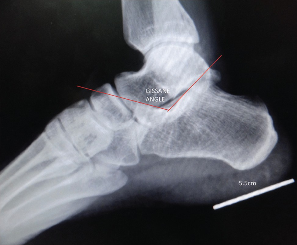
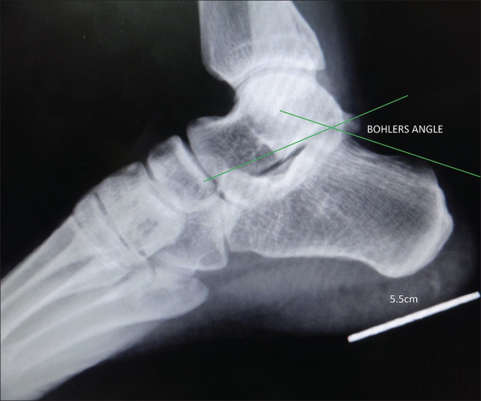
The study was conducted at Government Coimbatore Medical College Hospital, Coimbatore, after ethical committee clearance. The study was conducted on the feet (324 in number) of Indian adults. The study included both feet of 38 males and 82 females (total: 240 feet) and one foot of 32 males and 52 females (total: 84 feet). Patients were in the age range of 13-74 years (mean age = 43.5 years). The number of X-rays viewed of the right calcaneum was 180 and of the left calcaneum was 144. Lateral view of the ankle was taken using digital X-ray machine (Canon CXDI-1). All radiographs were of normal feet without congenital or acquired deformities and with no arthritic changes. Two parameters, namely Bohler's and Gissane angles, were measured [Figures 1 and 2]. Values were measured by two independent observers and were repeated after two weeks by the same observers to reduce the error of calculation. A 5-centimeter kirschner wire was used to ascertain the magnification in our study and was magnified 10%. The inter-observer agreement and variations were not assessed. Student's test (two sample t test), P value were analyzed and compared with published data. All P values less than 0.05 were considered significant.

- X-ray of the ankle in lateral view shows the angle of Gissane.

- X-ray of the ankle in lateral view shows Bohler's angle.
RESULTS
The Gissane (crucial) angle is formed by two strong cortical struts extending laterally, one along the lateral margin of the posterior facet and the other extending anterior to the beak of the calcaneus.[4] The average Gissane angle in our study is 126.79 degrees, whereas in the western population, it is 122.5 degrees.[4] The difference between the western and Indian value in Gissane angle is statistically significant (P < 0.05).
The Bohler's angle is composed of a line drawn from the highest point of the anterior process of the calcaneus to the highest point of the posterior facet and a line drawn tangential from the posterior facet to the superior edge of the tuberosity.[4] The average Bohler's angle in our study ranges from 18-43 degrees (mean = 30.62 degrees), whereas in the western population, it ranges from 20-40 degrees (mean = 30.00 degrees).[4] We didn’t get any statistical difference in this calcaneal parameter. The lowest Bohler's angle is 18 degrees in our study, which is less than the western value [Table 1].[4] Bohler's and Gissane angles are listed in [Table 2].


DISCUSSION
Calcaneal fracture is the most common tarsal bone injury. Intra-articular fractures represent 60-75% of calcaneal fractures and occur in individuals in the low socio-economic group.[3] Intra-articular fractures of the calcaneus require anatomical reduction to prevent post-operative complications.[4] In unilateral calcaneal fractures, the same angle on the contra-lateral extremity may be used as a reference value.[5] Some Asian countries like Saudi Arabia use reference calcaneal values when treating fractures.
The average Gissane angle in our study is 126.79 degrees, whereas in data that has been published, it is 122.5 degrees. The difference between previously published values and those for the Indian population is statistically significant (P < 0.05).
In India, the reference value for Bohler's and Gissane angles are as per the western text books, and no standard parameters are available for Indian patients.[4] From our study, it is evident that a separate reference value is required for the Indian population. This value will aid in assessing the intra-articular reduction of calcaneal fractures.
As both calcaneal angles generally decrease during calcaneal fractures, the lower limit of the angles should be of greater interest.[5] The lower limit of Bohler's angle in our study is 18 degrees, which is less than the western value of 20 degrees.[4]
A study of calcaneal angles in Turkish population by Aksel seyahi et al., did not reveal a statistically significant Bohler's angle difference between the sexes.[5] In our study also, there was no significant difference in Bohler's and Gissane angles between the sexes [Table 3].

When possible, angles should be compared between contralateral extremities on the same patient. In our study, there was no statistically significant difference in the Bohler's and Gissane angle between the right feet and the left feet [Table 4]. This result is in correlation with the study done in Turkish and Saudi Arabian populations.

The relation of the calcaneal angles with age should ideally be assessed on X-rays of the same individual, taken at different ages.[5] A study in the Turkish population showed no significant difference between the mean calcaneal angles of the different age groups.[5] These results suggest that an old X-ray of a patient with calcaneus fracture can be considered to assess the normal calcaneal angles for this individual.[5]
CONCLUSION
Our study reveals the importance of establishing calcaneal angle parameters when planning intra-articular reduction of calcaneal fractures in the Indian population. A larger multi-center study should be conducted in other regions of India to prepare more specific radiologic calcaneal guidelines. We hope this study will encourage future studies and aid in improving the management of calcaneal fractures in India.[7]
Available FREE in open access from: http://www.clinicalimagingscience.org/text.asp?2012/2/1/77/104310
Source of Support: Nil
Conflict of Interest: None declared.
REFERENCES
- Double vertical fractures of the pelvis.1859. Clin Orthop Relat Res. 2007;458:17-9.
- [Google Scholar]
- Egol, Kenneth A, Koval, Kenneth J, Zuckerman, Joseph D, eds. Handbook of fractures. Philadelphia PA: Lippincott Williams & Wilkins; 2010. p. :507-19.
- Rockwood and greens fractures in adults Vol 3. (7th ed). 2009. p. :2133-79.
- The calcaneal angles in the Turkish population. Acta Orthop Traumatol Turc. 2009;43:406-11.
- [Google Scholar]
- Böhler›s and Gissane›s angles of the calcaneus in the Saudi population. Saudi Med J. 2004;25:1967-70.
- [Google Scholar]


