More results...

Generic selectors
Exact matches only
Search in title
Search in content
Post Type Selectors
Filter by Categories
Abdominal Radiology, Original Research
Abdominal Radiology, Pictorial Essay
Anthropology and Dental Radiology, Original Research
Breast Imaging, Case Report
Breast Imaging, Original Research
Breast Imaging, Pictorial Essay
Breast Imaging, Review Article
Cardiopulmonary Imaging, Case Report
Cardiopulmonary Imaging, Case Series
Cardiopulmonary Imaging, Education
Cardiopulmonary Imaging, Original Research
Cardiopulmonary Imaging, Pictorial Essay
Cardiopulmonary Imaging, Review Article
Case Report
Case Report, Breast Imaging
Case Report, Cardiopulmonary Imaging
Case Report, Diagnostic Radiology
Case Report, Gastrointestinal Imaging
Case Report, Genitourinary and Gynecologic Imaging
Case Report, Neuroradiology/Head and Neck Imaging
Case Report, Vascular and Interventional Radiology
Case Series
Case Series, Abdominal Radiology
Case Series, Diagnostic Radiology
Case Series, Musculoskeletal Imaging
Case Series, Vascular and Interventional Radiology
DENTAL ARTICLE
Dental Radiology, Case Report
Dental Radiology, Case Series
Dental Radiology, Original Research
Diagnostic Radiology, Case Report
Diagnostic Radiology, Case Series
Diagnostic Radiology, Original Research
Diagnostic Radiology, Pictorial Essay
Diagnostic Radiology, Review Article
Editorial
Education
Education, Diagnostic Radiology
Education, Imaging science
Education, Original Research
Emergency, Original Research
Erratum
Gastrointestinal Imaging, Case Report
Gastrointestinal Imaging, Case Series
Gastrointestinal Imaging, Original Research
Gastrointestinal Imaging, Pictorial Essay
General and Emergency Radiology, Original Research
General and Emergency Radiology, Review Article
Genitourinary and Gynecologic Imaging, Case Report
Genitourinary and Gynecologic Imaging, Original Research
Imaging Science, Original Research
Interventional Radiology, Original Research
Letter to Editor
Musculoskeletal Imaging, Case Report
Musculoskeletal Imaging, Original Article
Musculoskeletal Imaging, Original Research
Musculoskeletal Imaging, Pictorial Essay
Musculoskeletal Imaging, Review Article
Neuroradiology , Review Article
Neuroradiology Head and Neck Imaging, Pictorial Essay
Neuroradiology, Case Report
Neuroradiology, Pictorial Essay
Neuroradiology/Head and Neck Imaging, Case Report
Neuroradiology/Head and Neck Imaging, Case Series
Neuroradiology/Head and Neck Imaging, Original Research
Neuroradiology/Head and Neck Imaging, Review Article
Neuroradiology/Spine Imaging, Original Research
Notice of Retraction
Nuclear Medicine, Case Report
Nuclear Medicine, Original Research
Nuclear Medicine, Pictorial Essay
Original Article
Original Article, Neuroradiology
Original Research
Original Research Article
Original Research, Cardiopulmonary Imaging
Original Research, Dental Radiology
Original Research, Descriptive Study
Original Research, Diagnostic Radiology
Original Research, Gastrointestinal Imaging
Original Research, Genitourinary and Gynecologic Imaging
Original Research, Musculoskeletal Imaging
Original Research, Neuroradiology/Head and Neck Imaging
Original Research, Nuclear Medicine
Original Research, Pediatric Imaging
Original Research, Vascular and Interventional Radiology
Pediatric Imaging, Case Report
Pediatric Imaging, Pictorial Essay
PICTORIAL ESSAY
Pictorial essay, Musculoskeletal Imaging
Pictorial essay, Neuroradiology/Head and Neck Imaging
PICTORIAL REVIEW
Radiologic-Pathologic Correlation
RADIOLOGICAL-PATHOLOGICAL CORRELATION
Radiology Business, Original Research
Research Article
Review Article
Review Article, Diagnostic Radiology
Review Article, Gastrointestinal imaging
Review Article, Musculoskeletal
Review Article, Musculoskeletal Imaging
Review Article, Neuroradiology/Head and Neck Imaging
Review Article, Vascular and Interventional Radiology
Technical Innovation
Technical Innovation, Cardiopulmonary Imaging
Technical Innovation, Gastrointestinal Imaging
Ultrasound, Case Report
Ultrasound, Original Research
Ultrasound, Review Article
Vascular and International Radiology, Case Report
Vascular and Interventional Radiology, Case Report
Vascular and Interventional Radiology, Case Series
Vascular and Interventional Radiology, Original Research
Vascular and Interventional Radiology, Pictorial Essay
Vascular and Interventional Radiology, Review Article
Vascular and Interventional Radiology, Short Communication
Vascular and Interventional, Original Research
View/Download PDF

Translate this page into:

ORIGINAL ARTICLE
2012
:2;
24
doi:
10.4103/2156-7514.96539

Arthroscopic and Low-Field MRI (0.25 T) Evaluation of Meniscus and Ligaments of Painful Knee

Department of MRI, Second Affiliated Hospital of Soochow University, San Xiang Road, 1055, Suzhou - 215004, Jiangsu Province, China

Address for correspondence: Dr. Harish S Lokannavar, Second Affiliated Hospital of Soochow University, Department of MRI, San Xiang Road, 1055, Suzhou - 215004, Jiangsu Province, China. E-mail: drharry4u@gmail.com

Licence

This is an open-access article distributed under the terms of the Creative Commons Attribution License, which permits unrestricted use, distribution, and reproduction in any medium, provided the original author and source are credited.

Disclaimer:
This article was originally published by Medknow Publications & Media Pvt Ltd and was migrated to Scientific Scholar after the change of Publisher.

Abstract

Objective:

Magnetic resonance imaging (MRI) is an accurate, non-invasive, cost-effective technique for examination of the soft tissue and osseous structures of the knee. The purpose of this study was to evaluate the accuracy of low-field MRI by comparing the results with subsequent arthroscopy.

Materials and Methods:

MR imaging study of 146 patients was done using 0.25 T ESTOATE G-SCAN and the sequence used were SE, FSE and GRE in all the three planes. The comparison was based on five parameters: accuracy, sensitivity, specificity, positive predictive value, and negative predictive value.

Result:

Our study showed high accuracy (98.08%) and negative predictive value (98.62%) for MRI in comparison with arthroscopy.

Conclusion:

Low-field MRI alleviates the need of arthroscopy for detection of meniscus tears and ligament tears.

Keywords

Arthroscopy
ligaments
low field magnetic resonance imaging
meniscus
painful knee

INTRODUCTION

MRI is the examination of choice in the evaluation of internal joint structures of the knee like menisci, cruciate ligaments, and articular cartilage. The diagnostic accuracy of MRI, although variable for different individual structures, compares well with arthroscopy, which is considered the Gold Standard, especially when assessing knee injury, in appropriately identifying patients who require arthroscopic therapy.[12] MRI of the knee joint has effectively replaced arthrography[3] and as the imaging modality of choice in the evaluation of both acute and chronic disorders causing pain in the knee.[4]

Low-field MRI is adequate for imaging the knee. Studies have shown that it is as effective as high-field MRI machines in evaluating meniscal tears[57] and ligaments especially Anterior Cruciate Ligament (ACL).[68] Low-field MRI has been readily accepted by both patients and referring clinicians. MR 0.25 T magnet is a safe and valuable adjunct to the clinical examination of the knee in preoperative planning[39] and in avoiding diagnostic arthroscopy.

MATERIALS AND METHODS

MRI studies of the knee were performed on 146 patients (87 females, 59 males with a history of painful knee joints) using a 0.25 T ESTOATE G-SCAN machine. The sequences used were SE, FSE, and GRE in all three planes: sagittal, coronal, and axial. The slice thickness of 4.5 mm and a 256×256 matrix were used with knee coil slightly flexed and internally rotated. The criteria for selection of patients were history of painful knee with no previous history of surgery. The study period was from January 2011 to October 2011. All patients underwent subsequent arthroscopy within the time frame of 12 days from the date of MRI examinations. Meniscus and the ligaments were Graded 1, 2, 3 [Tables 1 and 2]. Three radiologists analyzed all the images. We determined the reliability of magnetic resonance imaging for the medial meniscus, the lateral meniscus, the anterior cruciate ligament, and the posterior cruciate ligament (PCL). Five parameters were calculated: (i) accuracy - the percentage of patients for whom the diagnosis based on magnetic resonance imaging was correct, (ii) sensitivity - the percentage sensitivity - the percentage of patients in whom an arthroscopy confirmed tear preoperatively diagnosed on the basis of MRI, (iii)specificity - the percentage of patients who had no tear at arthroscopy and who had been found to have no tear on the basis of MRI, (iv) negative predictive value (NPV) - the percentage of patients who were diagnosed as having no tear on the basis of MRI and were subsequently found to have no tear at arthroscopy, and (v) positive predictive value (PPV) - the percentage of patients who were diagnosed as having a tear on the basis of MRI and were subsequently seen to have a tear at arthroscopy.
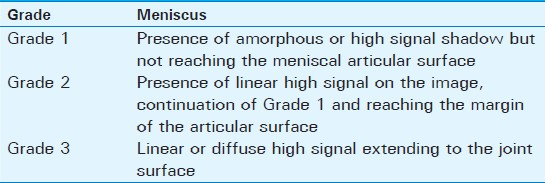
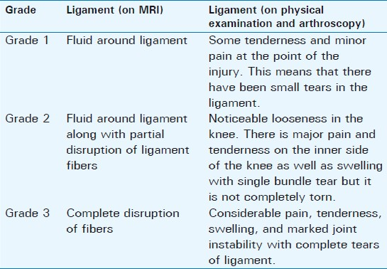
Table 1 Grading of meniscal tears
Table 2 Grading of ligament tears

RESULTS

The study population consisted of patients in the age group 11-78 years with a majority of the patients belonging to the age group of 40-60 years. This study also showed a female preponderance for knee pain, accounting for 59.6% of the cases. Of the 146 patients with knee pain, 45.9% were in the left knee and 54.1% were in the right knee.

In this study, 78 patients had meniscal tears with 36 belonging to anterior horn and 42 belonging to posterior horn. Of these, 38 had medial meniscal tears [Figure 1] of whom 4 had Grade 1 tear, 8 had Grade 2 tear, and 26 had Grade 3 tear. A total of 40 had lateral meniscal tears of whom 4 had Grade 1 tear, 6 had Grade 2 tear, and 30 had Grade 3 tear. In this study, 19 patients had ACL tears [Figure 2] of whom 2 had Grade 1 tear, 4 had Grade 2 tear, and 13 had Grade 3 tear. In this study 2 patients had PCL tears [Figure 3] of whom one was Grade 2 and other was Grade 3. The accuracy, sensitivity, specificity, PPV, and NPV of the above have been calculated and shown in Tables 3 and 4.
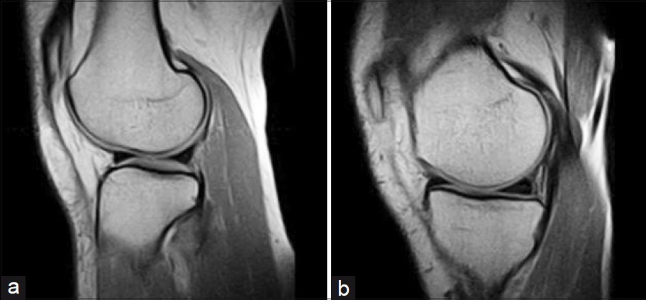
Sagittal view of the knee shows (a) normal meniscus, (b) medial meniscus posterior horn Grade 3.
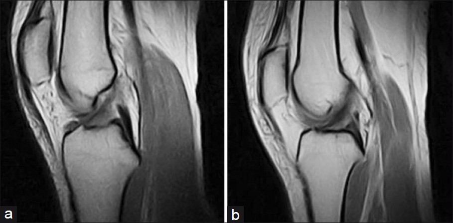
Figure 1
Sagittal view of the knee shows (a) normal meniscus, (b) medial meniscus posterior horn Grade 3.
Sagittal view of the knee shows (a) normal anterior cruciate ligament, and (b) ACL tear Grade 2.
Figure 2
Sagittal view of the knee shows (a) normal anterior cruciate ligament, and (b) ACL tear Grade 2.
Sagittal view of the knee shows (a) normal posterior cruciate ligament, and (b) PCL tear Grade 2.
Figure 3
Sagittal view of the knee shows (a) normal posterior cruciate ligament, and (b) PCL tear Grade 2.
Table 3 Number of patients with true and false, positive and negative results for meniscus and ligament tears
Table 4 Accuracy, sensitivity, specificity, positive predictive value, negative predictive value of MRI (as percentages) for diagnosing different types of meniscus and ligament tears

DISCUSSION

This study was conducted to determine the reliability and accuracy of low-field MRI diagnosis of meniscal and ligament lesions when compared to diagnostic arthroscopy. Although arthroscopy has been considered the Gold Standard in diagnosis of meniscal and ligament lesions, MRI remains a reliable, non-invasive modality, which can reduce the use of diagnostic arthroscopy. In this study on 146 patients, the accuracy of magnetic resonance imaging was 97.9% for the medial meniscus, 97.2% for the lateral meniscus, 97.9% for the anterior cruciate ligament, and 99.32% for the posterior cruciate ligament. In a prospective study of 561 patients who also underwent arthroscopy, Justice and Quinn[10] reported an accuracy rate of 93% for the lateral meniscus and 95% for the medial meniscus, which was comparable to results of our study. In a retrospective study by De Smet and colleagues, on 400 MRI examinations (800 menisci), there were 83 original diagnostic errors, yielding an accuracy rate of 90%. De Smet's group also provided an analysis of causative errors.[11] Errors were classified as unavoidable errors (40%), errors related to equivocal MRI findings (21%), and interpretation errors (21%). Unavoidable errors (discordant MRI-arthroscopic correlation) were represented by 21 false-negative and 12 false-positive diagnoses. Subtle or equivocal MRI findings resulted from inter-observer differences in interpretations. Of the interpretation errors, 38% were attributed to normal MRI variants mistaken for a meniscal tear. Even in retrospective review, 6% of the meniscal tears could not be diagnosed. A small (1.5%) false-positive diagnosis attributed to healed tears[12] or tears overlooked at arthroscopy. This study emphasizes that observer variation as represented in the equivocal MRI finding category can affect MRI accuracy rates in diagnosis of meniscal tears. Other studies have minimized differences in observer performance. Increased observer experience may improve accuracy with subtle or equivocal MRI findings, including Grade 1 versus Grade 2 signal intensities, flap tears, and peripheral tears. Correlating peripheral meniscal signal intensity on sagittal MR images with coronal plane images of the corresponding menisci may reduce false-positive MR interpretations, especially in the posterior horn of the medial meniscus. Behairy et al.,[13] in their prospective study of 70 patients showed 94% of accuracy in detecting ACL tears that is a little lower than in our study that showed 97.33% accuracy for ACL.

Vaz et al.,[14] in their study observed sensitivity of 97.5%, and specificity of 92.5% for medial meniscus; sensitivity of 91.9%, and specificity of 93.6% for lateral meniscus; sensitivity of 99%, specificity of 95.9%, and accuracy of 96.6% for ACL; and for PCL sensitivity of 100%, specificity of 99.6%, and accuracy of 84.6% for PCL, which is comparable to the results in our study.

Khandha et al.,[15] in their study of 50 patients observed sensitivity, specificity, and accuracy of 100%, 69.27%, and 92% for medial meniscus, 87.5%, 88.23%, and 88% for lateral meniscus, 86.67%, 91.43%, and 88% for ACL and 100%, 95.83%, and 96% for PCL which is lower than the results obtained in our study. The reason for this difference has been discussed earlier.

MRI is highly accurate and helps in indentifying patients needing surgery thus avoiding diagnostic arthroscopy.[1617] Various previously published studies also support our view that results obtained by using low-field MRI are comparable to results with high-field MRI.[1821]

Source of Support: Nil

Conflict of Interest: None declared.

REFERENCES

  1. , , , , . Is MRI effective in detecting intraarticular abnormalities of injured knee? J Med Liban. 2002;50:168-74.
    [Google Scholar]
  2. , , , , . The diagnosis of meniscal tears: The role of MRI and clinical examination. Clin Orthop Relat Res. 2007;455:123-33.
    [Google Scholar]
  3. , , , . Magnetic resonance imaging in the evulation of knee injuries. South Med J. 1991;84:1123-7.
    [Google Scholar]
  4. , , , , , . Imaging evaluation of meniscal injury of the knee joint: A comparative MR imaging and arthroscopic study. Clin Imaging. 2004;28:372-6.
    [Google Scholar]
  5. , , , , , , . MR imaging of meniscal tears on low field (0.35T) and high field (1.5T) MR units. Tani Girsim Radoyl. 2004;10:316-9.
    [Google Scholar]
  6. , , , , , , . MR imaging of knee at 0.2T and 1.5T:correlation with surgery. AJR Am J Roentgenol. 2000;174:1093-7.
    [Google Scholar]
  7. , , , , , . MR imaging before arthroscopy in knee joint disorders? Acta Radiol. 1996;37:602-9.
    [Google Scholar]
  8. , , , . Magnetic resonance imaging of cruciate ligament injuries of the knee. Can Assoc Radiol J. 2010;61:80-9.
    [Google Scholar]
  9. , , , , , . 0.2 Tesla magnetic resonance imaging of internal lesions of knee joint: A prospective arthroscopically controlled clinical study. Knee Surg Sports Traumatol Arthrosc. 1999;7:37-41.
    [Google Scholar]
  10. , , . Error patterns in the MR imaging evaluation of menisci of the knee. Radiology. 1995;196:617-21.
    [Google Scholar]
  11. , , , , . MR diagnosis of meniscal tears: Analysis of causes of errors. AJR Am J Roentgenol. 1994;163:1419.
    [Google Scholar]
  12. , , , , , . Clinical and MR findings associated with false-positive knee MR diagnoses of medial meniscus tears. AJR Am J Rogentgenol. 2008;191:93-9.
    [Google Scholar]
  13. , , , . Accuracy of routine magnetic resonance imaging in meniscal and ligamentous injuries of the knee: Comparison with arthroscopy. Int Orthop. 2009;33:961-7.
    [Google Scholar]
  14. , , , , . Accuracy of magnetic resonance in identifying traumatic intraarticular knee lesions. Clinics. 2005;60:445-50.
    [Google Scholar]
  15. , , , , . Assessment of menisci and ligamentous injuries of the knee on magnetic resonance imaging: Correlation with arthroscopy. J Pak Med Assoc. 2008;58:537-40.
    [Google Scholar]
  16. , , , , , . The efficacy of magnetic resonance imaging in acute knee injuries. Clin J Sport Med. 2000;10:34-9.
    [Google Scholar]
  17. , , , , . Magnetic resonance imagining as a screening procedure to avoid arthroscopy for meniscal tears. Arch Orthop Trauma Surg. 2000;120:14-6.
    [Google Scholar]
  18. , , , , , . Low Field MRI of knee of the knee joint: Results of a prospective arthroscopically controlled study. Rofo. 1999;170:35-40.
    [Google Scholar]
  19. , , , , , . Comparison of diagnostic sensitivity in meniscus diagnosis of MRI examinations with a 0.2 T low.field and a 1.5 T high field system. Radiologe. 1997;37:802-6.
    [Google Scholar]
  20. , , , , , , . MRI of the knee joint: First results of a comparison of 0,2-T specialized system and 1,5-T high field strength magnet. Rofo. 1995;162:390-5.
    [Google Scholar]
  21. , , , , , , . Low-field versus high-field MR imaging of the knee: A comparison of signal behaviour and diagnostic performance. Eur J Radiol. 1995;19:132-8.
    [Google Scholar]
Show Sections