More results...

Generic selectors
Exact matches only
Search in title
Search in content
Post Type Selectors
Filter by Categories
Abdominal Radiology, Original Research
Abdominal Radiology, Pictorial Essay
Anthropology and Dental Radiology, Original Research
Breast Imaging, Case Report
Breast Imaging, Original Research
Breast Imaging, Pictorial Essay
Breast Imaging, Review Article
Cardiopulmonary Imaging, Case Report
Cardiopulmonary Imaging, Case Series
Cardiopulmonary Imaging, Education
Cardiopulmonary Imaging, Original Research
Cardiopulmonary Imaging, Pictorial Essay
Cardiopulmonary Imaging, Review Article
Case Report
Case Report, Breast Imaging
Case Report, Cardiopulmonary Imaging
Case Report, Diagnostic Radiology
Case Report, Gastrointestinal Imaging
Case Report, Genitourinary and Gynecologic Imaging
Case Report, Neuroradiology/Head and Neck Imaging
Case Report, Vascular and Interventional Radiology
Case Series
Case Series, Abdominal Radiology
Case Series, Diagnostic Radiology
Case Series, Musculoskeletal Imaging
Case Series, Vascular and Interventional Radiology
DENTAL ARTICLE
Dental Radiology, Case Report
Dental Radiology, Case Series
Dental Radiology, Original Research
Diagnostic Radiology, Case Report
Diagnostic Radiology, Case Series
Diagnostic Radiology, Original Research
Diagnostic Radiology, Pictorial Essay
Diagnostic Radiology, Review Article
Editorial
Education
Education, Diagnostic Radiology
Education, Imaging science
Education, Original Research
Emergency, Original Research
Erratum
Gastrointestinal Imaging, Case Report
Gastrointestinal Imaging, Case Series
Gastrointestinal Imaging, Original Research
Gastrointestinal Imaging, Pictorial Essay
General and Emergency Radiology, Original Research
General and Emergency Radiology, Review Article
Genitourinary and Gynecologic Imaging, Case Report
Genitourinary and Gynecologic Imaging, Original Research
Imaging Science, Original Research
Interventional Radiology, Original Research
Letter to Editor
Musculoskeletal Imaging, Case Report
Musculoskeletal Imaging, Original Article
Musculoskeletal Imaging, Original Research
Musculoskeletal Imaging, Pictorial Essay
Musculoskeletal Imaging, Review Article
Neuroradiology , Review Article
Neuroradiology Head and Neck Imaging, Pictorial Essay
Neuroradiology, Case Report
Neuroradiology, Pictorial Essay
Neuroradiology/Head and Neck Imaging, Case Report
Neuroradiology/Head and Neck Imaging, Case Series
Neuroradiology/Head and Neck Imaging, Original Research
Neuroradiology/Head and Neck Imaging, Review Article
Neuroradiology/Spine Imaging, Original Research
Notice of Retraction
Nuclear Medicine, Case Report
Nuclear Medicine, Original Research
Nuclear Medicine, Pictorial Essay
Original Article
Original Article, Neuroradiology
Original Research
Original Research Article
Original Research, Cardiopulmonary Imaging
Original Research, Dental Radiology
Original Research, Descriptive Study
Original Research, Diagnostic Radiology
Original Research, Gastrointestinal Imaging
Original Research, Genitourinary and Gynecologic Imaging
Original Research, Musculoskeletal Imaging
Original Research, Neuroradiology/Head and Neck Imaging
Original Research, Nuclear Medicine
Original Research, Vascular and Interventional Radiology
Pediatric Imaging, Case Report
Pediatric Imaging, Pictorial Essay
PICTORIAL ESSAY
Pictorial essay, Musculoskeletal Imaging
Pictorial essay, Neuroradiology/Head and Neck Imaging
PICTORIAL REVIEW
Radiologic-Pathologic Correlation
RADIOLOGICAL-PATHOLOGICAL CORRELATION
Radiology Business, Original Research
Research Article
Review Article
Review Article, Diagnostic Radiology
Review Article, Gastrointestinal imaging
Review Article, Musculoskeletal
Review Article, Musculoskeletal Imaging
Review Article, Vascular and Interventional Radiology
Technical Innovation
Technical Innovation, Cardiopulmonary Imaging
Technical Innovation, Gastrointestinal Imaging
Ultrasound, Case Report
Ultrasound, Original Research
Ultrasound, Review Article
Vascular and International Radiology, Case Report
Vascular and Interventional Radiology, Case Report
Vascular and Interventional Radiology, Case Series
Vascular and Interventional Radiology, Original Research
Vascular and Interventional Radiology, Pictorial Essay
Vascular and Interventional Radiology, Review Article
Vascular and Interventional Radiology, Short Communication
Vascular and Interventional, Original Research
View/Download PDF

Translate this page into:

CASE REPORT
2012
:2;
4
doi:
10.4103/2156-7514.93036

An Unusual Case of Extraosseous Accumulation of Bone Scan Tracer in a Renal Calculus - Demonstration by SPECT-CT

Department of Nuclear Medicine and PET/CT, Jaslok Hospital and Research Center, Deshmukh Marg, Worli, Mumbai, India
Radiology Department, Jaslok Hospital and Research Center, Deshmukh Marg, Worli, Mumbai, India

Address for correspondence: Dr. Prathamesh Joshi, Department of Nuclear Medicine and PET/CT, Jaslok Hospital and Research Centre, Deshmukh Marg, Worli, Mumbai–400 026, India. drprathamj@gmail.com

Licence

This is an open-access article distributed under the terms of the Creative Commons Attribution License, which permits unrestricted use, distribution, and reproduction in any medium, provided the original author and source are credited.

Disclaimer:
This article was originally published by Medknow Publications & Media Pvt Ltd and was migrated to Scientific Scholar after the change of Publisher.

Abstract

Extraosseous localization of radioisotope, used in bone scan, in a variety of physiological and pathological conditions is a well-known phenomenon. The causes of extraosseous accumulation of bone-seeking radiotracers should be kept in mind when bone-imaging studies are reviewed to avoid incorrect interpretations. We report an extremely rare occurrence of extraosseous accumulation of bone scintigraphy tracer in a renal calculus, in a patient with adenocarcinoma of prostate, that was demonstrated by Single Photon Emission Computed Tomography and Computed Tomography (SPECT-CT) fusion imaging.

Keywords

Bone scan
extraosseous uptake
renal calculus
SPECT-CT
99mTc-MDP

INTRODUCTION

The purpose of bone scintigraphy is to portray areas of new bone formation within the skeleton. This is useful in imaging reaction of bone to tumor, fracture, and infection. Since approximately half of the administered radioisotope is excreted through renal filtration, abnormalities of the urinary system are also frequently noted during bone scintigraphy. In such cases, to reach an accurate diagnosis, the interpreting physician must first recognize which structures are involved in the uptake and the significance of the uptake.[1] We describe a rare case in which extraosseous bone scan tracer accumulation was noted in a renal calculus.

CASE REPORT

A 60-year-old man, who had difficulty in passing urine for the past 6 months and a complaint of lower back pain presented to our department. On ultrasound imaging, he was found to have an enlarged prostate. Transrectal ultrasound-guided biopsy revealed adenocarcinoma of prostate, Gleason's score 3+3=6. His PSA was normal (1.3 ng/ml) and serum Alkaline phosphatase was elevated 58 U/L (Normal range 30-50 U/L). A 99m Technitium methylene diphosphonate (99m Tc-MDP) bone scan was performed. The scan [Figures 1 and 2] showed mildly increased tracer uptake in lumbar vertebrae and focal accumulation of tracer in lower pole of the left kidney. Fusion imaging [Figure 3], using Single Photon Emission Computed Tomography along with X-ray Computed Tomography (SPECT-CT) of the lumbar spine was performed to characterize the vertebral tracer uptake. Lasix 40 mg was given 45 minutes prior to SPECT-CT to monitor the renal tracer accumulation. The scan demonstrated degenerative changes in lumbar vertebrae. The kidneys were included in the SPECT-CT imaging. High tracer uptake in a lower pole of the left kidney was found localized in a calculus in the lower calyx of kidney. The calculus measured 8 x 8 mm in size. The CT attenuation factor was 1060 Hounsfield unit, compatible with that of a renal calculus.
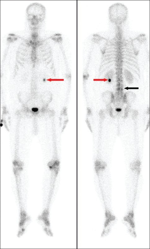
Whole body bone scan with 99Tc MDP of the patient. (a) Anterior and (b) posterior views, showing mildly increased tracer uptake in the lumbar vertebrae (black arrow) and intense, focal localization of the tracer in the lower pole of left kidney (red arrows).
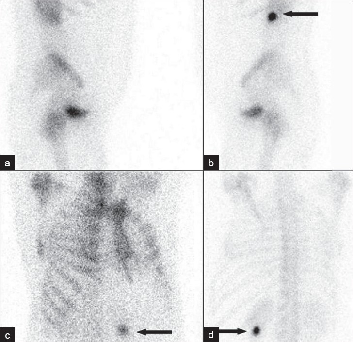
Figure 1
Whole body bone scan with 99Tc MDP of the patient. (a) Anterior and (b) posterior views, showing mildly increased tracer uptake in the lumbar vertebrae (black arrow) and intense, focal localization of the tracer in the lower pole of left kidney (red arrows).
(a-b) Lateral and (c-d) oblique views clearly demonstrate the focal tracer uptake outside the skeletal structures in left kidney (arrows).
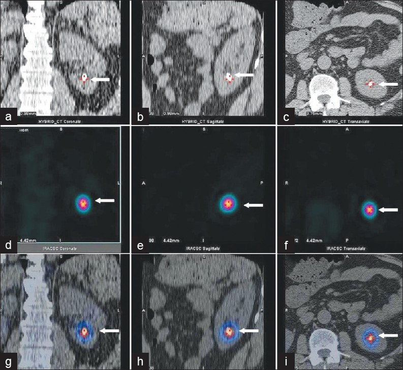
Figure 2
(a-b) Lateral and (c-d) oblique views clearly demonstrate the focal tracer uptake outside the skeletal structures in left kidney (arrows).
(a) Coronal, (b) sagittal and (c) transaxial CT images; (d) coronal (e) sagittal and (f) transaxial SPECT images; (g) Coronal, (h) sagittal and (i) transaxial SPECT-CT images. These revealed focal tracer accumulation in the left renal calculus (white arrows). The calculus measured 8 × 8 mm in size. The CT attenuation factor was 1060 Hounsfield unit, compatible with that of a renal calculus.
Figure 3
(a) Coronal, (b) sagittal and (c) transaxial CT images; (d) coronal (e) sagittal and (f) transaxial SPECT images; (g) Coronal, (h) sagittal and (i) transaxial SPECT-CT images. These revealed focal tracer accumulation in the left renal calculus (white arrows). The calculus measured 8 × 8 mm in size. The CT attenuation factor was 1060 Hounsfield unit, compatible with that of a renal calculus.

We concluded this as extra-skeletal bone scintigraphy tracer uptake in a renal calculus.

DISCUSSION

Bone scintigraphy is a valuable diagnostic tool in the evaluation of patients with a variety of osseous abnormalities. However, accumulation of bone scan tracer outside the skeleton can pose a difficulty in reporting for a nuclear medicine physician, especially if only planar imaging is performed and the tracer uptake is overlapping or is in close vicinity of the skeleton.

Localization of bone scan tracer in a renal calculus has been reported in the past and its use in preoperative in vivo localization of the renal calculus has been explored.[2] Retention of bone scan tracer on ureteric calculi and bladder calculus has been reported in the past.[35] In one of the studies, autoradiography of ureteric calculus demonstrated peripheral tracer distribution within the calculus. Accumulation of the radionuclide due to sluggish flow and its absorption onto the crystal surface, within the calculus, were suggested as possible mechanisms of tracer uptake.[4] The other common causes of urinary system localization of the bone scan tracer are dilation of urinary-collecting system, bladder diverticulum, and presence of an ureterostomy bag.[69]

CONCLUSIONS

Conditions causing extraskeletal accumulation of bone scan tracer must be kept in mind while reporting a bone scan. Our case demonstrates a rare occurrence of extraosseous bone tracer accumulation in a renal calculus. It also highlights important role played by SPECT-CT in localizing the extraskeletal tracer uptake. SPECT-CT can be used effectively when an extraskeletal uptake is encountered on planar bone imaging.

Source of Support: Nil

Conflict of Interest: None declared.

REFERENCES

  1. , , . Nonosseous, nonurologic uptake on bone scintigraphy: Atlas and analysis. Semin Nucl Med. 2010;40:242-56.
    [Google Scholar]
  2. , , , . In-vivo labelling of renal calculi with technetium 99m methylene diphosphonate. Br J Radiol. 1982;55:39-41.
    [Google Scholar]
  3. , , . Differential diagnosis of a persistent tracer uptake in the paraspinal lumbar area detected by SPET and CT: An ureteral calculus? Hell J Nucl Med. 2009;12:74-5.
    [Google Scholar]
  4. , , . Bone tracer uptake in a ureteric calculus. Australas Radiol. 1998;42:83-5.
    [Google Scholar]
  5. , , , , . Bone tracer uptake in urinary bladder stones. Clin Nucl Med. 2003;28:337-9.
    [Google Scholar]
  6. , , . Visualization of megacystis on bone scan. Clin Nucl Med. 1991;16:369-71.
    [Google Scholar]
  7. , . Evaluation of renal and urinary tract abnormalities noted on scintiscans. Mayo Clin Proc. 1975;50:370-8.
    [Google Scholar]
  8. , , . Differential diagnosis in nuclear medicine. New York, NY: McGraw Hill; . p. :305.
    [Google Scholar]
  9. , , , , . Atlas of nuclear medicine artifacts and variants: The skeletal system. Chicago, IL: Yearbook Medical; . p. :159-89.
    [Google Scholar]

Fulltext Views
10,143

PDF downloads
2,699
View/Download PDF
Download Citations
BibTeX
RIS
Show Sections