Translate this page into:
Acute Concomitant Anterior Cruciate Ligament and Patellar Tendon Tears in a Non-dislocated Knee
Address for correspondence: Dr. Robert D. Wissman, Department of Radiology, University of Cincinnati Medical Center, 234 Goodman Street, Cincinnati, OH 45267-0761, USA. Robert.Wissman@Healthall.com
-
Received: ,
Accepted: ,
This is an open-access article distributed under the terms of the Creative Commons Attribution License, which permits unrestricted use, distribution, and reproduction in any medium, provided the original author and source are credited.
This article was originally published by Medknow Publications & Media Pvt Ltd and was migrated to Scientific Scholar after the change of Publisher.
Abstract
Anterior cruciate ligament (ACL) tears are common and may occur in isolation or with other internal derangements of the joint. Tears of the patellar tendon (PT) occur less frequently and are rarely associated with intra-articular pathology. Acute combined tears of both the ACL and PT are known complications of high-energy traumatic knee dislocations. We present a case of an acute concomitant ACL and PT tears in a low-energy non-dislocated knee. To our knowledge, this injury has only been described in a limited number of case reports in the orthopedic literature. We present the imaging findings of this combined injury and discuss the importance of magnetic resonance (MR) in diagnosis.
Keywords
Anterior cruciate ligament
knee
magnetic resonance imaging
patellar tendon
INTRODUCTION
Magnetic resonance (MR) imaging is an effective and accurate diagnostic tool in the preoperative assessment of the multiligamentous injured knee.[1] Anterior cruciate ligament (ACL) tears may occur in isolation or in combination with other internal derangements of the joint.[2] Combined ACL and extensor mechanism injuries have been well documented in the dislocated knee.[3] Acute concomitant rupture of the ACL and the patellar tendon (PT) in a non-dislocated knee, however, is extremely rare and thus far limited mostly to case reports in the orthopedic literature.[4–8] To our knowledge, this injury pattern has not been reported in the radiology literature and the imaging features have not been fully described. The purpose of our case report is to introduce this combined injury to the radiology community and emphasize the role imaging plays to avoid misdiagnosis. We present a case of an acute concomitant ACL and PT tear in a nondislocated knee diagnosed at MR imaging with both clinical and surgical correlation.
CASE REPORT
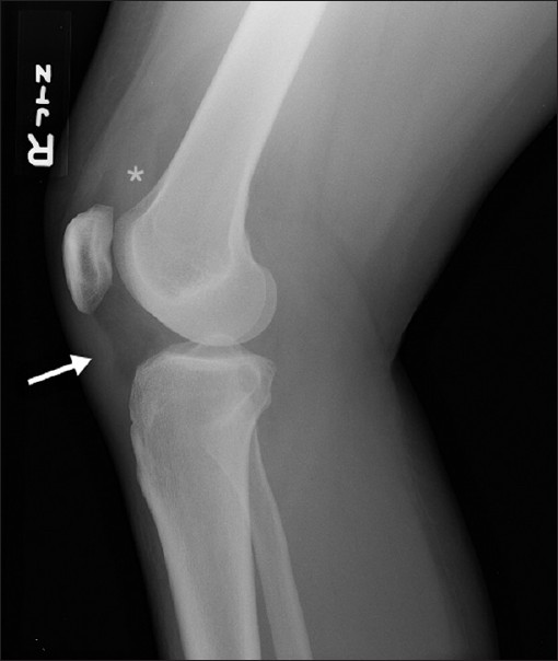
A 36-year-old female jumped from a four-foot high deck landing flatly on both feet. She felt a pop in the right knee and experienced extreme pain and inability to bear weight on the limb. She sought emergency medical attention where radiographs revealed a joint effusion, and a wavy appearance to the tendon [Figure 1]. There was no patellar elevation, fracture or malalignment. Clinically the patient was believed to have an ACL tear. The patient was treated with a knee brace and instructed to follow-up with an orthopedic surgeon and obtain an MR examination of the knee.

- Lateral radiograph of the right knee following the initial injury demonstrates a large suprapatellar joint effusion (asterisk) and undulating contour of the patellar tendon (arrow).
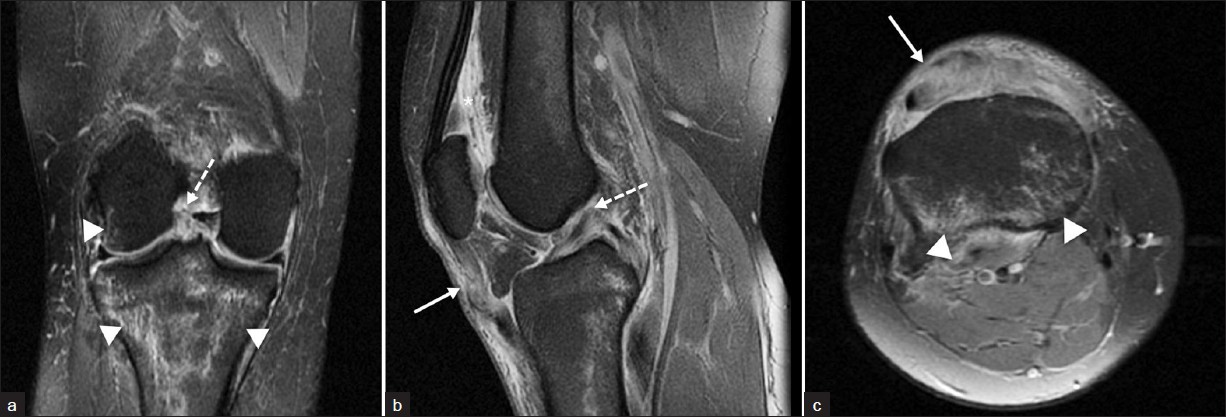
The MR examination of the right knee was obtained 10 days later and demonstrated abnormal increased T2 signal in the midsubstance of the ACL and abnormal orientation of the ligament consistent with a complete ACL disruption [Figure 2]. Bone contusions in the posterior medial and lateral tibial plateaus as well as the lateral femoral condyle were present. The patellar tendon was abnormal in morphology and signal. The tendon had an abnormal wavy contour at the junction of the middle and distal thirds of the tendon and had abnormally increased signal on T2-weighted images. The axial images demonstrated near complete disruption of the patellar tendon with only a few intact fibers.

- T2-weighted fast spin echo fat-suppressed images of the right knee. (a) Coronal image shows complete absence of anterior cruciate ligament fibers (broken arrow) consistent with a complete tear. There is also contusion within the posterior medial and lateral tibial plateau (arrowhead) and lateral femoral condyle (arrowhead). (b) Sagittal image with a large joint effusion (asterisk), undulation of the patellar tendon (arrow), and complete tear of the anterior cruciate ligament (dashed arrow). (c) Axial image demonstrates near complete tear of the patellar tendon (arrow) and edema within the posterior tibial plateau (arrowheads).
Orthopedic follow-up visit was delayed for nearly 2 months. At the time of presentation she had persistent pain, instability, and very limited range of motion. She had a positive Lachman test of the right knee. Although the patient had tenderness over the patellar tendon, there was no palpable tendon defect and she was able to hold a straight leg raise with minimal extensor lag. The patient was diagnosed with a complete ACL tear and high-grade partial patellar tendon tear. Physical therapy was instituted to help prevent postoperative fibrosis. A delayed ACL reconstruction was schedule 8 weeks later.
A diagnostic arthrogram at the time of surgery confirmed a complete midsubstance ACL tear. The ACL was reconstructed with an ipsilateral hamstring autograft. The patellar tendon was not directly inspected at the time of surgery and was treated clinically as a partial patellar tendon tear. The patient's postoperative course was complicated by complex regional pain syndrome requiring multiple nerve block injections. A year following the initial injury, the patient had continued stiffness, difficulty with walking and had developed quadriceps muscle atrophy.
DISCUSSION
Simultaneous ACL and PT tears have been described largely in a limited number of case reports with the largest series of six patients published by Levakos et al.[4–8] Five of these individuals had other associated injuries including medial collateral ligament (MCL) or meniscal tears. The most common reported location of PT rupture was the middle third of the tendon followed by the proximal patellar attachment. Misdiagnosis with a delay in treatment was not uncommon in this series. Two ACL and two PT ruptures were clinically undetected being found either at surgery or presenting during follow-up visits. Rae and Davies described a similar experience of a PT tear found incidentally during surgical repair for an acute ACL and MCL injury.[6] In this particular case, swelling disguised the defect in the PT and radiographs showed a normally positioned patella. Of these seven patients, preoperative MR examinations were obtained in only two individuals, correctly identifying the extent of their injuries.
Such outcomes inspired McKinney et al., to conduct a retrospective study of intra-articular injuries in patients with extensor mechanism ruptures.[9] During a 10-year period, 33 patellar tendon injuries requiring surgical intervention were identified. Ten (30%) patients had additional knee injuries which were most commonly diagnosed with MR imaging. Individuals were young (mean age 36 years) and usually injured during low-energy activities such as walking, noncontact sports, or a short distance fall. Although low energy injuries were three times more common, high-energy injuries were more likely to be associated with intra-articluar pathology. In both groups, ACL and medial meniscal tears were the most common intra-articular pathology. Patellar tendon failure occurred with nearly equal frequency from the inferior pole of the patella or the mid-substance of the tendon with avulsions from the tibial tubercle in only three individuals.
The mechanism of concurrent injury to the ACL and PT has not been clearly established but is usually the result of noncontact low-energy injuries.[67] A consistently reported activity is rapid deceleration of the flexed knee on a fixed foot which was also seen in our patient.[57] Stretching or failure of the PT tendon on an already flexed knee may destabilize the joint resulting in secondary ACL failure.
ACL tears are commonly the result of pivot shift injuries.[10] This noncontact injury occurs with external rotation of the tibia or internal rotation of the femur with a valgus stress applied to the flexed knee. Once the ACL is disrupted, anterior subluxation of the tibia relative to the femur occurs resulting in impaction of the lateral femoral condyle against the posterolateral margin of the lateral tibial plateau. Edema in the posterior medial tibial plateau is thought to result from contrecoup forces in the medial compartment at the resolution of valgus forces. A similar contusion pattern was seen in our patient and suggests a similar mechanism of ACL failure. A component of valgus stress is also supported by the relatively common occurrence of MCL tears in patients with concomitant ACL and PT tears.[67]
Because of their rarity, there is no general consensus in the literature regarding the most appropriate treatment options for these combined injuries.[45] In general, PT tears tend to be repaired as soon possible, while ACL tears are often reconstructed after the inflammatory stage of an acute injury. Rehabilitation regimens also differ with most patellar tendon injuries requiring more conservative rehabilitation than isolated ACL reconstructions.[5] Previously reported complications in patients treated for acute ACL and PT tears include patella baja, limitation of flexion, and arthrofibrosis.[4]
In conclusion, we present a case of acute concomitant ACL and PT tears in a non-dislocated knee following a low-energy injury. This combined injury is commonly the result of rapid deceleration of the flexed knee on a fixed foot and can easily elude detection. Diagnosis may be limited by soft tissue swelling and radiographs may not always reveal an elevated patella. MR imaging, therefore, is an invaluable diagnostic tool to avoid misdiagnosis and to institute appropriate therapy.
Source of Support: Nil
Conflict of Interest: None declared.
Available FREE in open access from: http://www.clinicalimagingscience.org/text.asp?2012/2/1/3/93035
REFERENCES
- Complete Dislocation of the Knee: Spectrum of Associated Soft-Tissue Injuries Depicted by MR imaging. AJR Am J Roentgenol. 1995;164:135-9.
- [Google Scholar]
- MR imaging of knees having isolated and combined ligament injuries. AJR Am J Roentgenol. 1998;170:1207-13.
- [Google Scholar]
- Extensor mechanism injuries in tibiofemoral dislocations. J Comput Assist Tomogr. 2009;33:145-9.
- [Google Scholar]
- Simultaneous rupture of the anterior cruciate ligament and the patellar tendon: Six case reports. Am J Sports Med. 1996;24:498-503.
- [Google Scholar]
- Acute rupture of the anterior cruciate ligament and patellar tendon in a collegiate athlete. Arthroscopy. 2007;23:112.e1-112.4.
- [Google Scholar]
- Simultaneous rupture of the ligamentum patellae, medial collateral and anterior cruciate ligaments: A case report. Am J Sports Med. 1991;19:529-30.
- [Google Scholar]
- Simultaneous ipsilateral ruptures of the anterior cruciate ligament and patellar tendon: A case report. Bull Hosp Jt Dis. 2005;62:134-6.
- [Google Scholar]
- Intra-articular knee injuries in patients with knee extensor mechanism ruptures. Knee Surg Sports Traumatol Arthrosc. 2008;16:633-8.
- [Google Scholar]
- Bone contusion patterns of the knee at MR imaging: Footprint of the mechanism of injury. Radiographics. 2000;20:S135-51.
- [Google Scholar]


