Translate this page into:
Periappendicular Abscess Presenting within an Inguinal Hernia
Address for correspondence: Dr. Norman Loberant, Department of Radiology, Galilee Medical Center, Nahariya, Israel. E-mail: nloberant@yahoo.com
-
Received: ,
Accepted: ,
This is an open access article distributed under the terms of the Creative Commons Attribution-NonCommercial-ShareAlike 3.0 License, which allows others to remix, tweak, and build upon the work non-commercially, as long as the author is credited and the new creations are licensed under the identical terms.
This article was originally published by Medknow Publications & Media Pvt Ltd and was migrated to Scientific Scholar after the change of Publisher.
Abstract
The presence of the appendix within an inguinal hernia is a rare finding. We present the case of an elderly woman who developed appendicitis within an inguinal hernia, complicated by a supervening periappendicular abscess. She was successfully treated with a combination of antibiotics and percutaneous drainage.
Keywords
Amyand's hernia
appendicitis
inguinal hernia
periappendicular abscess

INTRODUCTION
Inguinal hernia is a common condition, rarely complicated by the presence of the appendix within the hernia sac, known as Amyand's hernia. Even more rare are complications arising from the appendix. We report a case of an elderly female with Amyand's hernia who developed periappendicular abscess within the hernia sac.
CASE REPORT
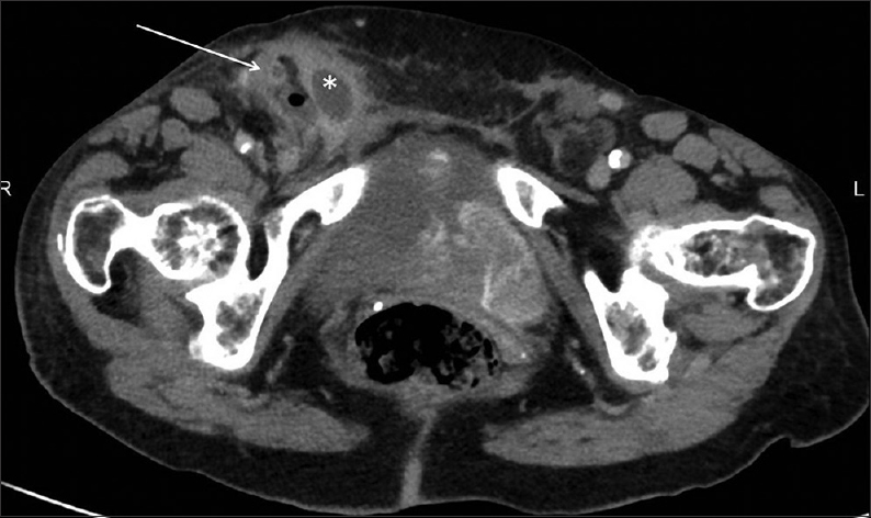
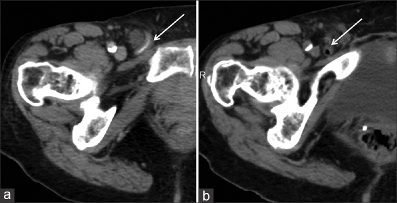
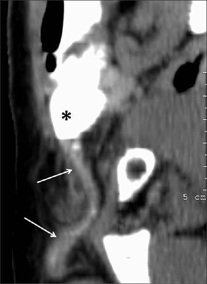
An 86-year-old female in good general health who was alert and independent presented to the emergency department because of a painful swelling in the right groin that had been present for 1 week. She was febrile with a temperature of 39°C and had leukocytosis of 18,000 cells/mm3 with 78% neutrophils. The patient was referred for emergency computed tomography (CT) of the abdomen and pelvis. The examination showed fluid collection within the right inguinal hernia; in addition, a thickened appendix was identified within the hernia sac [Figure 1]. On review of the patient's past imaging studies, two CTs of the abdomen from 2008 and 2010 showed the right inguinal hernia containing a normal appendix [Figure 2], a finding included in the CT report; the appendix could be traced on reformatted images to its origin from the cecum [Figure 3].

- 86-year-old female with fever and tender swelling in the groin caused by a periappendicular abscess and diagnosed with appendicitis within an inguinal hernia. Axial computed tomography (CT) image of the pelvis shows multilocular fluid collection (*) and gas bubble within a right inguinal hernia. The 8-mm thick appendix (arrow) is seen laterally.

- 86-year-old female with fever and tender swelling in the groin caused by a periappendicular abscess and diagnosed with appendicitis within an inguinal hernia. Axial CT images of the pelvis taken in 2008 and 2010 are presented. (a) The 2008 study shows a contrast-filled appendix in its long axis (arrow). (b) The 2010 scan shows the gas-filled appendix in its short axis (arrow).

- 86-year-old female with fever and tender swelling in the groin caused by a periappendicular abscess and diagnosed with appendicitis within an inguinal hernia Curved-plane reconstruction of the right groin from the 2008 CT study in Figure 2a shows the appendix (arrows) originating from the base of the contrast-filled cecum (*), extending caudally into the hernia sac.
The patient was admitted and started on intravenous antibiotics. After 2 days, there was no improvement in the swelling and tenderness of the right groin mass, and she was referred for ultrasound-guided percutaneous drainage. On ultrasound, a heterogeneous process was seen in the right inguinal hernia, consisting of moderately vascular soft tissue and a small amount of fluid. Under local anesthesia, an 18G needle was inserted into the fluid and 10 cc of pus were aspirated. Culture of the aspirate showed growth of Escherichia coli. Within 24 h of treatment, the patient's swelling and tenderness markedly improved and 5 days after aspiration, she was discharged from the hospital on oral antibiotics, with follow-up scheduled in the general surgery clinic.
DISCUSSION
The presence of the appendix within an inguinal hernia is relatively rare, occurring in less than 1% of hernias. This condition is known as Amyand's hernia, named after Claudius Amyand (1681–1740), who performed the first appendectomy in 1735.[1] His patient was an 11-year-old boy with appendicitis within an inguinal hernia sac.
With the widespread use of cross-sectional imaging, inguinal hernia is detected far more often at present than in the past. Most hernias are incidental findings, which contain fat. However, hernias may contain other organs including small and large intestines, urinary bladder, ovary, or as in our case, the vermiform appendix. Complications of inguinal hernias may result from their unique content, as in our case of periappendicular abscess.
The presence of the appendix within an inguinal hernia is estimated to be prevalent at a rate of about 0.5%, with male preponderance and a bimodal age distribution in neonates and individuals over 70 years of age.[2] The appendix within an Amyand's hernia may develop any of the complications associated with this organ, including acute inflammation, periappendicular abscess, mucocele, and benign and malignant tumors. An estimated 0.1% of the cases of acute appendicitis occur in the setting of Amyand's hernia.[2]
Differential diagnosis of a tubular structure within an inguinal hernia includes entities far less common than the appendix, including Meckel's diverticulum (Littre's hernia),[3] other small bowel diverticula, and enteric duplication. Tracing the structure to the base of the cecum confirms its identity as the appendix [Figure 3].
The incidental discovery of an Amyand's hernia introduces management options of surgery versus watchful waiting; the decision depends on the patient's symptomatology, age, and risk factors.[24] In our patient, watchful waiting had been a successfully documented option for at least 7 years. However, the ultimate complication of appendicitis and periappendicular abscess caused higher morbidity than would be expected in an uncomplicated hernia repair.
CONCLUSION
We have described a rare complication of Amyand's hernia with periappendicular abscess presenting within an inguinal hernia sac in an elderly female. The description of inguinal hernia in the radiologist's report must include visceral contents within the sac, so that the clinician may be aware of potential complications in order to ensure prompt and appropriate treatment.
Financial support and sponsorship
Nil.
Conflicts of interest
There are no conflicts of interest.
Available FREE in open access from: http://www.clinicalimagingscience.org/text.asp?2015/5/1/54/166356
REFERENCES
- Bowel obstruction due to Littre hernia: CT diagnosis. Abdom Imaging. 2005;30:682-4.
- [Google Scholar]
- Watchful waiting vs repair of inguinal hernia in minimally symptomatic men: A randomized clinical trial. JAMA. 2006;295:285-92.
- [Google Scholar]


