Translate this page into:
Intrapetrous Anastomosis between the Internal Jugular Vein and the Superior Petrosal Sinus: Cone Beam Computed Tomography Incidental Finding
Address for correspondence: Dr. Ilson Sepúlveda, Otorhinolaryngology, Head and Neck Surgery Service, General Hospital of Concepcion, San Martin AV, nº 1436, Concepción, Chile. E-mail: isepulvedaguilar@gmail.com
-
Received: ,
Accepted: ,
This is an open access article distributed under the terms of the Creative Commons Attribution-NonCommercial-ShareAlike 3.0 License, which allows others to remix, tweak, and build upon the work non-commercially, as long as the author is credited and the new creations are licensed under the identical terms.
This article was originally published by Medknow Publications & Media Pvt Ltd and was migrated to Scientific Scholar after the change of Publisher.
Abstract
This is a case report of a 62-year-old male who presented to the Ear, Nose, and Throat clinic for a follow-up exam for hearing loss stemming from a fall from a horse in his infancy. A Cone Beam Computed Tomography (CBCT) examination revealed an intrapetrous communication between the internal jugular vein bulb and the superior petrosal sinus. Three-dimensional bone and soft tissues volume renderings were generated to demonstrate this incidental anatomical variant.
Keywords
Anastomosis
cone beam computed tomography
jugular
petrosal
sinus
vein

INTRODUCTION
The internal jugular vein (IJV) is a major vessel that collects blood from the head and neck areas and is connected with ipsilateral cavernous sinuses through the inferior petrosal sinus (IPS). The superior petrosal sinus (SPS) is a dural sinus that communicates with the cavernous sinus anteriorly and with the transverse sinus posteriorly. The SPS receives blood from the anterior cerebellar and complex superior petrosal vein. Communication between the IJV and SPS is not normally present.
CASE REPORT
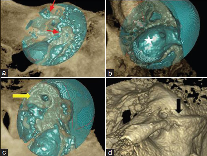
A 62-year-old male bus driver with a history of hearing loss in the right ear presented to the Ear, Nose, and Throat (ENT) clinic for a follow-up exam. The hearing loss resulted from a fracture of the temporal bone following a fall from a horse in his infancy. The otolaryngological exam was normal, but a Cone Beam Computed Tomography (CBCT) showed an intrapetrous communication between the IJV and the SPS, revealing an ascendant tortuous path passing through the middle of the ipsilateral superior semicircular canal. Three-dimensional bone and soft tissues volume renderings were generated to demonstrate this anatomical variant [Figures 1 and 2]. Magnetic resonance imaging (MRI) was not performed because the sensorineural hearing loss is not an acute complication of his fracture. Presently, the patient does not have any other complication and continues with his normal life and will be monitored with follow-up examinations.

- 62-year-old male patient with clinical history of unilateral sensorineural hearing loss and fracture of the right temporal bone in his infancy. CBCT of temporal bone: (a) Sagittal, (b) axial, (c) coronal, and (d) three-dimensional images with osseous volume rendering show the intrapetrous communication between the IJV and the SPS (red arrows) with an ascendant tortuous path, passing through the middle of the superior semicircular canal.

- 62-year-old male patient with clinical history of unilateral sensorineural hearing loss and fracture of the right temporal bone in his infancy. 3D soft tissues volume rendering: (a) shows intrapetrous communicationpath (red arrows); (b) inferior view shows IJV bulb, (white star); (c) superior semicircular canal (yellow arrow); (d) 3D osseous volume rendering shows SPS canal (black arrow).
DISCUSSION
Development of the intracerebral veins and their extracranial drainage is a complex process in humans. The superficial vessels drain into the external jugular vein and the middle and deep vessels into the IJV.[1] The IJV is a major vein that collects blood from the head and neck region and is also a clinically important vein.[2] The IJV is formed by the union of the lateral sinus and the inferior petrous vein.[1] The size of the IJV is directly affected by the drainage pattern of the dural sinuses, particularly the superior sagittal sinus (SSS) and rectal sinus (RS).[2] The IJV is connected to ipsilateral cavernous sinuses through the IPS, a dural venous sinus running along the intracranial surface of the petroclivial fissure.[3] Actually, in the literature, there are no reports about anastomosis between IJV and SPS in humans and animals.
The SPS is also known as a dural sinus which communicates with the cavernous sinus anteriorly and with the transverse sinus posteriorly. It runs over the trigeminal nerve in the lateral margin of the tentorium and in the superior petrosal sulcus of the temporal bone. The SPS normally works as a drainage route receiving blood from the anterior cerebellar and brain stem, venous systems, and does not work as a normal drainage route from the cavernous sinus.[4] In addition, the SPS receives different drainages from the complex superior petrosal vein: Type I above and lateral to the facial nerve, Type II between the lateral limit of the trigeminal nerve and the medial limit of the facial nerve, and Type III empties above and medial to the trigeminal nerve.[5]
Evaluation of the temporal bone region requires higher-resolution imaging as demonstrated in this particular case. High-resolution imaging should be used in the periodic assessment of patients with history of temporal bone fractures. This is due to the risk of meningitis resulting from a middle ear infection disseminating through membranous consolidation of the fracture.[67] Several studies have been published on the use of CBCT technology for imaging of the temporal bone, specifically the middle and inner ear structures.[8] CBCT offers lower radiation dose, thinner slices, and provides reliable morphologic assessment of the temporal bone resulting from an increase in spatial resolution, when compared to Multidetector Computed Tomography (MDCT).[910] The Computed Tomographic Dose Index (CTDI) of an MDCT scan of the middle ear is around 170 mGy, compared to 15–30 mGy from CBCT imaging. This makes CBCT the ideal technique of choice for ENT applications.[11]
CONCLUSION
An intrapetrous anastomosis between the IJV and the SPS is an abnormal finding. This is a clear anatomical variant discovered as an incidental finding in a routine follow-up CBCT examination. CBCT imaging plays a highly important role for the surgeon when planning surgical intervention in the temporal bone which may be required either because of complications due to underlying disease or for other reasons.
Financial support and sponsorship
Nil.
Conflicts of interest
There are no conflicts of interest.
Available FREE in open access from: http://www.clinicalimagingscience.org/text.asp?2015/5/1/46/163990
REFERENCES
- The petrosquamosal sinus: CT and MR findings of a rare emissary vein. AJNR Am J Neuroradiol. 2001;22:1186-93.
- [Google Scholar]
- Relation between bilateral differences in internal jugular vein caliber and flow patterns of dural venous sinuses. Anat Sci Int. 2013;88:141-50.
- [Google Scholar]
- Morphologic evaluation of the caudal end of the inferior petrosal sinus using 3D rotational venography. AJNR Am J Neuroradiol. 2007;28:1179-84.
- [Google Scholar]
- Superior petrosal sinus: Hemodynamic features in normal and cavernous sinus dural arteriovenous fistulas. AJNR Am J Neuroradiol. 2013;34:609-15.
- [Google Scholar]
- Anatomical variation of superior petrosal vein and its management during surgery for cerebellopontine angle meningiomas. Acta Neurochir (Wien). 2013;155:1871-8.
- [Google Scholar]
- Meningitis de causa otica. Otorhinolaryngology and Head and Neck Surgery Magazine. 1988;48:26-30.
- [Google Scholar]
- MSCT versus CBCT: Evaluation of high-resolution acquisition modes for dento-maxillary and skull-base imaging. Eur Radiol. 2015;25:505-15.
- [Google Scholar]
- Classification and volumetric analysis of temporal bone pneumatization using cone beam computed tomography. Oral Surg Oral Med Oral Pathol Oral Radiol. 2014;117:376-84.
- [Google Scholar]
- Evaluation of the superior semicircular canal morphology using cone beam computed tomography: A possible correlation for temporomandibular joint symptoms. Oral Surg Oral Med Oral Pathol Oral Radiol. 2014;117:e280-8.
- [Google Scholar]
- Morphologic examination of the temporal bone by cone beam computed tomography: Comparison with multislice helical computed tomography. Eur Ann Otorhinolaryngol Head Neck Dis. 2011;128:230-5.
- [Google Scholar]
- Cone-beam imaging: Applications in ENT. Eur Ann Otorhinolaryngol Head Neck Dis. 2011;128:65-78.
- [Google Scholar]


