Translate this page into:
Intrauterine Volvulus of Terminal Ileum Without Malrotation
Address for correspondence: Dr. Suheil Artul, Department of Radiology, Nazareth Hospital, Edinburgh Medical Missionary Society, Nazareth, Israel. E-mail: Suheil_artul@hotmail.com
-
Received: ,
Accepted: ,
This is an open-access article distributed under the terms of the Creative Commons Attribution License, which permits unrestricted use, distribution, and reproduction in any medium, provided the original author and source are credited.
This article was originally published by Medknow Publications & Media Pvt Ltd and was migrated to Scientific Scholar after the change of Publisher.
Abstract
Neonatal terminal ileum volvulus in the absence of malrotation has never been reported before in English literature. However, another similar rare entity known as neonatal primary segmental volvulus without malrotation has been reported before. Volvulus, in general, is an extreme emergency and cases not diagnosed in time lead to death. The main diagnosis is based on radiological features seen on imaging. We present a case of volvulus of terminal ileum that was diagnosed and surgically treated at age of 15 h ensuring the newborn survived. The definitive diagnosis was based mainly on ultrasonographic findings.
Keywords
Malrotation
ultrasound
volvulus terminal ilium
INTRODUCTION

Majority of neonatal midgut volvulus cases are due to malrotation. Some other causes can be due to postoperative adhesions, duplication cyst, meconium “ileus”, and internal hernia.[1] According to our knowledge, this is the first report of intrauterine volvulus of terminal ileum without malrotation that was diagnosed by ultrasound.
Our case deals with terminal ileum torsion around its axis with twisting of mesenteric vessels. Torsion, in general, leads to necrosis of the involved bowel segment. Therefore, it is of medical importance to recognize the radiological signs of bowel torsion as soon as possible. We present a case of volvulus of terminal ileum which was diagnosed and treated surgically in a neonate just 15 h after delivery.
CASE REPORT
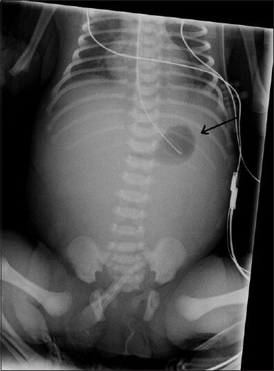
A female baby was born preterm at 35-weeks, with parameters appropriate for her gestational age, weighing 2666 g, with APGAR 9-10 at 1 and 10 min, respectively. Maternal history provided a previous missed abortion, otherwise the mother was a healthy woman who had a normal pregnancy and normal fetal screening follow-up findings. On physical examination of the preterm baby done immediately after delivery revealed a distended abdomen and some grunting. Investigational work-up was started including, chest X-ray which was normal, arterial blood gases showed mild metabolic acidosis, blood and urine culture were obtained, and wide spectrum intravenous antibiotics were started. The baby was referred to our radiological department for plain abdominal X-ray at age of 2 h. The X-ray showed a distended and gasless abdomen, except for a small amount of air in the stomach [Figure 1].

- Female baby at 2 h after birth with distended abdomen diagnosed with intrauterine volvulus of terminal ileum without malrotation. X-ray of abdomen shows distended and gasless abdomen except a small amount of air in the stomach (black arrow).
This radiological finding might be normal at this age, but since the baby was crying it was assumed to have more air in the intestine. The radiological finding raised the possibility of intestinal obstruction. Thus, we started an upper gastrointestinal study to rule out any cause of intestinal obstruction.
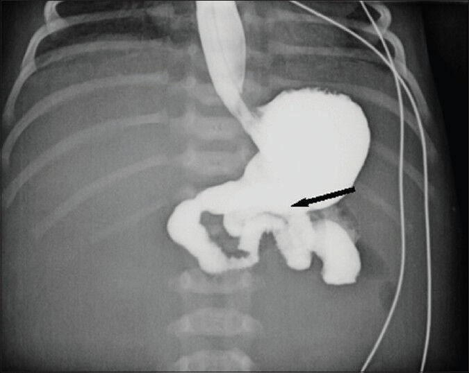
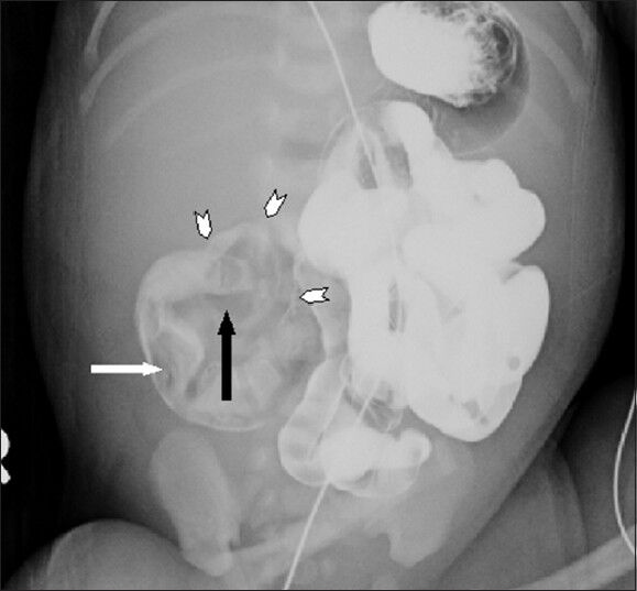
The barium study ruled out a malrotation showing a normal gastric, duodenal, and normal location of the Treitz ligament [Figure 2]. Two hours after the start of the barium study, we noticed lack of propagation of the barium at the level of right lower quadrant of the abdomen. This led us to suspect ileal atresia, which is the most common cause of obstruction after malrotation.[2] After 13 h, a small bowel loop showed a “bird beak” sign with filling defect and a round structure around this end loop [Figure 3]. This radiological finding suggested ileal obstruction due to volvulus with presumed duplication cyst as the leading point.

- Female baby at 2 h after birth with distended abdomen diagnosed with intrauterine volvulus of terminal ileum without malrotation. Barium study shows normal location of Trieitz ligament (black arrow).

- Female baby at 13 h after birth with distended abdomen diagnosed with intrauterine volvulus of terminal ileum without malrotation. At 13 h, the barium study shows stopage of barium with a beak-like shape of the pre-obstructed bowel (black arrow) with filling defect(white arrow) and a structure containing air around the end loop (arrowheads)
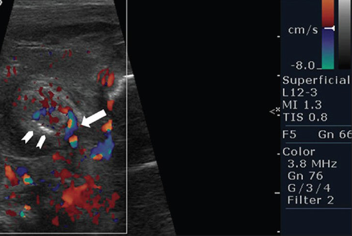
Immediate ultrasonographic examination of the abdomen was done to confirm the diagnosis. Ultrasound scan showed a target lesion consistent of two loops of bowel, pneumatosis of the bowel wall, and twisting of vessels [Figure 4], suggesting a diagnosis of ileal volvulus with intestinal ischemia. The baby was operated on immediately. Intraoperative findings showed volvulus of terminal ileum with intestinal ischemia and perforation with free meconium in the abdomen. We think that meconium in this case was the main predisposing factor for the volvulus of this baby. A resection of the suffered terminal ileum was done with protective ileostomy. The postoperative follow-up revealed a satisfactory clinical course.

- Female baby at 13 h after birth with distended abdomen diagnosed with intrauterine volvulus of terminal ileum without malrotation. Ultrasonographic examination of the abdomen shows a target lesion consistent with two loops of bowel, pneumatosis of the bowel wall (arrowheads) and twisting of vessels (white arrow).
DISCUSSION
According to our knowledge, this is the first case of intrauterine volvulus of terminal ileum without malrotation, that was diagnosed definitively by ultrasound and operated on within a few hours after birth thus saving the life of the baby.
Congenital volvulus is a life-threatening condition for the newborn. Neonatal volvulus is secondary to malrotation, and involves twisting of bowel loop with its feeding vessels that leads to ischemia, necrosis, and perforation with fatal complication, if not recognized and treated in time. Intestinal volvulus without malrotation occurs in 19%-20% of the small bowel volvulus in children.[23] The etiology of neonatal volvulus without malrotation remains unknown. However, other rare predisposing causes are Meckel's diverticulum, duplication cyst, and meconium plug. Delayed diagnosis can lead to significant morbidity and mortality.[4]
Diagnosis of volvulus in any suspected neonatal intestinal obstruction by plain film and barium study is extremely difficult especially when malrotation is ruled out. The most common causes of these cases of intestinal obstruction are intestinal atresia that is fatal in the first hours of life if not treated immediately.
CONCLUSION
Based on our case, we suggest: In any case of suspected intestinal obstruction, first we have to rule out intestinal malrotation with an upper gastrointestinal barium study. Once malrotation is ruled out, we suggest immediately performing an ultrasound examination of the abdomen to rule out an ileal volvulus.
Late diagnosis in volvulus could be fatal. Thus, ultrasound examination that shows a target lesion “bird beak” sign of the involved bowel allows to quickly identify this morbid entity without loss of precious time.
Available FREE in open access from: http://www.clinicalimagingscience.org/text.asp?2013/3/1/50/122317
Source of Support: Nil
Conflict of Interest: None declared.
REFERENCES
- Malrotation and midgut volvulus: A historical review and current controversies in diagnosis and management. Pediatr Radiol. 2009;39:359-66.
- [Google Scholar]
- Segmental small bowel volvulus not associated with malrotation in childhood. Pediatr Surg Int. 1995;10:335-8.
- [Google Scholar]
- Congenital anomalies of the small intestine, colon, and rectum. Radiographics. 1999;19:1219-36.
- [Google Scholar]
- Congenital midgut volvulus associated with fetal anemia. Fetal Diagn Ther. 2010;28:119-22.
- [Google Scholar]


