More results...

Generic selectors
Exact matches only
Search in title
Search in content
Post Type Selectors
Filter by Categories
Abdominal Radiology, Original Research
Abdominal Radiology, Pictorial Essay
Anthropology and Dental Radiology, Original Research
Breast Imaging, Case Report
Breast Imaging, Original Research
Breast Imaging, Pictorial Essay
Breast Imaging, Review Article
Cardiopulmonary Imaging, Case Report
Cardiopulmonary Imaging, Case Series
Cardiopulmonary Imaging, Education
Cardiopulmonary Imaging, Original Research
Cardiopulmonary Imaging, Pictorial Essay
Cardiopulmonary Imaging, Review Article
Case Report
Case Report, Breast Imaging
Case Report, Cardiopulmonary Imaging
Case Report, Diagnostic Radiology
Case Report, Gastrointestinal Imaging
Case Report, Genitourinary and Gynecologic Imaging
Case Report, Neuroradiology/Head and Neck Imaging
Case Report, Vascular and Interventional Radiology
Case Series
Case Series, Abdominal Radiology
Case Series, Diagnostic Radiology
Case Series, Musculoskeletal Imaging
Case Series, Vascular and Interventional Radiology
DENTAL ARTICLE
Dental Radiology, Case Report
Dental Radiology, Case Series
Dental Radiology, Original Research
Diagnostic Radiology, Case Report
Diagnostic Radiology, Case Series
Diagnostic Radiology, Original Research
Diagnostic Radiology, Pictorial Essay
Diagnostic Radiology, Review Article
Editorial
Education
Education, Diagnostic Radiology
Education, Imaging science
Education, Original Research
Emergency, Original Research
Erratum
Gastrointestinal Imaging, Case Report
Gastrointestinal Imaging, Case Series
Gastrointestinal Imaging, Original Research
Gastrointestinal Imaging, Pictorial Essay
General and Emergency Radiology, Original Research
General and Emergency Radiology, Review Article
Genitourinary and Gynecologic Imaging, Case Report
Genitourinary and Gynecologic Imaging, Original Research
Imaging Science, Original Research
Interventional Radiology, Original Research
Letter to Editor
Musculoskeletal Imaging, Case Report
Musculoskeletal Imaging, Original Article
Musculoskeletal Imaging, Original Research
Musculoskeletal Imaging, Pictorial Essay
Musculoskeletal Imaging, Review Article
Neuroradiology , Review Article
Neuroradiology Head and Neck Imaging, Pictorial Essay
Neuroradiology, Case Report
Neuroradiology, Pictorial Essay
Neuroradiology/Head and Neck Imaging, Case Report
Neuroradiology/Head and Neck Imaging, Case Series
Neuroradiology/Head and Neck Imaging, Original Research
Neuroradiology/Head and Neck Imaging, Review Article
Neuroradiology/Spine Imaging, Original Research
Notice of Retraction
Nuclear Medicine, Case Report
Nuclear Medicine, Original Research
Nuclear Medicine, Pictorial Essay
Original Article
Original Article, Neuroradiology
Original Research
Original Research Article
Original Research, Cardiopulmonary Imaging
Original Research, Dental Radiology
Original Research, Descriptive Study
Original Research, Diagnostic Radiology
Original Research, Gastrointestinal Imaging
Original Research, Genitourinary and Gynecologic Imaging
Original Research, Musculoskeletal Imaging
Original Research, Neuroradiology/Head and Neck Imaging
Original Research, Nuclear Medicine
Original Research, Pediatric Imaging
Original Research, Vascular and Interventional Radiology
Pediatric Imaging, Case Report
Pediatric Imaging, Pictorial Essay
PICTORIAL ESSAY
Pictorial essay, Musculoskeletal Imaging
Pictorial essay, Neuroradiology/Head and Neck Imaging
PICTORIAL REVIEW
Radiologic-Pathologic Correlation
RADIOLOGICAL-PATHOLOGICAL CORRELATION
Radiology Business, Original Research
Research Article
Review Article
Review Article, Diagnostic Radiology
Review Article, Gastrointestinal imaging
Review Article, Musculoskeletal
Review Article, Musculoskeletal Imaging
Review Article, Neuroradiology/Head and Neck Imaging
Review Article, Vascular and Interventional Radiology
Technical Innovation
Technical Innovation, Cardiopulmonary Imaging
Technical Innovation, Gastrointestinal Imaging
Ultrasound, Case Report
Ultrasound, Original Research
Ultrasound, Review Article
Vascular and International Radiology, Case Report
Vascular and Interventional Radiology, Case Report
Vascular and Interventional Radiology, Case Series
Vascular and Interventional Radiology, Original Research
Vascular and Interventional Radiology, Pictorial Essay
Vascular and Interventional Radiology, Review Article
Vascular and Interventional Radiology, Short Communication
Vascular and Interventional, Original Research
View/Download PDF

Translate this page into:

Pictorial Essay
2018
:8;
28
doi:
10.4103/jcis.JCIS_10_18

Tomosynthesis-Guided Core Biopsy of the Breast: Why and How to Use it

Department of Diagnostic Radiology, Division of Diagnostic Imaging, The University of Texas MD Anderson Cancer Center, Houston TX 77030, USA

Address for correspondence: Dr. Kyungmin Shin, Department of Diagnostic Radiology, Division of Diagnostic Imaging, The University of Texas MD Anderson Cancer Center, 1515 Holcombe BLVD, Houston TX 77030, USA. E-mail: kshin1@mdanderson.org

Licence

This is an open access journal, and articles are distributed under the terms of the Creative Commons Attribution-NonCommercial-ShareAlike 4.0 License, which allows others to remix, tweak, and build upon the work non-commercially, as long as appropriate credit is given and the new creations are licensed under the identical terms.

Disclaimer:
This article was originally published by Medknow Publications & Media Pvt Ltd and was migrated to Scientific Scholar after the change of Publisher.

Abstract

Digital breast tomosynthesis (DBT) has become an important tool in breast imaging. It decreases the call-back rate while increasing the cancer detection rate on screening mammography and is useful for diagnostic examination of noncalcified lesions and for the evaluation of patients presenting with clinical symptoms. Management challenges and dilemmas that are encountered with abnormalities detected on DBT and lacking a sonographic correlate can now be addressed with tomosynthesis-guided core biopsy.

Keywords

Biopsy
breast
cancer
stereotactic biopsy
tomosynthesis

INTRODUCTION

Digital mammography remains the screening modality of choice for the detection of breast cancer in women over the age of 40 years. With continued advances in technology, multiple studies have demonstrated that compared with digital mammography alone, digital mammography plus the recently developed technique of digital breast tomosynthesis (DBT) significantly increases the cancer detection rate while decreasing the call-back rate.[1234567] Following the 2011 US Food and Drug Administration approval of the use of DBT in combination with standard digital mammography for breast cancer screening and the recent publication of the current procedural terminology code for the use of DBT as a diagnostic imaging modality, the implementation of DBT in clinical practice as a screening and diagnostic tool continues to grow.[8] With increasing use of DBT in clinical practice, management challenges related to noncalcified DBT-detected abnormalities without a sonographic correlate are being encountered more frequently. Here, we discuss the benefits and challenges of tomosynthesis-guided core biopsy of the breast and review the steps in the procedure.

MANAGEMENT OF DIGITAL BREAST TOMOSYNTHESIS-DETECTED ABNORMALITIES WITHOUT A SONOGRAPHIC CORRELATE

The initial step in the workup of an abnormality detected on DBT should be ultrasonography to look for a sonographic correlate. If a sonographic correlate is seen, management of the abnormality is similar, if not identical, to management of an abnormality seen on conventional two-dimensional (2D) digital mammography with a sonographic correlate. However, if no sonographic correlate is seen, the question of how to manage the abnormality arises.

When DBT was first introduced into clinical practice, tomosynthesis-guided biopsy capability was not available, and for noncalcified abnormalities without a sonographic correlate, tomosynthesis-guided wire localization was performed before surgical excisional biopsy. However, surgical excisional biopsy is more invasive and costly than conventional percutaneous image-guided core biopsy. Furthermore, if pathologic examination of the surgical excisional biopsy specimen shows malignancy, the patient may require additional imaging for proper staging and possibly additional surgery, such as re-excision for positive margins and surgical management of the axilla. These additional procedures may substantially increase treatment time and cost. The introduction of tomosynthesis-guided core biopsy technology allowed for tissue diagnosis without more invasive surgical intervention, an approach generally preferred by patients, surgeons, and radiologists.

BENEFITS OF TOMOSYNTHESIS-GUIDED CORE BIOPSY

Tomosynthesis-guided core biopsy is becoming more readily available, replacing traditional prone stereotactic biopsy in many facilities. Tomosynthesis-guided core biopsy requires initial training of the radiologists and the technologists; however, the transition is often smooth, especially when trainees are already proficient in traditional 2D mammography-guided stereotactic biopsy.

Tomosynthesis-guided core biopsy offers a number of benefits over traditional prone stereotactic biopsy. Since tomosynthesis-guided core biopsy is easily performed by trained radiologists, it allows patients with benign concordant biopsy findings to avoid unnecessary surgery. Tomosynthesis-guided core biopsy facilitates tissue diagnosis for mammographic abnormalities seen on 2D and 3D mammograms, including asymmetries, focal asymmetries, masses, architectural distortions, and calcifications. Tomosynthesis-guided core biopsy also facilitates stereotactic biopsy of noncalcified abnormalities, which is demonstrated in the case examples provided later in this article.

Tomosynthesis-guided core biopsy may also be more comfortable for patients. For tomosynthesis-guided core biopsies, the patient may be sitting or be in the right or left lateral decubitus position [Figure 1]. In our experience, this range of options has improved patient comfort, especially for patients with back pain, respiratory problems, or difficulty with prone positioning, because it allows positioning according to the patient's needs and physical limitations.
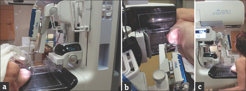
Patient positioning options for tomosynthesis-guided core biopsy of the breast. (a) The patient is in a lateral decubitus position with the biopsy needle positioned for a lateral to medial approach. (b) The patient is in a lateral decubitus position with the biopsy device at a 90-degree angle for an caudocranial approach. (c) The patient is in a seated position with the biopsy needle positioned for a craniocaudal approach.
Figure 1
Patient positioning options for tomosynthesis-guided core biopsy of the breast. (a) The patient is in a lateral decubitus position with the biopsy needle positioned for a lateral to medial approach. (b) The patient is in a lateral decubitus position with the biopsy device at a 90-degree angle for an caudocranial approach. (c) The patient is in a seated position with the biopsy needle positioned for a craniocaudal approach.

In addition, tomosynthesis-guided core biopsy may be helpful in sampling of the very far posterior lesions that are difficult to get to with the traditional prone 2D stereotactic biopsy table. This is largely felt to be related to the loss of the table thickness of the prone table as well as the angle of the needle in the upright tomosynthesis-guided core biopsy units.

It has been reported that tomosynthesis-guided core biopsy may be faster than traditional prone stereotactic biopsy.[910] Our experience suggests that the faster biopsy time is due to faster targeting of the abnormality since there is no need for 15-degree stereo pair images. Confirmation of clip placement and the biopsy site is also easier and improved with tomosynthesis-guided core biopsy, especially for noncalcified findings or when there is complete removal of the calcifications. With tomosynthesis-guided core biopsy, the final postclip placement mammograms are obtained with the same unit that is used for the biopsy. In contrast, with traditional prone stereotactic biopsy, the patient is usually transferred to another mammography unit, which is usually in a different room, for postclip placement mammography.

PROCEDURE FOR TOMOSYNTHESIS-GUIDED CORE BIOPSY

The basic steps in tomosynthesis-guided core biopsy are similar to those in traditional 2D mammography-guided stereotactic biopsy; the biggest difference between the two techniques is in lesion targeting. First, the biopsy approach is planned. The options are craniocaudal, caudocranial, lateral to medial, and medial to lateral, and the approach is chosen on the basis of the location of the abnormality. Then, once the patient is in position and the breast is under compression, tomosynthesis imaging of the targeted area is performed, which replaces the scout and the conventional 15-degree stereo pair images obtained for z-coordinate/depth calculation in 2D mammography-guided stereotactic biopsy. Once tomosynthesis imaging of the targeted area is complete, the abnormality is targeted on the image slice that best shows the abnormality [Figure 2]. Once the abnormality is targeted, the remaining tomosynthesis-guided core biopsy steps, including the biopsy, obtaining a specimen radiograph, and placing the biopsy clip, are identical to the steps of 2D mammography-guided stereotactic biopsy. The mammogram to confirm proper clip placement before the full-view mammograms is obtained may be performed with tomosynthesis or 2D techniques. In our experience, visualization of the biopsy cavity and clip under tomosynthesis was helpful in some cases of noncalcified abnormalities to confirm that the biopsy cavity was in the area of the abnormality; this technique was especially helpful if clip migration was suspected. Different types of targets for tomosynthesis-guided core biopsy are demonstrated in [Figures 37]. Cine clips of craniocaudal tomosynethesis-guided targeting [Video 1] and postbiopsy clip placement [Video 2], for the same patient as figure 3, demonstrate a marker clip at the targeted site, confirming biopsy of the targeted architectural distortion. Pathology showed invasive lobular carcinoma.

Image of the targeting screen with coordinates and the pictorial demonstration of the coordinates (x, y, and z) for targeting.
Figure 2
Image of the targeting screen with coordinates and the pictorial demonstration of the coordinates (x, y, and z) for targeting.
A 56-year-old woman with architectural distortion seen on mammography without a sonographic correlate. (a-d) Craniocaudal full-field digital mammography (a) and tomosynthesis (b) views and mediolateral oblique full-field digital mammography (c) and tomosynthesis (d) views demonstrate that the finding is not well seen on two-dimensional images (a and c, solid circles) but is readily visible on tomosynthesis images (b and d, dashed circles).
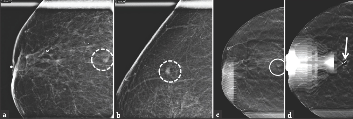
Figure 3
A 56-year-old woman with architectural distortion seen on mammography without a sonographic correlate. (a-d) Craniocaudal full-field digital mammography (a) and tomosynthesis (b) views and mediolateral oblique full-field digital mammography (c) and tomosynthesis (d) views demonstrate that the finding is not well seen on two-dimensional images (a and c, solid circles) but is readily visible on tomosynthesis images (b and d, dashed circles).
A 85-year-old woman called back for additional imaging because screening mammography revealed a 0.6-cm focal asymmetry in the right breast at 12 o'clock. (a and b) Craniocaudal (a) and lateromedial (b) spot magnification views demonstrate a 0.6-cm irregular, high-density mass with indistinct margins in the right breast at 12 o'clock (dashed circles). No sonographic correlate was found, and tomosynthesis-guided core biopsy was performed. (c) Tomosynthesis image shows the targeted mass (circle). (d) Postbiopsy mammogram demonstrates the marker clip in the targeted area (arrow). Pathology result showed invasive ductal carcinoma.
Figure 4
A 85-year-old woman called back for additional imaging because screening mammography revealed a 0.6-cm focal asymmetry in the right breast at 12 o'clock. (a and b) Craniocaudal (a) and lateromedial (b) spot magnification views demonstrate a 0.6-cm irregular, high-density mass with indistinct margins in the right breast at 12 o'clock (dashed circles). No sonographic correlate was found, and tomosynthesis-guided core biopsy was performed. (c) Tomosynthesis image shows the targeted mass (circle). (d) Postbiopsy mammogram demonstrates the marker clip in the targeted area (arrow). Pathology result showed invasive ductal carcinoma.
A 38-year-old woman who presented for evaluation of nonfocal pain in the right breast. (a) On the craniocaudal view, only a 0.9-cm asymmetry was seen in the central right breast (circle). No sonographic correlate was found, and tomosynthesis-guided core biopsy was performed. (b) Tomosynthesis image shows the targeted lesion (circle). (c) Postbiopsy mammogram demonstrates the marker clip in the targeted area (arrow). Pathology result showed mild stromal fibrosis and focal fibroadenomatoid changes.
Figure 5
A 38-year-old woman who presented for evaluation of nonfocal pain in the right breast. (a) On the craniocaudal view, only a 0.9-cm asymmetry was seen in the central right breast (circle). No sonographic correlate was found, and tomosynthesis-guided core biopsy was performed. (b) Tomosynthesis image shows the targeted lesion (circle). (c) Postbiopsy mammogram demonstrates the marker clip in the targeted area (arrow). Pathology result showed mild stromal fibrosis and focal fibroadenomatoid changes.
A 79-year-old woman who presented with a recent diagnosis of ductal carcinoma in situ in the left breast. (a and b) Craniocaudal (a) and mediolateral oblique (b) views of the right breast show a 1.6-cm focal asymmetry (circle and oval). No sonographic correlate was found, and tomosynthesis-guided core biopsy was performed. (c) Tomosynthesis image shows the targeted focal asymmetry on the craniocaudal view (circle). (d) Postbiopsy mammogram shows the marker clip (arrow). The clip had migrated 2 cm medially from the targeted focal asymmetry (circle). Pathology result showed benign fibrocystic changes.
Figure 6
A 79-year-old woman who presented with a recent diagnosis of ductal carcinoma in situ in the left breast. (a and b) Craniocaudal (a) and mediolateral oblique (b) views of the right breast show a 1.6-cm focal asymmetry (circle and oval). No sonographic correlate was found, and tomosynthesis-guided core biopsy was performed. (c) Tomosynthesis image shows the targeted focal asymmetry on the craniocaudal view (circle). (d) Postbiopsy mammogram shows the marker clip (arrow). The clip had migrated 2 cm medially from the targeted focal asymmetry (circle). Pathology result showed benign fibrocystic changes.
A 55-year-old woman with a screening call-back for calcifications in the left breast. (a) Lateromedial magnification view shows new grouped fine pleomorphic calcifications in the lower inner quadrant of the left breast at 8 o'clock at middle depth (dashed circle). (b) Tomosynthesis images clearly demonstrate the calcifications (circle). (c) Specimen radiograph shows the calcifications (arrows). (d) Postbiopsy lateromedial mammogram demonstrates the marker clip in the targeted area with an associated small postbiopsy hematoma (dashed circle). Pathology result showed ductal carcinoma in situ.
Figure 7
A 55-year-old woman with a screening call-back for calcifications in the left breast. (a) Lateromedial magnification view shows new grouped fine pleomorphic calcifications in the lower inner quadrant of the left breast at 8 o'clock at middle depth (dashed circle). (b) Tomosynthesis images clearly demonstrate the calcifications (circle). (c) Specimen radiograph shows the calcifications (arrows). (d) Postbiopsy lateromedial mammogram demonstrates the marker clip in the targeted area with an associated small postbiopsy hematoma (dashed circle). Pathology result showed ductal carcinoma in situ.

CHALLENGES AND PITFALLS OF TOMOSYNTHESIS-GUIDED CORE BIOPSY

The most significant challenge with tomosynthesis-guided stereotactic biopsy is obtaining clearance, which may require quite a bit of patient cooperation because the machine has more moving parts that are present in traditional prone 2D mammography-guided stereotactic biopsy machines. However, this challenge has been addressed with newer prone tomosynthesis-guided stereotactic biopsy tables. Some patients may experience vasovagal reactions due to upright positioning, although we rarely see such reactions with proper patient coaching before the procedure. The incidence of vasovagal reactions is also diminished with the new prone 3D stereotactic biopsy table. Covering the patient's eyes with a small towel so that the biopsy needle is not visible to the patient may also reduce the risk of a vasovagal reaction.

One very important concern is that subtle faint calcifications may be difficult to visualize on the tomosynthesis targeting images, in which case the procedure may need to be converted to a 2D mammography-guided stereotactic biopsy.

CONCLUSION

Tomosynthesis-guided stereotactic core biopsy has several benefits over traditional 2D mammography-guided stereotactic biopsy. With increased utilization of tomosynthesis for both screening and diagnostic examinations, understanding this new technology and tomosynthesis-guided biopsy techniques is important for both radiologists and technologists, which will likely replace traditional 2D mammography-guided stereotactic biopsy over time.

Declaration of patient consent

The authors certify that they have obtained all appropriate patient consent forms. In the form the patient(s) has/have given his/her/their consent for his/her/their images and other clinical information to be reported in the journal. The patients understand that their names and initials will not be published and due efforts will be made to conceal their identity, but anonymity cannot be guaranteed.

Financial support and sponsorship

Nil.

Conflicts of interest

There are no conflicts of interest.

Video Available on: www.clinicalimagingscience.org

REFERENCES

  1. , , , , , , . Breast cancer screening using tomosynthesis in combination with digital mammography. JAMA. 2014;311:2499-507.
    [Google Scholar]
  2. , , , , , , . Detection and classification of calcifications on digital breast tomosynthesis and 2D digital mammography: A comparison. AJR Am J Roentgenol. 2011;196:320-4.
    [Google Scholar]
  3. , , , , , , . Assessing radiologist performance using combined digital mammography and breast tomosynthesis compared with digital mammography alone: Results of a multicenter, multireader trial. Radiology. 2013;266:104-13.
    [Google Scholar]
  4. , , , , , , . Implementation of breast tomosynthesis in a routine screening practice: An observational study. AJR Am J Roentgenol. 2013;200:1401-8.
    [Google Scholar]
  5. , , , , , , . Comparison of digital mammography alone and digital mammography plus tomosynthesis in a population-based screening program. Radiology. 2013;267:47-56.
    [Google Scholar]
  6. , , , , , , . Integration of 3D digital mammography with tomosynthesis for population breast-cancer screening (STORM): A prospective comparison study. Lancet Oncol. 2013;14:583-9.
    [Google Scholar]
  7. , , , , , , . Comparison of tomosynthesis plus digital mammography and digital mammography alone for breast cancer screening. Radiology. 2013;269:694-700.
    [Google Scholar]
  8. US Food and Drug Administration. Selenia Dimensions 3D System–P080003. Available from: http://www.accessdata.fda.gov/scripts/cdrh/cfdocs/cftopic/pma/pma.cfm?num=p080003
  9. , , , , , , . Digital breast tomosynthesis-guided vacuum-assisted breast biopsy: Initial experiences and comparison with prone stereotactic vacuum-assisted biopsy. Radiology. 2015;274:654-62.
    [Google Scholar]
  10. , , , , , , . Tomosynthesis-guided vacuum-assisted breast biopsy: A feasibility study. Eur Radiol. 2016;26:1582-9.
    [Google Scholar]
Show Sections