
Translate this page into:
Surgical Management of Aggressive Central Giant Cell Granuloma of Maxilla through Le Fort I Access Osteotomy
Address for correspondence: Dr. G. Siva Prasad Reddy, Department of Oral and Maxillofacial Surgery, Panineeya Institute of Dental Sciences, Road No.5, Kamala Nagar, Dilsukhnagar, Hyderabad – 500 060, Andhra Pradesh, India. E-mail: gspreddy@yahoo.com
-
Received: ,
Accepted: ,
This is an open-access article distributed under the terms of the Creative Commons Attribution License, which permits unrestricted use, distribution, and reproduction in any medium, provided the original author and source are credited.
This article was originally published by Medknow Publications & Media Pvt Ltd and was migrated to Scientific Scholar after the change of Publisher.
Abstract
Giant cell granuloma (GCG) is an uncommon bony lesion in the head and neck region, most commonly affecting the maxilla and mandible and has a female predilection. The clinical behavior of central GCG ranges from a slowly growing asymptomatic swelling to an aggressive lesion. The clinical, radiological, histological features and management of an aggressive GCG of maxilla in an 18-year-old female patient are described and discussed. It is emphasized that surgery is the traditional and still the most accepted treatment for GCG. Le Fort I osteotomy has been advocated as one of the access osteotomy for the surgical management of aggressive and extensive GCG involving the maxilla. The postoperative morbidity and recurrence have been discussed.
Keywords
Giant cell granuloma
Le Fort I access osteotomy
maxillary sinus tumor

INTRODUCTION

Giant cell granuloma (GCG) was first described by Jaffe in 1953 and is characterized by proliferation of fibroblasts and multinucleated giant cells.[1] GCG is a relatively uncommon non-odontogenic tumor, accounting for less than 7% of all benign facial bone lesions. GCG affects children and adults, and may occur at any age, but is most commonly seen in the first three decades.[2] It is twice as frequent in females.[3] The mandible to maxilla ratio is 2:1 to 3:1.[4] The etiology of GCG still remains controversial. However, it is thought to be a reactive, inflammatory, infective, or neoplastic process.[5] The clinical behavior of GCG ranges from a slowly growing asymptomatic swelling to an aggressive lesion.[6] Radiographic appearance of GCG may be unilocular or multilocular, with either well-defined or less-defined margins. Root resorption and tooth displacement may also be evident.[7] Histologically, multinucleated giant cells, in a cellular vascular stroma, and often new bone formations are detected. Ultrastructurally, the proliferating cells include spindle-shaped fibroblasts, myofibroblasts, and inflammatory mononuclear cells.[89] The treatment of GCG involves surgical removal, varying from curettage to en block resection.[6]
CASE REPORT
An 18-year-old female presented with a painful slowly expanding swelling of left maxilla that had started approximately 3 months earlier [Figure 1]. She also complained of restricted mouth opening, nasal discharge, and lacrimation on the left side. On extra-oral examination there was an obvious swelling on the left side of the cheek and mild proptosis of the left eye. Intra-oral examination showed an expansive mass in the upper left premolar and molar region with sulcus obliteration. The overlying mucosa was slightly bluish purple in color and firm in consistency. There was also expansion of the palate. Clinically Grade II mobility of teeth 25, 26, and 27 were noted.
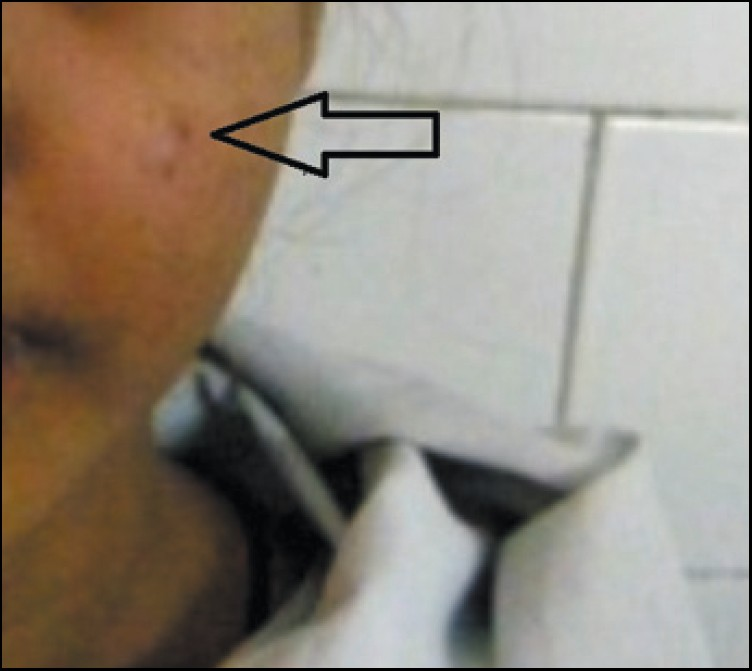
- Preoperative photograph shows swelling on the leftside of the face.
Radiographic features
Computed tomography scan revealed a soft tissue mass completely obliterating the left maxillary sinus. The mass extended inferiorly into the body of the maxilla up to the alveolus, medially it obliterated the nasal cavity. Superiorly, it extended up to the floor of the orbit and posteriorly, it reached the pterygoid plates. Root resorption of teeth 25, 26, and 27 was evident indicating the aggressiveness of the lesion [Figures 2a and 2b].
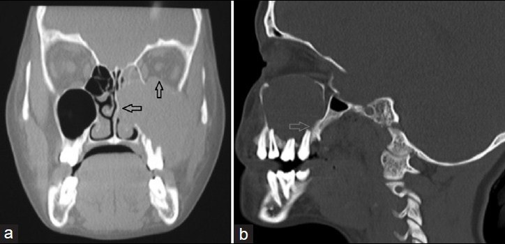
- (a) Coronal CT scan section shows the lesion completely obliterating the maxillary sinus, extending in to the nasal cavity and floor of the orbit. (b) Sagittal CT scan section shows the extension of the lesion toward the posterior surface of maxilla and pterygoid plates.
Histopathology
An incisional biopsy was performed under local anesthesia. Tissue was removed from intraosseouss lesion located in the alveolar ridge with a surgical curette and hemostasis was achieved and primary closure done. The specimen was submitted to the Oral and Maxillofacial pathology department. The histopathplogical examination showed diffused distribution of giant cells and confirmed an aggressive GCG [Figure 3].
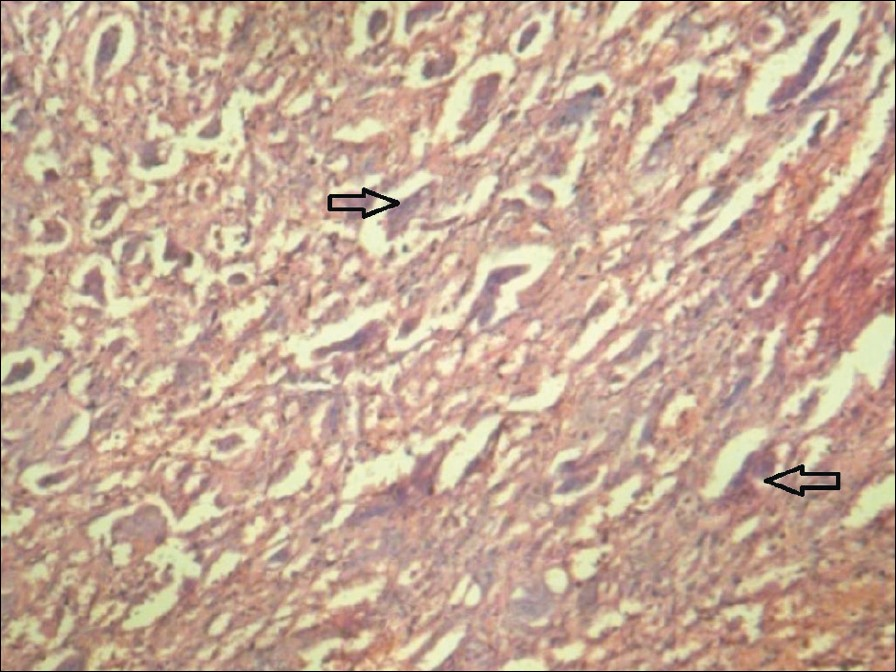
- Histopathological examination using hematoxylin and eosin stain at 10X magnification shows diffused distribution of giant cells.
Surgery
The patient was an young unmarried female and as aesthetics was a major concern, an intra-oral approach was planned as opposed to the conventional extra-oral approach. Since the lesion was quite extensive involving the whole of maxillary antrum extending upto left ethmoidal sinus, orbital floor and pterygoid plates, it was decided to perform an Unilateral (left) Le Fort I osteotomy to gain complete access to the entire lesion. An intra-oral vestibular incision was made extending from the midline to the left second molar region. The underlying bone was exposed and Le Fort I cuts were performed, followed by mid-palatine split and down fracture of the left side maxilla [Figure 4]. This provided excellent visibility and access to the entire lesion.The lesion was excised with the help of surgical curettes and the entire bony margins were visualized for any remnants [Figure 5]. All the mobile teeth on that side were extracted. Hemostasis was achieved and then the ostetomised maxilla was reduced and fixed with the help of a 4-hole ‘L’ plate and screws made of Titanium [Figure 6]. Patient was checked at regular intervals with radiographs [Figure 7] and CT scans [Figure 8]. Patient had no recurrence of the lesion during a 2-year follow-up period.
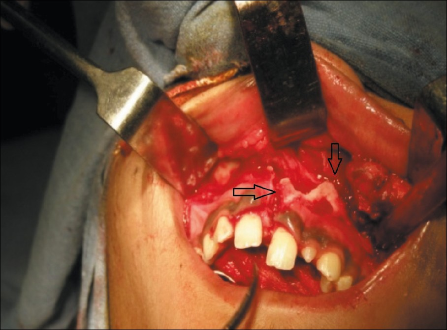
- Unilateral Le fort I osteotomy with midpalatal split to access the lesion.
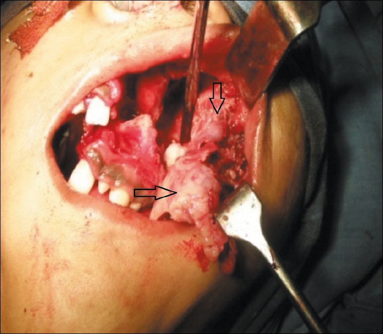
- Curettage and complete removal of the lesion.
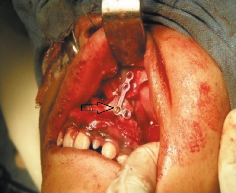
- Stabilization of the Le Fort I segment with L–shaped miniplate and screws.
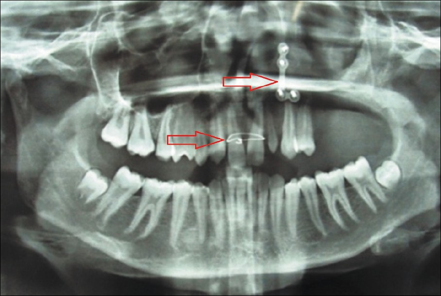
- Postoperative orthopantomograph after 1 year.

- Two-year postoperative follow-up CT scan.
DISCUSSION
World Health Organization defines central GCG as an intra-osseous lesion consisting of cellular fibrous tissue that contains multiple foci of hemorrhage, aggregations of multinucleated giant cells and occasionally trabeculae of woven bone. The clinical behavior of GCG ranges from a slowly growing asymptomatic swelling to an aggressive lesion. Chuong et al., believe that the term “nonaggressive” and “aggressive” should be used based on clinical behavior. When GCG is a slow-growing lesion, it can be asymptomatic and discovered on a routine radiographs, while the rapidly expanding aggressive variety is characterized by pain and facial swelling.[610] Radiographic appearance of GCG can be unilocular or multilocular, with either well-defined or less-defined margins. Root resorption and tooth displacement may also be evident.[7] In this case, all the features were of a more aggressive type. Histologically, multinucleated giant cells in a cellular vascular stroma, and often new bone formations are detected. Ultrastructurally, the proliferating cells include spindle-shaped fibroblasts, myofibroblasts, and inflammatory mononuclear cells.[89] Clinically aggressive GCGs demonstrate a more dense distribution of mononuclear cells and giant cells with less fibrovascular tissue. Focal areas of new bone formation are common. In our case the histological features were of the aggressive type.
Surgery is the most accepted and traditional form of treatment for GCGs. However, the extent of tissue removal ranges from simple curettage to en bloc resection. Nonsurgical approaches to avoid disfigurement have been used, including daily doses of calcitonin and intra-lesional injections with corticosteroids.[6] Osteoprotegrin, which inhibits the osteoclastic bone resorption, has been used in the treatment of central GCGs. More recently, a combined surgical and medical treatment protocol consisting of curettage, with preservation of vital structures, followed by subcutaneous α-interferon that has anti-angiogenic effect has been introduced.
Nonsurgical treatment of central GCGs is probably a valued treatment option for small, slowly enlarging lesions, but successful treatment of painful, large and rapidly growing lesions is more likely to be achieved by surgical removal. Aggressive maxillary lesion are removed en bloc by partial maxillectomy.
The Le Fort I osteotomy is now a commonly performed procedure, that has become the workhorse of orthognathic surgery. These osteotomies should be used more frequently by surgeons trained in these techniques to obtain surgical access and removal of pathological lesions involving mid-face and inaccessible areas. Le Fort I osteotomy has also been explored as an adjunct to skull base tumor surgery. Advantages of Le Fort I osteotomy are many. This is an intraoral procedure, it gives excellent visibility and accessibility to the surgical area, it is easy to perform, with low complication rate and the absence of disfiguring facial incisions provides good cosmesis.
The complications of Le Fort I osteotomy include malocclusion, bleeding, perfusion deficiencies, devitalized teeth or periodontium. These can be avoided or minimized by careful manipulation of soft and hard tissues with good surgical skills.
Extra-oral Weber Fergusson's incision is usually indicated in partial maxillectomy procedures to treat malignant lesions. Facial disfigurement with unacceptable scars is a disadvantage of this incision.
In this case a conservative management of an aggressive maxillary lesion was accomplished by intraoral curettage assisted by unilateral Le Fort I osteotomy with mid-palatine split. No postsurgical morbidity except for a transient infraorbital parasthesia was noticed. The patient did not have any disfigurement postoperatively [Figure 9]. Incidence of recurrence after surgery is 4-20% and it usually occurs due to incomplete removal of the tumor. However, in this case no recurrence was noticed during the 2 year follow-up period, indicating complete removal of the lesion.

- Postoperative photograph.
CONCLUSIONS
GCGs occuring in maxillary sinus may sometimes be aggressive and involve deeper sturctures making surgical approach to the lesion difficult. In our case, as the patient was a young unmarried female, an intra-oral approach was planned as opposed to the conventional extra-oral approach through Weber Fergussion incision, to preserve aeshtetics. Le Fort I osteotomy procedure is commonly used to correct the dentofacial deformities in orthognathic surgeries. It can also be used to gain access to inaccessable lesions as it was used in our case. To conclude, Le Fort I osteotomy procedure provides excellent visibility and access to extensive maxillary lesions and can be advocated as one of the access osteotomies for such lesions.
Source of Support: Nil
Conflict of Interest: None declared.
Available FREE in open access from: http://www.clinicalimagingscience.org/text.asp?2012/2/1/28/96543
REFERENCES
- Giant cell reparative granuloma, traumatic bone cyst and fibrous (fibro-osseous) dysplasia of the jawbones. Oral Surg Oral Med Oral Pathol. 1953;6:159-75.
- [Google Scholar]
- Radiologic features of central giant cell granuloma of the jaws. Oral Surg Oral Med Oral Pathol Oral Radiol Endod. 1996;81:720-6.
- [Google Scholar]
- Cawson's Essentials of Oral Medicine and Pathology. (7th ed). Philadelphia: Churchill Livingstone; 2002. p. :135-6.
- [Google Scholar]
- Central giant cell lesions of the jaws.A Clinical, radiologic, and histopahtologic study. Oral Surg Oral Med Oral Pathol. 1993;75:199-208.
- [Google Scholar]
- Giant cell granuloma of the jawbones- -a proliferative vascular lesion? Immunochemical study with vascular endothelial growth factor and basic fibroblast growth factor. J Oral Pathol Med. 2006;35:613-9.
- [Google Scholar]
- The surgical treatment of central giant cell granuloma of the mandible. J Oral Maxillofac Surg. 2002;60:756-61.
- [Google Scholar]
- Central giant cell granuloma of the jaws: A clinical and radiologic study. J Contemp Dent Pract. 2003;4:87-97.
- [Google Scholar]
- Giant Cell Granuloma of the Maxilla. In: Rosai J, ed. Ackerman's surgical pathology (8th ed). St. Louis. MO: CV Mosby Co; 1996.
- [Google Scholar]
- Expansile Lesions Arising from Structures and Spaces Adjacent to the Paranasal Sinuses. In: Neville BW, Damm DD, Allen CM, eds. Oral and Maxillofacial Pathology. Philadelphia: WB Saunders Co; 2004.
- [Google Scholar]
- Central giant cell lesions of the jaws: A clinicopathologic study. J Oral Maxillofac Surg. 1986;44:708-13.
- [Google Scholar]


