More results...

Generic selectors
Exact matches only
Search in title
Search in content
Post Type Selectors
Filter by Categories
Abdominal Radiology, Original Research
Abdominal Radiology, Pictorial Essay
Anthropology and Dental Radiology, Original Research
Breast Imaging, Case Report
Breast Imaging, Original Research
Breast Imaging, Pictorial Essay
Breast Imaging, Review Article
Cardiopulmonary Imaging, Case Report
Cardiopulmonary Imaging, Case Series
Cardiopulmonary Imaging, Education
Cardiopulmonary Imaging, Original Research
Cardiopulmonary Imaging, Pictorial Essay
Cardiopulmonary Imaging, Review Article
Case Report
Case Report, Breast Imaging
Case Report, Cardiopulmonary Imaging
Case Report, Diagnostic Radiology
Case Report, Neuroradiology/Head and Neck Imaging
Case Report, Vascular and Interventional Radiology
Case Series
Case Series, Diagnostic Radiology
Case Series, Vascular and Interventional Radiology
DENTAL ARTICLE
Dental Radiology, Case Report
Dental Radiology, Case Series
Dental Radiology, Original Research
Diagnostic Radiology, Case Report
Diagnostic Radiology, Case Series
Diagnostic Radiology, Original Research
Diagnostic Radiology, Pictorial Essay
Diagnostic Radiology, Review Article
Editorial
Education, Original Research
Emergency, Original Research
Erratum
Gastrointestinal Imaging, Case Report
Gastrointestinal Imaging, Case Series
Gastrointestinal Imaging, Original Research
Gastrointestinal Imaging, Pictorial Essay
General and Emergency Radiology, Original Research
General and Emergency Radiology, Review Article
Genitourinary and Gynecologic Imaging, Case Report
Genitourinary and Gynecologic Imaging, Original Research
Imaging Science, Original Research
Interventional Radiology, Original Research
Letter to Editor
Musculoskeletal Imaging, Case Report
Musculoskeletal Imaging, Original Article
Musculoskeletal Imaging, Original Research
Musculoskeletal Imaging, Pictorial Essay
Musculoskeletal Imaging, Review Article
Neuroradiology , Review Article
Neuroradiology Head and Neck Imaging, Pictorial Essay
Neuroradiology, Case Report
Neuroradiology, Pictorial Essay
Neuroradiology/Head and Neck Imaging, Case Report
Neuroradiology/Head and Neck Imaging, Case Series
Neuroradiology/Head and Neck Imaging, Original Research
Neuroradiology/Head and Neck Imaging, Review Article
Neuroradiology/Spine Imaging, Original Research
Nuclear Medicine, Case Report
Nuclear Medicine, Original Research
Nuclear Medicine, Pictorial Essay
Original Article
Original Article, Neuroradiology
Original Research
Original Research Article
Original Research, Dental Radiology
Original Research, Descriptive Study
Original Research, Diagnostic Radiology
Original Research, Gastrointestinal Imaging
Original Research, Genitourinary and Gynecologic Imaging
Original Research, Musculoskeletal Imaging
Original Research, Neuroradiology/Head and Neck Imaging
Original Research, Nuclear Medicine
Original Research, Vascular and Interventional Radiology
Pediatric Imaging, Case Report
Pediatric Imaging, Pictorial Essay
PICTORIAL ESSAY
PICTORIAL REVIEW
Radiologic-Pathologic Correlation
RADIOLOGICAL-PATHOLOGICAL CORRELATION
Radiology Business, Original Research
Research Article
Review Article
Review Article, Diagnostic Radiology
Review Article, Gastrointestinal imaging
Review Article, Musculoskeletal
Review Article, Vascular and Interventional Radiology
Technical Innovation
Technical Innovation, Gastrointestinal Imaging
Ultrasound, Case Report
Ultrasound, Original Research
Ultrasound, Review Article
Vascular and International Radiology, Case Report
Vascular and Interventional Radiology, Case Report
Vascular and Interventional Radiology, Case Series
Vascular and Interventional Radiology, Original Research
Vascular and Interventional Radiology, Pictorial Essay
Vascular and Interventional Radiology, Review Article
Vascular and Interventional Radiology, Short Communication
Vascular and Interventional, Original Research
View/Download PDF

Translate this page into:

PICTORIAL ESSAY
2011
:1;
19
doi:
10.4103/2156-7514.78528

Sonographic Upper Gastrointestinal Series in the Vomiting Infant: How We Do It

Department of Diagnostic Imaging, Warren Alpert School of Medicine, Brown University, RI, USA
Address for correspondence: Dr. Thaddeus W. Herliczek, Department of Diagnostic Imaging, Warren Alpert School of Medicine, Brown University, 355 Eddy Street, Providence, RI 02903, USA. E-mail: thaddeus_herliczek@brown.edu
Licence

This is an open-access article distributed under the terms of the Creative Commons Attribution License, which permits unrestricted use, distribution, and reproduction in any medium, provided the original author and source are credited.

Disclaimer:
This article was originally published by Medknow Publications & Media Pvt Ltd and was migrated to Scientific Scholar after the change of Publisher.

Abstract

Sonography (ultrasound) is used routinely to assess an infant with nonbilious projectile emesis. Fluoroscopic upper gastrointestinal (UGI) series has been the standard method to evaluate infants with bilious emesis. We use sonographic UGI routinely to assess infants with nonbilious emesis as well as infants with bilious emesis. This essay illustrates our technique, the results obtained using this technique for normal anatomy, and the commonly encountered pathology.

Keywords

Malrotation
pediatric
sonographic UGI
ultrasound
hypertrophic pyloric stenosis

INTRODUCTION

Sonography (US) is the imaging modality recommended by the American College of Radiology (ACR) to assess an infant with nonbilious projectile emesis.[1]

As of 2008, the ACR recommends fluoroscopic upper gastrointestinal (UGI) series rather than US to evaluate infants with bilious emesis.[1]

More recently, Yousefzadeh described US as an assessment protocol for infants with bilious emesis.[23] His US technique remains controversial as neither prospective nor retrospective analyses of the technique's sensitivity and specificity are available. Nevertheless, we have used sonographic UGI for years and find it offers direct assessment of the gastroesophageal junction, stomach, pylorus, duodenum, and midgut fixation.

We use sonographic UGI routinely at our institution to assess infants with nonbilious emesis as well as infants with bilious emesis. We use it also to assess infants with persistent or progressive gastroesophageal reflux (GER) or GER complicated by failure to thrive, apnea, or aspiration.

The difference in cost between fluoroscopic UGI and sonographic UGI (US) at our institution is less than 2%. Although US is more expensive, this technique is quick, readily available, nonionizing, and portable, therefore it is valuable to us and we believe it is an alternative assessment tool that can be used by experienced pediatric sonologists.

Sonographic upper gastrointestinal series: Technique

Infants are not given any feed by mouth for 1–2 h prior to examination. However, no preparation or delay is prescribed for an infant with bilious emesis given the possibility of malrotation and midgut volvulus, a surgical emergency.

All sonographic UGI at our institution are performed either by a pediatric radiologist or performed initially by an experienced pediatric sonographer with confirmatory scanning done by the pediatric radiologist.

We use a linear high-frequency probe (9–17 MHz) to insonate two sonographic windows: (1) left and right hepatic lobes, and (2) epigastrum with graded compression.

Images are taken in sagittal and transverse planes. We find a transverse plane with craniocaudal obliquity to give the best results.

Luminal contrast medium is not always necessary, but has proved to be helpful in our experience. We give 30 mL 5% glucose water by mouth or 30 mL sterile water via nasogastric tube. The exam requires real-time assessment to demonstrate the entire course of the duodenum.

We spend 5 min or less performing sonographic UGI. If sonographic UGI for the infant with bilious emesis is equivocal at 5 min, fluoroscopic UGI is performed immediately.

Anatomy to assess

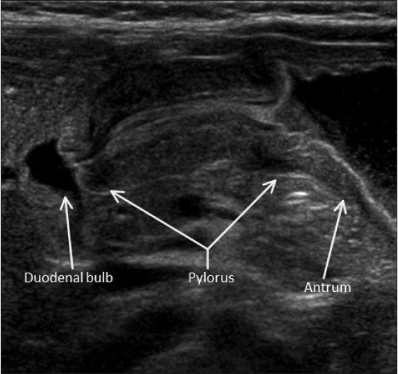
We assess the gastroesophageal junction [Figure 1], stomach, pylorus [Figure 2], and the entire course of the duodenum from pylorus to the duodenojejunal junction (DJJ) [Figures 35].

Figure 1
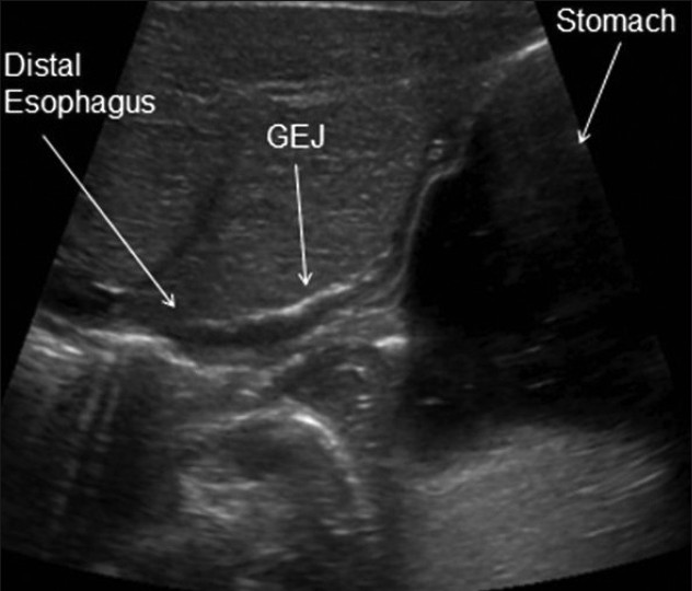
Sonographic image of a healthy infant shows the gastroesophageal junction (GEJ) in an oblique sagittal plane.
Figure 2
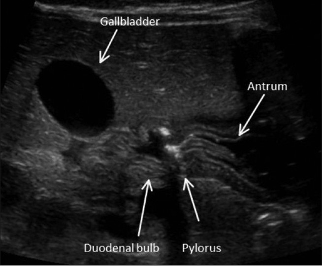
Sonographic image of a healthy infant shows the gastroduodenal junction in a transverse plane.
Figure 3
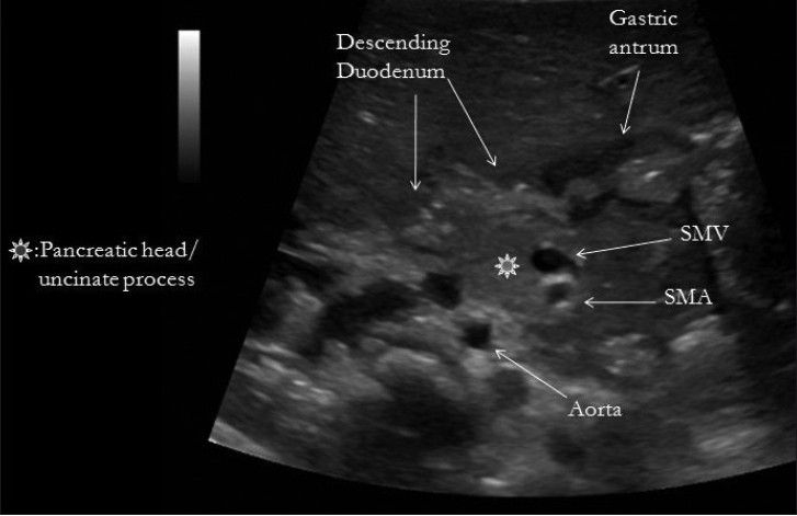
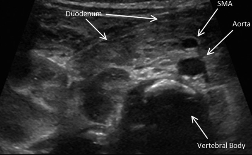
Sonographic image of a healthy infant shows the retroperitoneum in a transverse plane.
Figure 4
Sonographic image of a healthy infant shows the transverse portion of the duodenum positioned normally between superior mesenteric artery (SMA) and abdominal aorta.
Figure 5
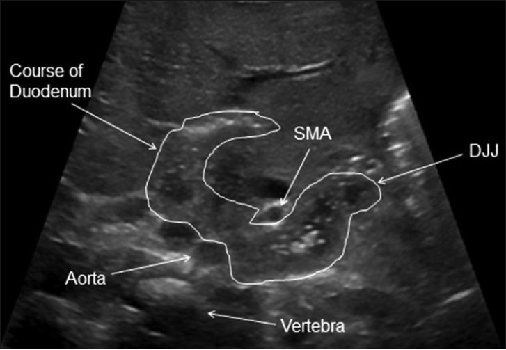
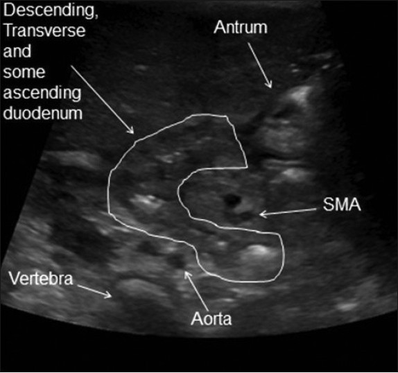
Sonographic image of a healthy infant shows the normal course of the entire duodenum in a transverse plane with craniocaudal obliquity.

Particular attention is given to the relationship of the transverse, or third portion of duodenum (D3) to the superior mesenteric artery (SMA) and abdominal aorta as described by Yousefzadeh.[23] D3 normally courses between SMA and aorta [Figures 4 and 6].

Figure 6
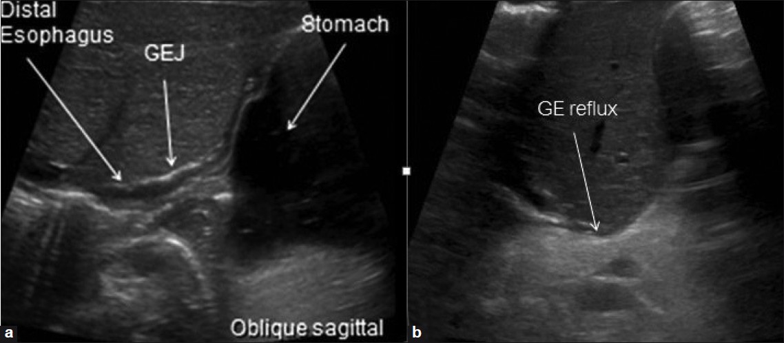
Gastroesphageal reflux. US in an oblique sagittal plane shows (a) closed GEJ, (b) open GEJ with reflux of gastric contents (arrow).

We confirm if the DJJ is to the left of the vertebral column and reaches the height of the duodenal bulb.

While we routinely assess the normal mesenteric vascular relationship, it neither confirms nor excludes malrotation, as one third of infants with malrotation have a normal mesenteric vascular relationship. Also, an abnormal mesenteric vascular relationship can be seen in cases without malrotation.[4]

COMMON PATHOLOGY

Gastroesophageal reflux

While gastroesophageal reflux (GER) is a common entity in infants less than 6 months of age and often diagnosed clinically, our referring pediatricians and gastroenterologists appreciate real-time confirmation of this entity through observation of the infant's gastroesophageal junction [Figure 6].

Gastritis

While often diagnosed clinically or endoscopically, sonographic UGI can demonstrate diffuse gastritis and document its improvement [Figure 7].

Figure 7
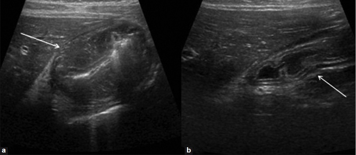
Gastritis. US in a transverse plane shows (a) marked mural thickening (arrow) and (b) Resolution following appropriate therapy (arrow).

Hypertrophic pyloric stenosis

Sonography is the preferred imaging modality to assess an infant with hypertrophic pyloric stenosis (HPS). Objective criteria for HPS vary. We use a mural muscular thickness of more than 3 mm and a pyloric channel length of more than 16 mm. Real-time observation will demonstrate little, if any, transit of gastric contents into the duodenum. Additionally, real-time observation aids confident distinction of HPS from transient antral spasm [Figure 8].

Figure 8
Hypertrophic pyloric stenosis. Sonographic image in a transverse plane

Technical tip: We suggest the infant be placed in a right lateral decubitus position for imaging. However, the pylorus may orient posteriorly and be difficult to image if the stomach is markedly distended. In such a case, using the left lateral decubitus position will improve visualization.

Duodenal duplication cyst

Enteric duplication cysts may occur in any location from the mouth to the anus and are most commonly encountered in the ileum. They can be found in the duodenum where they may produce symptoms of duodenal obstruction [Figure 9].

Figure 9
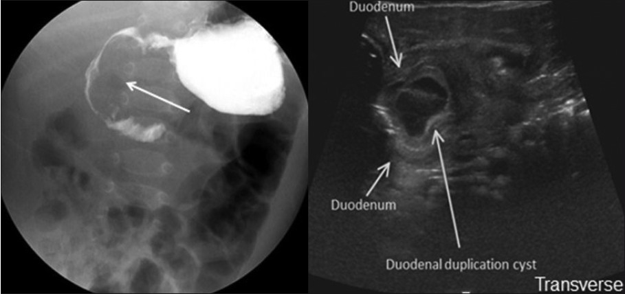
Duodenal duplication cyst. (a) Frontal fluoroscopic projection demonstrates effacement of the medial aspect of the descending portion of the duodenum (arrow). (b) Sonographic UGI in a transverse plane demonstrates descending portion of duodenum and duplication cyst.

Annular pancreas

Annular pancreas is an uncommon developmental variant that often results in emesis in the neonate [Figure 10].

Figure 10
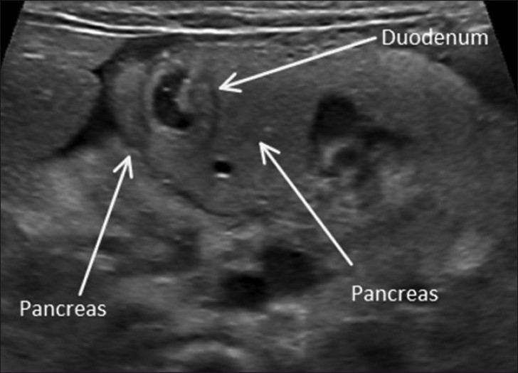
Annular pancreas. Sonographic image in a transverse plane.

Malrotation

An aberrant midgut fixation predisposes to midgut volvulus. A majority of these infants present with bilious emesis in the neonatal period. Aberrant course of the duodenum with D3 anterior to the SMA, proximal duodenal obstruction, and a whirlpool sign can be seen [Figure 11].

Figure 11
Malrotation. Sonographic image in a transverse plane shows the transverse portion of the duodenum courses anterior to the superior mesenteric artery (SMA). No small bowel is seen interposed between SMA and abdominal aorta.

CONCLUSION

Sonographic UGI offers direct assessment of the gastroesophageal junction, stomach, pylorus, duodenum, and retroperitoneal anatomy in the vomiting infant.[23] Various pathologic entities of the upper gastrointestinal tract, including malrotation, are well demonstrated or excluded with sonographic UGI investigation of the vomiting infant. While the role of the sonographic UGI is controversial, we and the referring pediatricians and pediatric surgeons at our institution find it a useful tool in the assessment of a vomiting infant.

Source of Support: Nil

Conflict of Interest: None declared.

REFERENCES

  1. American College of Radiology Appropriateness Criteria: Vomiting in Infants Up to 3 Months of Age. Available from: http://www.acr.org/SecondaryMainMenuCategories/quality_safety/app_criteria/pdf/ExpertPanelonPediatricImaging/VomitinginInfantsupto3MonthsofAgeDoc11.aspx
    [Google Scholar]
  2. , . The position of the duodenojejunal junction: the wrong horse to bet on in diagnosing or excluding malrotation. Pediatr Radiol. 2009;39:S172-7.
    [Google Scholar]
  3. , , , . Assessment of retromesenteric position of the third portion of the duodenum: an us feasibility study. Pediatr Radiol. 2010;40:1476-84.
    [Google Scholar]
  4. , , , , , . Midgut malrotation, the reliability of a sonographic diagnosis. Pediatr Radiol. 1991;22:21-3.
    [Google Scholar]
Show Sections