More results...

Generic selectors
Exact matches only
Search in title
Search in content
Post Type Selectors
Filter by Categories
Abdominal Radiology, Original Research
Abdominal Radiology, Pictorial Essay
Anthropology and Dental Radiology, Original Research
Breast Imaging, Case Report
Breast Imaging, Original Research
Breast Imaging, Pictorial Essay
Breast Imaging, Review Article
Cardiopulmonary Imaging, Case Report
Cardiopulmonary Imaging, Case Series
Cardiopulmonary Imaging, Education
Cardiopulmonary Imaging, Original Research
Cardiopulmonary Imaging, Pictorial Essay
Cardiopulmonary Imaging, Review Article
Case Report
Case Report, Breast Imaging
Case Report, Cardiopulmonary Imaging
Case Report, Diagnostic Radiology
Case Report, Neuroradiology/Head and Neck Imaging
Case Report, Vascular and Interventional Radiology
Case Series
Case Series, Abdominal Radiology
Case Series, Diagnostic Radiology
Case Series, Vascular and Interventional Radiology
DENTAL ARTICLE
Dental Radiology, Case Report
Dental Radiology, Case Series
Dental Radiology, Original Research
Diagnostic Radiology, Case Report
Diagnostic Radiology, Case Series
Diagnostic Radiology, Original Research
Diagnostic Radiology, Pictorial Essay
Diagnostic Radiology, Review Article
Editorial
Education, Diagnostic Radiology
Education, Original Research
Emergency, Original Research
Erratum
Gastrointestinal Imaging, Case Report
Gastrointestinal Imaging, Case Series
Gastrointestinal Imaging, Original Research
Gastrointestinal Imaging, Pictorial Essay
General and Emergency Radiology, Original Research
General and Emergency Radiology, Review Article
Genitourinary and Gynecologic Imaging, Case Report
Genitourinary and Gynecologic Imaging, Original Research
Imaging Science, Original Research
Interventional Radiology, Original Research
Letter to Editor
Musculoskeletal Imaging, Case Report
Musculoskeletal Imaging, Original Article
Musculoskeletal Imaging, Original Research
Musculoskeletal Imaging, Pictorial Essay
Musculoskeletal Imaging, Review Article
Neuroradiology , Review Article
Neuroradiology Head and Neck Imaging, Pictorial Essay
Neuroradiology, Case Report
Neuroradiology, Pictorial Essay
Neuroradiology/Head and Neck Imaging, Case Report
Neuroradiology/Head and Neck Imaging, Case Series
Neuroradiology/Head and Neck Imaging, Original Research
Neuroradiology/Head and Neck Imaging, Review Article
Neuroradiology/Spine Imaging, Original Research
Nuclear Medicine, Case Report
Nuclear Medicine, Original Research
Nuclear Medicine, Pictorial Essay
Original Article
Original Article, Neuroradiology
Original Research
Original Research Article
Original Research, Dental Radiology
Original Research, Descriptive Study
Original Research, Diagnostic Radiology
Original Research, Gastrointestinal Imaging
Original Research, Genitourinary and Gynecologic Imaging
Original Research, Musculoskeletal Imaging
Original Research, Neuroradiology/Head and Neck Imaging
Original Research, Nuclear Medicine
Original Research, Vascular and Interventional Radiology
Pediatric Imaging, Case Report
Pediatric Imaging, Pictorial Essay
PICTORIAL ESSAY
PICTORIAL REVIEW
Radiologic-Pathologic Correlation
RADIOLOGICAL-PATHOLOGICAL CORRELATION
Radiology Business, Original Research
Research Article
Review Article
Review Article, Diagnostic Radiology
Review Article, Gastrointestinal imaging
Review Article, Musculoskeletal
Review Article, Vascular and Interventional Radiology
Technical Innovation
Technical Innovation, Gastrointestinal Imaging
Ultrasound, Case Report
Ultrasound, Original Research
Ultrasound, Review Article
Vascular and International Radiology, Case Report
Vascular and Interventional Radiology, Case Report
Vascular and Interventional Radiology, Case Series
Vascular and Interventional Radiology, Original Research
Vascular and Interventional Radiology, Pictorial Essay
Vascular and Interventional Radiology, Review Article
Vascular and Interventional Radiology, Short Communication
Vascular and Interventional, Original Research
View/Download PDF

Translate this page into:

CASE REPORT
2015
:5;
42
doi:
10.4103/2156-7514.161858

Sonographic Hair-on-end Sign in Osteosarcoma

Department of Radiology, Galilee Medical Center, Nahariya, Israel
Department of Medical Imaging, Bar Ilan University Faculty of Medicine, Galilee, Safed, Israel
Address for correspondence: Dr. Norman Loberant, Department of Radiology, Galilee Medical Center, Nahariya, Israel. E-mail: nloberant@yahoo.com
Licence

This is an open access article distributed under the terms of the Creative Commons Attribution-NonCommercial-ShareAlike 3.0 License, which allows others to remix, tweak, and build upon the work non-commercially, as long as the author is credited and the new creations are licensed under the identical terms.

Disclaimer:
This article was originally published by Medknow Publications & Media Pvt Ltd and was migrated to Scientific Scholar after the change of Publisher.

Abstract

This report is a case of osteosarcoma in a young female whose initial examination was sonography. This examination demonstrated a femoral tumor and included a unique finding corresponding to the radiographic hair-on-end sign of malignant new bone formation.

Keywords

Hair-on-end
osteosarcoma
sonography
sunburst

INTRODUCTION

This report presents the case of a young female with knee pain whose ultrasound examination revealed a suspicious femoral cortical defect with associated soft tissue mass. In addition, a unique finding was seen, correlating with the hair-on-end radiographic sign occasionally seen in aggressive tumors.

CASE REPORT

A 12-year-old female was referred for ambulatory sonography of the knee because of several weeks of knee pain. Routine sonography of the knee was normal, but on questioning, the child's complaints were localized to the anterolateral distal thigh. Insonation of this area showed a cortical defect with a saucerized appearance and an oval hypoechoic tissue filling the defect and protruding into the adjacent soft tissues [Figure 1a]. Extended field of view showed the defect in relation to the more distal epiphyseal plate [Figure 1b]. In the depths of the soft tissue within the cortical defect, parallel vertical echogenic structures 2.5 to 4.5 mm in height were identified, and color Doppler demonstrated blood vessels coursing between these structures [Figure 1c and d].

Figure 1
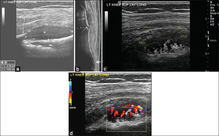
Femoral lesion in a 12-year-old girl who presented with leg pain. (a) Longitudinal gray-scale ultrasound image of the distal femur demonstrates a saucerized cortical defect (the length of the cortical defect is indicated by ultrasound cursor 1), peripheral periosteal reaction (arrow), and oval hypoechoic soft tissue mass (*). (b) Panoramic gray-scale ultrasound image shows the cortical defect (thin arrows) in relation to the femoral shaft and distal epiphyseal plate (thick arrow). (c) Grayscale ultrasound image of the femoral soft tissue mass shows parallel linear hyperechoic structures in the depth of the lesion (dotted lines). (d) Color Doppler image shows blood vessels surrounding and partly obscuring the hyperechoic structures.

Subsequent X-ray of the knee confirmed the cortical defect in distal femoral diaphysis with inhomogeneity of the medullary cavity and periosteal reaction. A complete imaging workup was performed [Figure 2] and the child was referred to a pediatric oncology center where the diagnosis of telangiectatic osteosarcoma was established. A hair-on-end pattern was not identified in either the radiographs or the computed tomographic images of the knee [Figure 3].

Figure 2
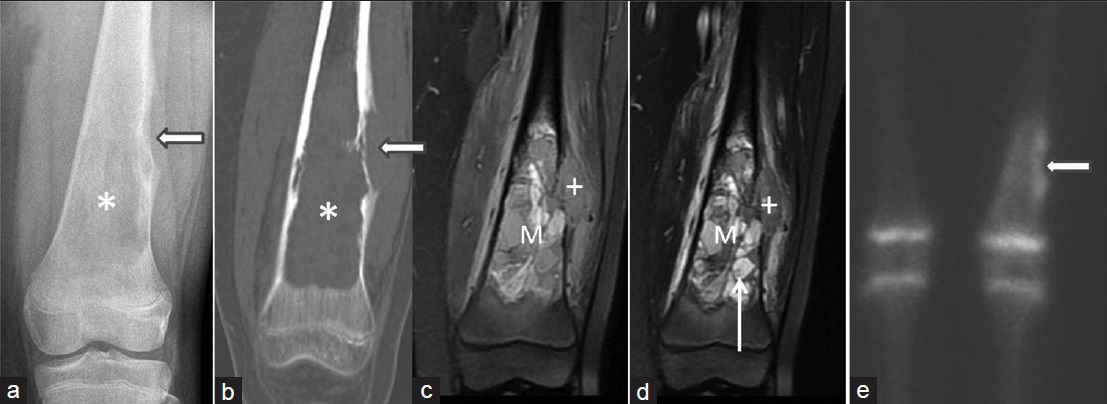
Femoral lesion in a 12-year-old girl who presented with leg pain. Comprehensive imaging of the femur, from left to right, includes (a) plain film and (b) coronal reformatted computed tomography image, both of which show a saucerized cortical lesion (arrow) and a central lytic lesion in the metadiaphysis of the femur (*); (c) coronal proton density fat-saturated magnetic resonance image and (d) coronal T1-weighted contrast enhanced magnetic resonance image which show a soft tissue mass within the cortical lesion (+) and the heterogeneous medullary tumor (M), with post-contrast enhancing nodules (arrow); (e) technetium 99m bone scan which shows tracer uptake at the margins of the cortical lesion (arrow).
Figure 3
(a-c) Femoral lesion in a 12-year-old girl who presented with leg pain. Enlarged images of radiograph and CT of the distal femur. (a) anteroposterior radiograph, (b) axial and (c) coronal CT images do not show a hair-on-end pattern. Codman triangles are indicated by arrows.

DISCUSSION

Osseous abnormalities can be evaluated using multiple imaging modalities. The utility of sonography to evaluate osseous abnormalities has been well documented in trauma,[1] as well as in benign[2] and malignant bone lesions.[3456] Discontinuities in the normal smooth cortical contour of long bones are well seen on ultrasound. The soft tissue component accompanying the bony defect can be characterized on both gray-scale and Doppler examinations.[6] Ultrasound is also useful in providing guidance for percutaneous biopsy.[4]

In the case reported here, ultrasound identified the large defect in the contour of the distal femur, as well as the accompanying oval soft tissue component. The aggressive nature of the lesion was suggested by the irregular and hypervascular tissue in the depth of the lesion. The parallel hyperechoic linear foci suggest the hair-on-end appearance occasionally seen on radiographs, in which periosteal new bone formation is represented by bone spicules perpendicularly oriented to the cortical contour. The hair-on-end pattern consists of parallel bone spicules originating from a broad-based lesion, whereas the similar sunburst pattern consists of radiating bone spicules originating from a narrower surface of bone destruction.[7]

The color Doppler finding of vascularity surrounding the bony spicules is consistent with vascular reparative tissue.

CONCLUSION

Gray-scale ultrasound can be an effective tool in the discovery of destructive cortical bone lesions and their associated soft tissue masses. Doppler ultrasound is useful in characterizing the vascularity of these findings.

The combination of gray-scale and Doppler findings can demonstrate both the parallel spicules of the hair-on-end finding in new bone formation and the vascularized soft tissue between these spicules.

It is important to emphasize the necessity of examining the area of the patient's complaint if it is outside the area of the ultrasound request. In the case presented here, “knee” ultrasound was normal, but the patient's pain and sonographic abnormality were located in her femoral metadiaphysis.

Financial support and sponsorship

Nil.

Conflicts of interest

There are no conflicts of interest.

REFERENCES

  1. , , , , . Ultrasound diagnosis of either an occult or missed fracture of an extremity in pediatric-aged children. Korean J Radiol. 2010;11:84-94.
    [Google Scholar]
  2. , , , . Gray-scale and Doppler characteristics of fibrous cortical defects in a child. J Clin Ultrasound. 2003;31:369-74.
    [Google Scholar]
  3. , , . Ultrasound in musculoskeletal tumors with emphasis on its role in tumor follow-up. Radiol Clin North Am. 1999;37:753-66.
    [Google Scholar]
  4. , , , . Imaging of periosteal reactions associated with focal lesions of bone. Clin Radiol. 2005;60:439-56.
    [Google Scholar]
  5. , , , . Pictorial review: Ultrasonography of primary bone tumours. Clin Radiol. 1998;53:239-46.
    [Google Scholar]
  6. , , , , , , . Ultrasonographic evaluation of osteosarcomas. J Huazhong Univ Sci Technolog Med Sci. 2006;26:629-32.
    [Google Scholar]
  7. , , . Tumor and tumor-like lesions of bone: Radiologic principles. In: , , eds. Bone and Joint Imaging (3rd ed). Philadelphia: Elsevier-Saunders; . p. :1112-6.
    [Google Scholar]
Show Sections