More results...

Generic selectors
Exact matches only
Search in title
Search in content
Post Type Selectors
Filter by Categories
Abdominal Radiology, Original Research
Abdominal Radiology, Pictorial Essay
Anthropology and Dental Radiology, Original Research
Breast Imaging, Case Report
Breast Imaging, Original Research
Breast Imaging, Pictorial Essay
Breast Imaging, Review Article
Cardiopulmonary Imaging, Case Report
Cardiopulmonary Imaging, Case Series
Cardiopulmonary Imaging, Education
Cardiopulmonary Imaging, Original Research
Cardiopulmonary Imaging, Pictorial Essay
Cardiopulmonary Imaging, Review Article
Case Report
Case Report, Breast Imaging
Case Report, Cardiopulmonary Imaging
Case Report, Diagnostic Radiology
Case Report, Gastrointestinal Imaging
Case Report, Genitourinary and Gynecologic Imaging
Case Report, Neuroradiology/Head and Neck Imaging
Case Report, Vascular and Interventional Radiology
Case Series
Case Series, Abdominal Radiology
Case Series, Diagnostic Radiology
Case Series, Musculoskeletal Imaging
Case Series, Vascular and Interventional Radiology
DENTAL ARTICLE
Dental Radiology, Case Report
Dental Radiology, Case Series
Dental Radiology, Original Research
Diagnostic Radiology, Case Report
Diagnostic Radiology, Case Series
Diagnostic Radiology, Original Research
Diagnostic Radiology, Pictorial Essay
Diagnostic Radiology, Review Article
Editorial
Education
Education, Diagnostic Radiology
Education, Imaging science
Education, Original Research
Emergency, Original Research
Erratum
Gastrointestinal Imaging, Case Report
Gastrointestinal Imaging, Case Series
Gastrointestinal Imaging, Original Research
Gastrointestinal Imaging, Pictorial Essay
General and Emergency Radiology, Original Research
General and Emergency Radiology, Review Article
Genitourinary and Gynecologic Imaging, Case Report
Genitourinary and Gynecologic Imaging, Original Research
Imaging Science, Original Research
Interventional Radiology, Original Research
Letter to Editor
Musculoskeletal Imaging, Case Report
Musculoskeletal Imaging, Original Article
Musculoskeletal Imaging, Original Research
Musculoskeletal Imaging, Pictorial Essay
Musculoskeletal Imaging, Review Article
Neuroradiology , Review Article
Neuroradiology Head and Neck Imaging, Pictorial Essay
Neuroradiology, Case Report
Neuroradiology, Pictorial Essay
Neuroradiology/Head and Neck Imaging, Case Report
Neuroradiology/Head and Neck Imaging, Case Series
Neuroradiology/Head and Neck Imaging, Original Research
Neuroradiology/Head and Neck Imaging, Review Article
Neuroradiology/Spine Imaging, Original Research
Notice of Retraction
Nuclear Medicine, Case Report
Nuclear Medicine, Original Research
Nuclear Medicine, Pictorial Essay
Original Article
Original Article, Neuroradiology
Original Research
Original Research Article
Original Research, Cardiopulmonary Imaging
Original Research, Dental Radiology
Original Research, Descriptive Study
Original Research, Diagnostic Radiology
Original Research, Gastrointestinal Imaging
Original Research, Genitourinary and Gynecologic Imaging
Original Research, Musculoskeletal Imaging
Original Research, Neuroradiology/Head and Neck Imaging
Original Research, Nuclear Medicine
Original Research, Vascular and Interventional Radiology
Pediatric Imaging, Case Report
Pediatric Imaging, Pictorial Essay
PICTORIAL ESSAY
Pictorial essay, Musculoskeletal Imaging
Pictorial essay, Neuroradiology/Head and Neck Imaging
PICTORIAL REVIEW
Radiologic-Pathologic Correlation
RADIOLOGICAL-PATHOLOGICAL CORRELATION
Radiology Business, Original Research
Research Article
Review Article
Review Article, Diagnostic Radiology
Review Article, Gastrointestinal imaging
Review Article, Musculoskeletal
Review Article, Musculoskeletal Imaging
Review Article, Vascular and Interventional Radiology
Technical Innovation
Technical Innovation, Cardiopulmonary Imaging
Technical Innovation, Gastrointestinal Imaging
Ultrasound, Case Report
Ultrasound, Original Research
Ultrasound, Review Article
Vascular and International Radiology, Case Report
Vascular and Interventional Radiology, Case Report
Vascular and Interventional Radiology, Case Series
Vascular and Interventional Radiology, Original Research
Vascular and Interventional Radiology, Pictorial Essay
Vascular and Interventional Radiology, Review Article
Vascular and Interventional Radiology, Short Communication
Vascular and Interventional, Original Research
View/Download PDF

Translate this page into:

CASE REPORT
2015
:5;
57
doi:
10.4103/2156-7514.168710

Sinonasal Adenoid Cystic Carcinoma with Intracranial Invasion and Perineural Spread: A Case Report and Review of the Literature

Department of ENT-Head and Neck Surgery Service, General Hospital of Concepcion, Santiago, Chile
Department of Oral and Maxillofacial Radiology, Finis Terrae University School of Dentistry, Santiago, Chile
Department of Oral and Maxillofacial Radiology, University of North Carolina School of Dentistry, Chapel Hill, North Carolina, USA
Department of Pathology, General Hospital of Concepción, University of Concepcion School of Medicine, Concepcion, Chile

Address for correspondence: Dr. Ilson Sepúlveda, Department of ENT-Head and Neck Surgery Service, General Hospital of Concepcion, San Martin AV, n° 1436, Concepcion, Postal Code: 4030000, Chile. E-mail: isepulvedaguilar@gmail.com

Licence

This is an open access article distributed under the terms of the Creative Commons Attribution-NonCommercial-ShareAlike 3.0 License, which allows others to remix, tweak, and build upon the work non-commercially, as long as the author is credited and the new creations are licensed under the identical terms.

Disclaimer:
This article was originally published by Medknow Publications & Media Pvt Ltd and was migrated to Scientific Scholar after the change of Publisher.

Abstract

We present the case of a 51-year-old patient with sinonasal adenoid cystic carcinoma (SACC). Computed tomography (CT) and magnetic resonance imaging (MRI) exams revealed an expansive process in the right nostril accompanied with perineural spread and invasion to the floor of the middle cranial fossa. Due to the size of the tumor and brain involvement, the Head and Neck Tumor Board (HNTB) recommended radiochemotherapy treatment to decrease the size of the lesion. Presently, the patient is undergoing treatment without major complications.

Keywords

Adenoid
carcinoma
computer tomography
cystic
magnetic resonance imaging
perineural
radiochemotherapy
spread

INTRODUCTION

Adenoid cystic carcinomas (ACCs) are rare salivary gland tumors that predominantly occur in women, between the fifth and sixth decades of life. On computed tomography (CT) and magnetic resonance imaging (MRI), ACCs may present as large irregular masses with bone destruction and heterogeneous density or high signal intensity. Another characteristic of ACC is perineural spread (PNS).

CASE REPORT

A 51-year-old male presented with right facial pain of 2-years duration with no significant medical history. Atfirst, it was treated as a dental condition and multiple maxillary tooth extractions were performed to relive the pain. After dental treatment, the pain persisted and the patient was then diagnosed as having trigeminal neuralgia. The patient developed nocturnal right epistaxis of 6 months duration and, consequently, was referred to the Ear Nose and Throat (ENT) clinic for consultation. Nasofibroscopy was performed, which revealed a purple tumor occupying almost the entire right nostril. The left nostril was intact. CT and MRI imaging studies were performed to identify the nature of the mass.

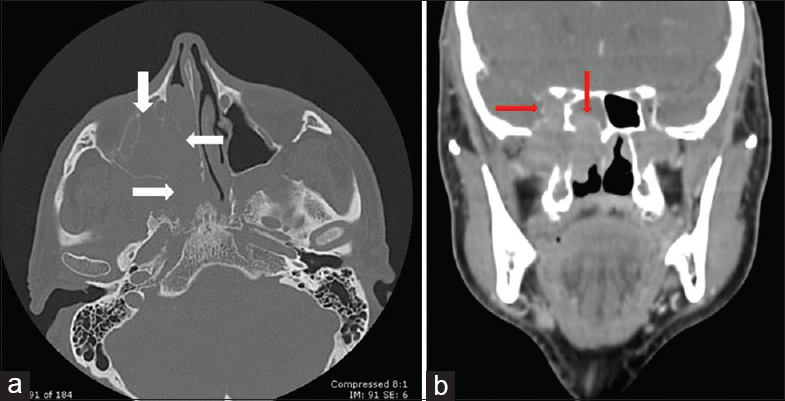
CT imaging revealed a large expansive and infiltrative process involving the right nasal cavity, maxillary, sphenoidal, and ethmoidal sinuses [Figure 1]. The mass crossed the sphenopalatine foramen and extended to the pterygopalatine and pterygomaxillary fossa. The lesion also involved the inferior orbital fissure and oval foramen, reaching the ipsilateral parasellar region. At its largest diameter, the lesion measured 6 cm. There was moderate enhancement post intravenous administration. There was no evidence of regional lymphadenopathy.
51-year-old male presented with right facial pain of 2 years duration with no significant medical history and was later diagnosed with sinonasal adenoid cystic carcinoma. (a) CT bone window reveals a large expansive and infiltrative process involving the right nasal cavity, maxillary, and pterygomaxillary fossa (with arrows). (b) CT soft tissue window post intravenous contrast injection demonstrates moderate enhancement and, intracranial invasion involving sphenoidal sinuses and the right parasellar region (red arrows).
Figure 1
51-year-old male presented with right facial pain of 2 years duration with no significant medical history and was later diagnosed with sinonasal adenoid cystic carcinoma. (a) CT bone window reveals a large expansive and infiltrative process involving the right nasal cavity, maxillary, and pterygomaxillary fossa (with arrows). (b) CT soft tissue window post intravenous contrast injection demonstrates moderate enhancement and, intracranial invasion involving sphenoidal sinuses and the right parasellar region (red arrows).

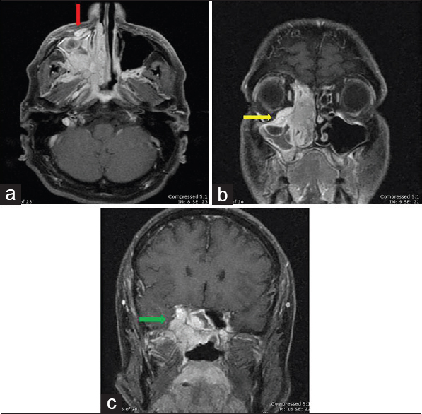
Post intravenous contrast administration, MRI revealed a hyperintense soft tissue expansive process and intracranial invasion of the middle temporal fossa involving the cavernous sinuses [Figure 2]. The mass covered the major sphenoidal fissure involving the eyeball muscles, infiltrating the extra-conal orbital floor and showed PNS through maxillary nerve (CN V2) in the infraorbital foramen.
51-year-old male presented with right facial pain of 2 years duration with no significant medical history and was later diagnosed with sinonasal adenoid cystic carcinoma. Fat-saturated T1-weighted and gadolinium-weighted MRI images: (a) axial view shows perineural spread through CN V2 in the infraorbital foramen (red arrow); (b) coronal view shows the mass involving the eyeball muscles, infiltrating the extraconal orbital floor (yellow arrow); and (c) coronal view shows intracranial invasion to the middle temporal fossa involving the cavernous sinuses (green arrow).
Figure 2
51-year-old male presented with right facial pain of 2 years duration with no significant medical history and was later diagnosed with sinonasal adenoid cystic carcinoma. Fat-saturated T1-weighted and gadolinium-weighted MRI images: (a) axial view shows perineural spread through CN V2 in the infraorbital foramen (red arrow); (b) coronal view shows the mass involving the eyeball muscles, infiltrating the extraconal orbital floor (yellow arrow); and (c) coronal view shows intracranial invasion to the middle temporal fossa involving the cavernous sinuses (green arrow).

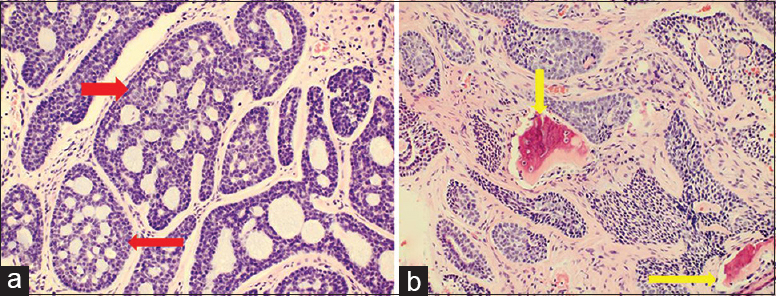
Following the results of the imaging studies, incisional biopsy was recommended confirming a “fragment of nasal adenoid cystic carcinoma” [Figure 3]. Based on the size and intracranial invasion of the tumor, the Head and Neck Tumor Board (HNTB) recommended palliative chemo-radiation therapy. Currently, the patient remains in treatment without any complications.
51-year-old male presented with right facial pain of 2 years duration with no significant medical history and was later diagnosed with sinonasal adenoid cystic carcinoma. Biopsy tissue stained with hematoxylin and eosin, magnification 20×, shows (a) classic cribiform pattern, epithelial cells nests that form cylindrical patterns (red arrows) and (b) bone tissue infiltration by carcinoma (yellow arrows).
Figure 3
51-year-old male presented with right facial pain of 2 years duration with no significant medical history and was later diagnosed with sinonasal adenoid cystic carcinoma. Biopsy tissue stained with hematoxylin and eosin, magnification 20×, shows (a) classic cribiform pattern, epithelial cells nests that form cylindrical patterns (red arrows) and (b) bone tissue infiltration by carcinoma (yellow arrows).

DISCUSSION

ACC is a rare malignant neoplasm that accounts for 1-2% of all head and neck malignancies and approximately 10% of all salivary gland neoplasms.[1] A little less than 5% of sinonasal malignancies are ACC.[2]

Thefirst case of ACC was described by Robin, Lorain, and Laboulbene in two articles published in 1853 and 1854.[2] Peak incidence occurs predominantly among women, between the fifth and sixth decades of life.[3]

ACCs of the minor glands have been reported to have a worse prognosis.[4] Adenocarcinomas of the sinonasal tract mainly arise from the respiratory epithelium or the underlying mucoserous glands (60%). Specifically, ACC is thought to arise from the minor mucoserous glands which lie within the mucosa, below the respiratory-type epithelium of the nasal cavity and paranasal sinuses.[5]

These tumors are made up of basaloid cells with primarily myoepithelial/basal cell differentiation. The three recognized morphologic patterns are tubular, cribriform, and solid.[6] The tubular- and cribriform-type ACCs are lower-grade tumors, whereas solid-type ACC is a higher-grade tumor.[7]

These tumors show slow but relentless growth, and are typically diagnosed late by their proximity to vital structures (e.g., dura, brain, orbit, and central nerves) which makes adequate oncological resection less likely. Another characteristic of ACC of the paranasal sinuses is PNS, with a reported incidence of over 50%. The overall frequency of intracranial invasion of ACC has been reported to be between 4 and 22%.[8]

PNS across the skull base is common in ACC of the head and neck. The maxillary, mandibular, and vidian nerves are the most frequently involved and allow PNS of sinonasal ACC through the foramina rotundum, ovale, and the vidian canal. This probably reflects the more frequent occurrence of ACC in the palate and sinonasal region. Detection is critical in planning the proper surgical approach, so that complete resection may be achieved. For this, the MRI is more sensitive and specific than CT (100% and 85%, and 88% and 89%, respectively).[9]

Surgical excision has been described as the main treatment over decades. However, the extensive local infiltration and PNS related to this malignancy often cause difficulty to achieve high tumor control. The effect of adjuvant radiation therapy on survival in patients with ACC is much debated.[10]

Tumor stage is an import indicator of overall survival and early cancer recurrence. The overall 5-, 10-, and 15-year survival rates for head and neck patients suffering from ACC have been reported to be relatively high: 90.3%, 79.9%, and 62.2%, respectively.[1] However, ACCs of the skull base and paranasal sinuses represent a disease with distinct clinical implications, where the status of surgical margins, the presence of perineural invasion, and the histologic grade of the tumor do not demonstrate correlation with recurrence, metastases, or survival.[10]

CONCLUSION

ACCs are rare malignant salivary gland tumors in the sinonasal tract. One the most important characteristics is PNS. They occur predominantly among women, with the highest incidence occurring between the fifth and sixth decades of life. Imaging features include irregular masses with bone destruction and heterogeneous density or high signal intensity. MRI is more sensitive than CT for detection of perineural tumor invasion. The treatment of choice is complete surgical excision and adjuvant radiation therapy for the extensive local infiltration and PNS related to this malignancy.

Financial support and sponsorship

Nil.

Conflicts of interest

There are no conflicts of interest.

REFERENCES

  1. , , , , , . Adenoid cystic carcinoma of the base of the tongue: Late metastasis to the pancreas. Int J Surg Case Rep. 2011;2:1-3.
    [Google Scholar]
  2. , , , , , , . Unusual presentations of adenoid cystic carcinoma in extra-salivary gland subsites in head and neck region: A case series. Indian J Otolaryngol Head Neck Surg. 2014;66(Suppl 1):S286-90.
    [Google Scholar]
  3. , , , , , . Adenoid cystic carcinoma of external auditory canal: A case report. EJENTAS. 2013;14:41-4.
    [Google Scholar]
  4. , , , , , . Adenoid cystic carcinoma of buccal mucosa. J Maxillofac Oral Surg. 2010;9:273-6.
    [Google Scholar]
  5. , , . Sinonasal tract adenoid cystic carcinoma ex-pleomorphic adenoma: A clinicopathologic and immunophenotypic study of 9 cases combined with a comprehensive review of the literature. Head Neck Pathol. 2012;6:409-21.
    [Google Scholar]
  6. , , , . High grade transformation in adenoid cystic carcinoma of the parotid: Report of a case with cytologic, histologic and immunohistochemical study. Head Neck Pathol. 2009;3:310-4.
    [Google Scholar]
  7. , , , , , , . Adenoid cystic carcinoma of the maxillary sinus with gradual histologic transformation to high-grade adenocarcinoma: A comparative report with dedifferentiated carcinoma. Virchows Arch. 2006;448:204-8.
    [Google Scholar]
  8. , , , . Cytodiagnosis of intracranial metastatic adenoid cystic carcinoma: Spread from a primary tumor in the lacrimal gland. J Cytol. 2011;28:200-2.
    [Google Scholar]
  9. , , , , , , . The sensitivity and specificity of high-resolution imaging in evaluating perineural spread of adenoid cystic carcinoma to the skull base. J Arch Otolaryngol Head Neck Surg. 2007;133:541-5.
    [Google Scholar]
  10. , , , , , , . The role of skull base surgery for the treatment of adenoid cystic carcinoma of the sinonasal tract. Head Neck. 1999;21:402-7.
    [Google Scholar]
Show Sections