Translate this page into:
Rupture of Plantaris Muscle - A Mimic: MRI Findings
Address for correspondence: Dr. Gopinath TN, Dr. Shajis MRI and Research Centre, Mini By-pass Road, Puthiyara, Kozhikode, Kerala – 673 001, India. E-mail: gopi_tn@yahoo.com
-
Received: ,
Accepted: ,
This is an open-access article distributed under the terms of the Creative Commons Attribution License, which permits unrestricted use, distribution, and reproduction in any medium, provided the original author and source are credited.
This article was originally published by Medknow Publications & Media Pvt Ltd and was migrated to Scientific Scholar after the change of Publisher.
Abstract
Calf muscle trauma commonly involves the gastrocnemius and soleus muscles. Plantaris muscle is a vestigial muscle coursing through the calf. Similar clinical features may be seen with injury to the plantaris muscle. It can also mimic other conditions like deep vein thrombosis, rupture of Baker's cyst, and tumors. MRI is helpful in identifying and characterizing it. We report two cases of ruptured plantaris muscle seen on MRI.
Keywords
Calf trauma
MRI
plantaris
rupture
INTRODUCTION

Plantaris muscle is a vestigial structure coursing through the calf. It is absent in 10% of the population.[1] Calf muscle trauma commonly involves gastrocnemius and soleus muscles. Injury to plantaris muscle can present with similar clinical features. Conditions like deep vein thrombosis, rupture of Baker's cyst, and tumors can also mimic these findings. MRI is accurate in identifying and characterizing plantar muscle injury. Here, we report two cases with MR imaging findings.
CASE REPORTS
Case 1
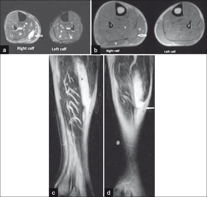
A 59-year-old male presented with a history of sudden snapping pain in the calf region of the right leg after a fall. MRI showed an elongated lesion in the calf between the gastrocnemius and soleus muscles, toward the medial head of gastrocnemius muscle [Figure 1] in the plantaris muscle. The lesion showed hyperintense signal on Short Tau Inversion Recovery (STIR) image and isointense signal on T1-weighted image indicating hemorrhage [Figures 1a and b]. The muscle showed avulsion at musculotendinous junction with proximal retraction of the torn end which was seen in the mid leg region [Figures 1c and 1d]. The gastrocnemius and soleus muscles were normal.

- A 59-year year-old male with avulsion injury of plantaris tendon at musculotendinous junction. (a) Axial short tau inversion recovery (STIR) shows hyperintense plantaris tendon (open white arrow) beneath the gastrocnemius (G) and superficial to soleus (S) in the right calf. Normal left calf is seen. (b) Axial T1 shows isointense plantaris tendon (open white arrow) beneath the gastrocnemius (G) and superficial to soleus (S) in the right calf. (c,d) Serial Sagittal T2 images of leg show the plantaris tendon (P) avulsion and proximal retraction of torn end (arrow in figure d).
Case 2
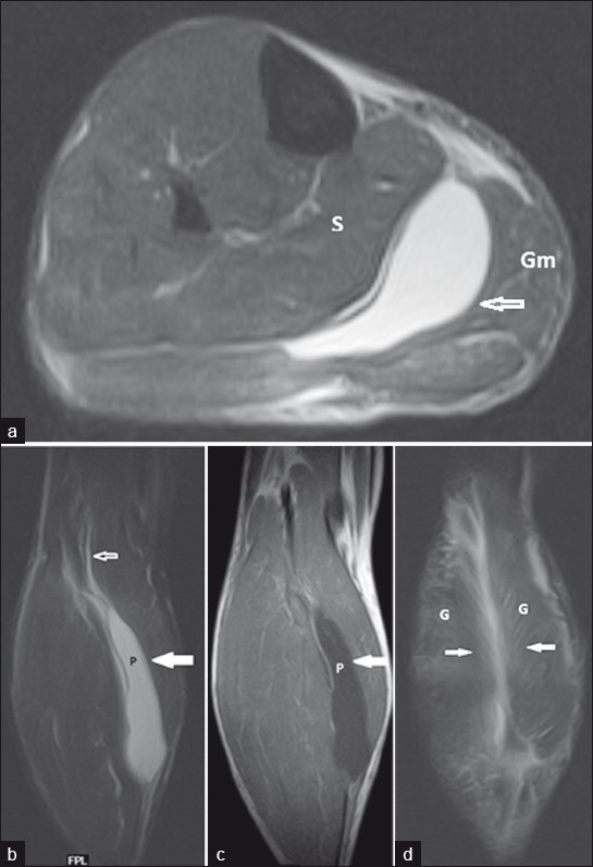
A 40-yr-old male presented with calf pain after injury to the leg. MRI revealed an elongated region of fluid collection that showed hyperintense signal on STIR [Figure 2a] and hypointense signal on T1-weighted image [Figure 2c]. This lesion was in the calf between the gastrocnemius and soleus muscles, toward the medial head of gastrocnemius muscle and represented a torn plantaris muscle. Superiorly the lesion extended up to the lateral condyle of the femur [Figure 2b], the site of origin of the plantaris muscle. Hyperintense signal seen in a part of the gastrocnemius muscles in the calf, represented a Grade 1 injury to the muscle [Figure 2d].

- A 40-year year-old male with rupture of plantaris muscle and Grade 1 injury of gastrocnemius muscles in the right calf. (a) Axial short tau inversion recovery (STIR) of calf shows fluid collection (open white arrow) hyperintense on short tau inversion recovery (STIR) between the gastrocnemius medial head (Gm) and soleus (S) muscles which represents the torn plantaris muscle. (b) Coronal short tau inversion recovery (STIR) of calf shows a hyperintense fluid collection (solid arrow) between the gastrocnemius and soleus (S) muscles which represents the torn plantaris muscle (P). Superiorly it extends upto the lateral condyle of femur (open white arrow), the site of origin of plantaris tendon. (c) Coronal T1 (figure c) of calf shows fluid collection (solid arrow) hypointense on T1 between the gastrocnemius and soleus muscles which represents the torn plantaris muscle. (d) Coronal short tau inversion recovery (STIR) of calf that shows hyperintense signal (solid arrows) in a part of the gastrocnemius muscles (G) in the calf, represents Grade 1 injury.
DISCUSSION
The plantaris muscle is a vestigial structure, absent in 7-20% of population.[1] The plantaris comprises of a slender rope-like tendon. The muscle originates from the lateral supracondylar line just superior and medial to the lateral head of the gastrocnemius muscle.[2] As it courses distally, the plantaris muscle belly lies just deep to the lateral head of the gastrocnemius muscle in the posterior aspect of the proximal leg. The long and slender plantaris tendon continues distally, following an oblique course, to the medial aspect of the lower extremity, where it is located between the medial head of the gastrocnemius muscle and the soleus muscle in the mid-portion of the calf. Distally, the plantaris tendon is located near the medial border of the Achilles tendon, inserting on the calcaneus anteromedial to the Achilles tendon. In some cases, it fuses with the Achilles tendon.[2] Trauma in the calf region commonly involves gastrocnemius, soleus, and Achilles tendon. Injury to the plantaris muscle can present with similar clinical features. Various grades of injury can be seen from contusion, partial tear, and avulsion. Complete plantaris rupture, which most often occurs at the myotendinous junction, results in proximal retraction of the muscle, which may appear as a mass located between the popliteus tendon anteriorly and the lateral head of the gastrocnemius posteriorly.[3] Fluid collections associated with the plantaris are sometimes seen even when no muscle or tendon tear is seen. Helms et al., demonstrated using MR imaging that fluid may be associated with plantaris strains[3] and further to this, Leekham et al., concluded that when such fluid collections are seen in patients with a strong clinical suspicion for plantaris injury, a diagnosis of plantaris strain is appropriate if no tear is seen.[34]
Rupture of the plantaris muscle may occur at the myotendinous junction with or without an associated hematoma or partial tear of the medial head of the gastrocnemius muscle or soleus.[5] Injury to gastrocnemius, soleus, Achilles tendon and ACL may be associated with plantaris tear.[3] Plantaris injury has been described as part of a clinical condition, “tennis leg” that can be caused by plantaris tear, medial head of gastrocnemius tear, soleus tear, or a combination of these.[5] The role of plantaris tendon in tennis leg has been questioned by some.[6] Clinically, the torn plantaris is considered a less severe injury than gastrocnemius muscle tears and is conservatively treated with ice, rest, and antiinflammatory medications.[3] Differential considerations also include deep vein thrombosis,[7] ruptured Baker's cyst, and tumors.[45] The lower component of Baker's cyst rupture may cause fluid collection between gastrocnemius and soleus muscles and mimic plantaris rupture.[4]
In our study, the first case showed rupture at musculotendinous junction, a common site of rupture, with proximal retraction of the torn end. In the second case, there was fluid collection associated with disruption of tendon in mid and proximal part which represents a Grade II injury. Also there was associated Grade I strain of both heads of gastrocnemius muscles.
Since, plantaris tendon rupture can mimic other conditions, like deep vein thrombosis, injury to gastrocnemius and soleus muscles, it is important to identify it in patients with calf pain.
Source of Support: Nil
Conflict of Interest: None declared.
Available FREE in open access from: http://www.clinicalimagingscience.org/text.asp?2012/2/1/19/95433
REFERENCES
- Tennis leg: Clinical US study of 141 patients and anatomic investigation of four cadavers with MR imaging and US. Radiology. 2002;224:112-9.
- [Google Scholar]
- Using sonography to diagnose injury of plantaris muscles and tendons. AJR Am J Roentgenol. 1999;172:185-9.
- [Google Scholar]
- The plantaris muscle: Anatomy, injury, imaging, and treatment. J Can Chiropr Assoc. 2007;51:158-65.
- [Google Scholar]
- Calf pain and swelling (pseudothrombophlebitis) caused by rupture of the plantaris muscle/tendon. J Clin Rheumatol. 1996;2:147-51.
- [Google Scholar]


