
Translate this page into:
Radiofrequency interference in magnetic resonance imaging: Identification and rectification


*Corresponding author: Wingchi Edmund Kwok, Department of Imaging Sciences, University of Rochester, Rochester, United States. edmund_kwok@urmc.rochester.edu
-
Received: ,
Accepted: ,
How to cite this article: Kwok WE. Radiofrequency interference in magnetic resonance imaging: Identification and rectification. J Clin Imaging Sci. 2024;14:33. doi: 10.25259/JCIS_74_2024
Abstract
Radiofrequency (RF) interference artifact is a common type of magnetic resonance imaging (MRI) artifacts caused by the presence of unwanted RF field inside the scanner room. The artifact has the appearance of parallel bright lines or bands that resemble zippers, which can mimic pathology, obstruct the viewing of underlining tissues, and lower image signal-to-noise ratio, affecting the diagnostic evaluation of the image and sometimes even rendering it non-diagnostic. Due to the presence of multiple possible sources of RF interference in MRI and potential nonrelated MRI artifacts that resemble RF interference artifact, it may be difficult to effectively and timely resolve the artifact problem. The objective of this paper is to provide a review of RF interference in MRI and to offer guidance in the prompt and correct identification of the associated image artifacts as well as efficient approaches to resolve and prevent RF interference problems. This article should serve as a useful educational reference to magnetic resonance (MR) technologists and radiologists in dealing with MR image artifacts that may be caused by RF interference.
Keywords
Magnetic resonance imaging
Radiofrequency interference
Image artifact
Magnetic resonance imaging artifact
Image quality

INTRODUCTION
Radiofrequency (RF) interference artifact is a type of image artifacts encountered in magnetic resonance imaging (MRI).[1-9] As the name implies, the artifact is caused by the presence of unwanted RF electromagnetic field [Table 1] inside the scanner room which interferes with the magnetic resonance (MR) signal. It typically has the appearance of one or multiple parallel bright lines or bands in the image that resemble zippers and so it is also called “zipper artefact.” Depending on the severity and the location of the artifact in an image, it may affect the diagnostic evaluation of the image, sometimes even rendering the image non-diagnostic. RF interference artifact is one of the most commonly seen image artifacts in clinical MRI examinations[3] due to the multiple different possible sources of RF interference. The objective of this article is to promote a better understanding of RF interference in MRI to promptly and correctly identify and remove the associated artifact to avoid its adverse effect to the image quality and minimize the potential interruption to patient scanning.
| Abbreviations/Technical Terms | Full terminology | Description |
|---|---|---|
| RF | Radiofrequency | Frequency range between 20 kHz and 300 GHz. MRI signal is within this frequency range |
| SNR | Signal-to-noise ratio | Signal intensity divided by its standard deviation in an image. For MRI, it is a measure of image quality |
| RF shielding | Radiofrequency shielding | Hardware installation to prevent unwanted radiofrequency electromagnetic field from entering the MRI system and interfering with the MRI signal |
| RF leakage | Radiofrequency leakage | Deficiency in radiofrequency shielding allowing unwanted radiofrequency field to enter the MRI system and interfere with the MRI signal |
| Waveguide | A tubular shaped device that guides the passage of electromagnetic wave. For MRI, it is constructed with a certain diameter to block RF field from entering into the MR scan room while allowing non-metallic tubing and fiber optic cable through an opening in the RF shielded room |
|
| Magnetic field gradient | Spatial varying magnetic field applied to spatially encode MRI signal for image formation |
|
| FID | Free induction decay | Decay of MRI signal after its generation by the application of a radiofrequency electromagnetic field |
| FSE | Fast spin echo | A commonly used MRI technique that allows time-efficient scanning |
| RF pulse | Radiofrequency pulse | Electromagnetic field applied at radiofrequency to generate or alter an MRI signal |
| T1 | Longitudinal relaxation time | A measure of how fast the magnetization of a body tissue return to its ground state after MRI signal generation by a radiofrequency pulse. It is one of the MRI characteristics for individual body tissues |
RF: Radiofrequency, SNR: Signal-to-noise ratio, FID: Free induction decay, FSE: Fast spin echo, MRI: Magnetic resonance imaging
This article consists of five main sections: (1) Characteristics of RF interference artifact, (2) common sources of RF interference, (3) unrelated MRI artifacts that resemble RF interference artifact, and (4) Identification and removal of RF interference artifact, followed by conclusion.
CHARACTERISTICS OF RF INTERFERENCE ARTIFACT
RF interference artifact usually appears as bright parallel lines or bands resembling zippers along the phase encoding direction of an image [Figure 1a]. The positions of the individual artifact lines/bands in the frequency encoding direction correspond to the frequency components of the interfering RF field. If the frequency and phase encoding directions are swapped, the orientation of the artifacts in the image changes accordingly [Figure 1b]. If the RF interference is very broadband,[6] the artifact is manifested as increased background noise, resulting in lower image signal-to-noise ratio (SNR) [Table 1] and lower image quality [Table 2]. RF interference artifact can mimic pathologies and obstruct the viewing of underlining tissues, affecting diagnostic evaluation and may even cause the image to be non-diagnostic in serve cases.
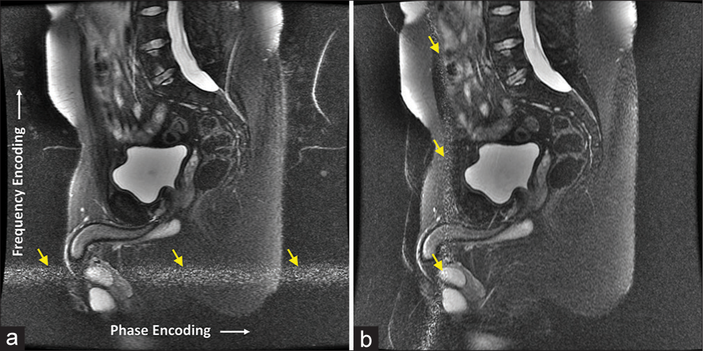
- (a) Abdomen magnetic resonance (MR) image with radiofrequency interference artifact (labeled by yellow arrows) caused by damaged shielding in the MR scanner room door. (b) The direction of the artifact (labeled by yellow arrows) changed with the swapping of frequency and phase encoding directions in the image acquisition.
| Type of RF interference artifact | Appearance |
|---|---|
| Single or multiple frequencies | Bright line or parallel lines |
| Range(s) of frequencies | Bright band or parallel bands |
| Broadband | Increased image noise, leading to reduced image SNR |
RF: Radio frequency, SNR: Signal-to-noise ration
COMMON SOURCES OF RF INTERFERENCE
The electromagnetic field causing RF interference often comes from outside of the MR scanner room via RF leakage, but it may also originate from equipment within the scanner room [Table 3].
| Source | Remedy |
|---|---|
| Scanner room door open or not properly closed | Close door tightly |
| Damage to RF shielding in wall, window or door of scanner room |
Check and fix by MR system manufacturer or shield company |
| Introduction of RF field into scanner room through waveguide | Make sure no electrically conductive objects, such as electric cable or power cord, pass through the waveguide |
| Equipment inside MR scanner room | Turn off and unplug the equipment that causes the RF interference |
RF: Radio frequency, MR: Magnetic resonace
RF shielding deficiency
To prevent unwanted RF field from entering the MR scanner room, it is necessary to install RF shielding [Table 1] that covers the entire room including all walls, ceiling, floor, window, and door[10] [Figure 2a-c]. RF shielding usually consists of copper, steel, or aluminum panels/sheets.[10] The typical shielding requirement is 100 decibels (dB) attenuation at 100 MHz for 1.5 Tesla (T) and 100 dB attenuation at 150–170 MHz for 3.0 T.
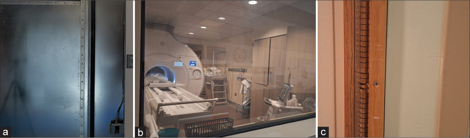
- Photographs of (a) wall shielding, (b) screened window shielding, and (c) door shielding in the magnetic resonance scanner room.
While RF leakage [Table 1] may be caused by a simple oversight of not closing the scanner room door properly during MR scanning, it can also be a result of RF shielding impairment. Due to routine repetitive opening and closing of the scanner room door, its shielding is prone to damage, especially if it consists of copper fingers that are prone to wearing-off and physical damage over time [Figure 2c]. Figure 1a shows an image with RF interference artifact caused by impaired door shielding. The artifact problem was rectified after the MR scanner room door was found to be the location of RF leakage by a shielding company and the door shielding was subsequently repaired. Other potential causes of shield impairment include physical damage to the screened window and puncturing of wall shielding, e.g., from improper drilling. Besides, flooding or water damage may damage the shielding under the floor and inside the walls, which can also lead to RF leakage.
RF field introduced by electrically conductive objects passing through waveguide
Waveguide [Table 1] is a device commonly installed in a MR scanner room to allow the passage of fiber optic cables and hoses for pneumatic or hydraulic system to the control room [Figure 3a]. It is, however, not intended for the passage of electrically conductive objects, such as electrical cable or power cord, which can introduce unwanted RF field into the scanner room. For electrical cable or power cord, RF electrical filtered port/outlet should be used instead [Figure 3b and c]. In recent years, some newer MRI rooms are designed with a notch by the side of the scanner room door [Figure 3d] to allow small non-metallic tubing such as intravenous lines to conveniently pass through without resorting to the waveguide that takes more time and effort to use.
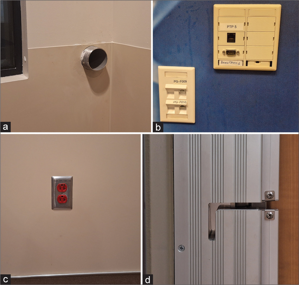
- Photographs of (a) waveguide, (b) electrically filtered ports, (c) power outlets inside the magnetic resonance scanner room, and (d) a notch for passing non-metallic tubing.
Figure 4a shows RF interference artifact appeared in a breast MRI examination. The MR technologists involved in the examination recognized the image artifact as being related to RF interference but failed to identify the source. Subsequent phantom testing by a site MRI physicist revealed that the artifact was caused by an MR-conditional movable lamp [Figure 4b] positioned near the scanner table to provide better lighting for MR-assisted biopsy procedure. The artifact appeared when the power cord of the lamp was connected to an outlet in the control room through the waveguide of the scanner room [Figure 3a], and the problem persisted even when the lamp was turned off [Figure 4c]. However, the artifact was eliminated once the power cord was pulled back completely inside the scanner room [Figure 4d].
- (a) A breast image with radiofrequency interference artifact (yellow arrows) caused by (b) a lamp inside the magnetic resonance (MR) scanner room with the power cord fed through the waveguide and connected to a power outlet in the control room. Phantom testing verified that the (c) artifact (yellow arrows) was caused by the power cord and (d) the artifact was eliminated after the power cord was pulled back completely into the MR scanner room.
In another patient MRI examination, RF interference artifact was observed in the abdomen images [Figure 5a]. The artifact was found to stem from an internet cable connecting an anesthesia machine [Figure 5b] in the scanner room to a computer in the control room through the waveguide [Figure 3a]. The artifact problem was resolved by first connecting the internet cable from the anesthesia machine to an electrically filtered port inside the MR scanner room [Figure 3b] and then to the computer using another internet cable plugged into the port on the other side of the wall in the control room [Figure 5c].
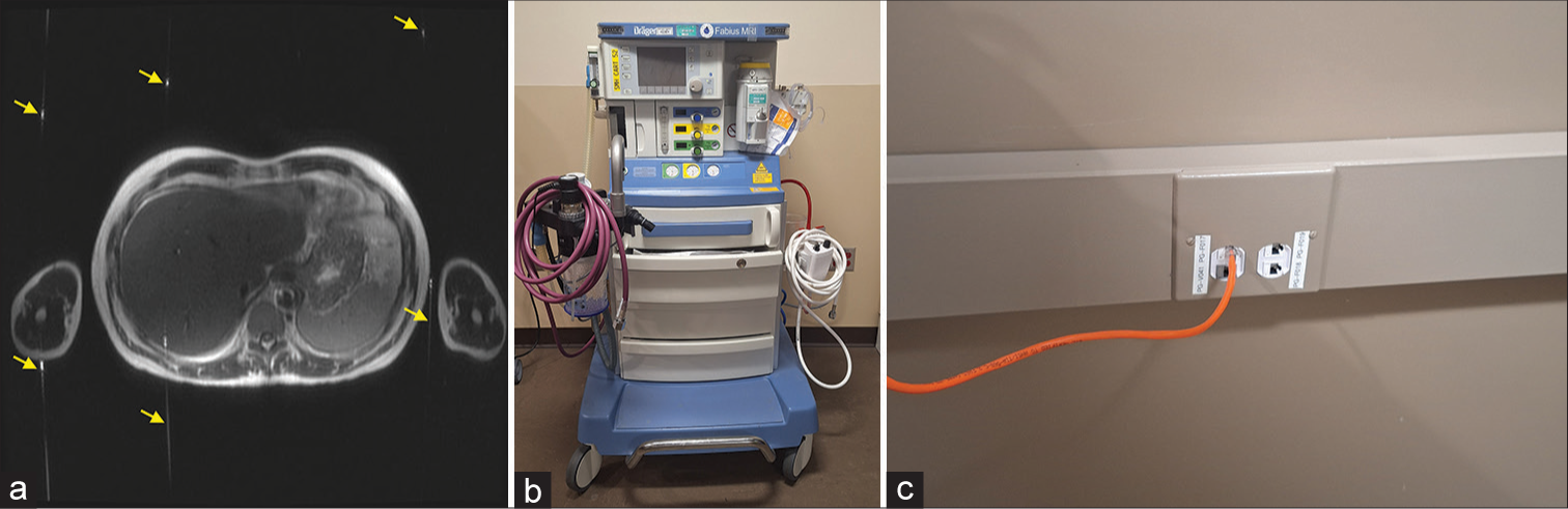
- (a) Abdomen magnetic resonance (MR) image with radiofrequency interference artifact (labeled by yellow arrows) caused by (b) an anesthesia machine in the MR scanner room. External radiofrequency field was introduced into the MR scanner room through an internet cable connected to a computer in the control room through the waveguide, leading to the artifact. The artifact problem was solved by first connecting the internet cable from the anesthesia machine to an electrically filtered port inside the MR scanner room and then to the computer using another internet cable plugged into (c) the port on the other side of the wall in the control room.
Equipment inside MR scanner room
Equipment inside the MR scanner room, such as patient-supporting equipment, may also be a source of RF interference. An example is a case of leg MRI acquired using an extremity RF coil [Figure 6a]. At first, the MR technologist thought that the image artifact was caused by the extremity coil since the artifact was not observed when she switched to use the scanner body coil for the MRI exam.[11] However, later investigation by an MRI physicist discovered that the artifact was actually caused by a power injector inside the room [Figure 6b]. Phantom testing showed that the artifact was present on images obtained by the extremity coil when the power injector was plugged to an outlet inside the scanner room, regardless of whether the injector was turned on or off. However, the artifact disappeared when the injector was unplugged from the power outlet. The fact that the artifact was not observed with the body coil earlier was due to low-signal sensitivity of the body coil, resulting in the RF artifact hidden in the background noise.
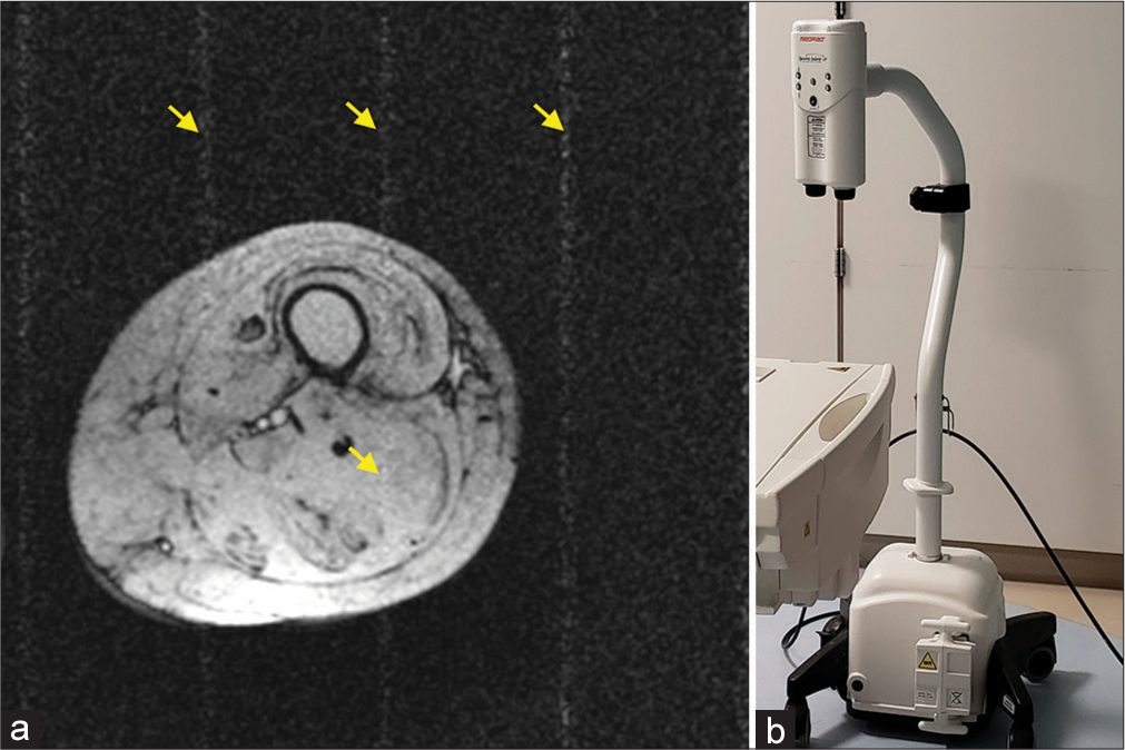
- (a) Radiofrequency interference artifact (labeled by yellow arrows) on a leg magnetic resonance (MR) image caused by (b) a power injector inside the MR scan room.
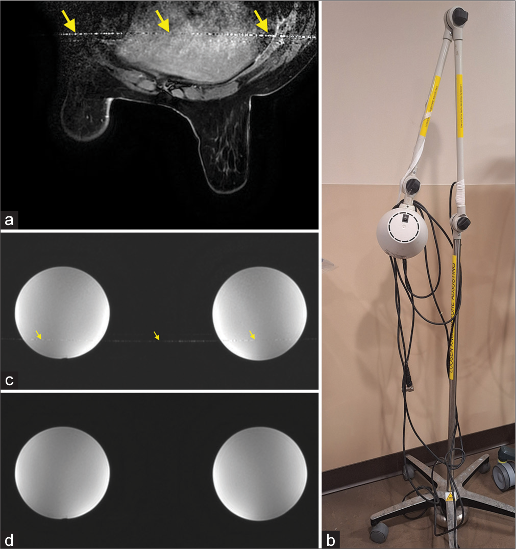
Besides line/band artifacts, equipment inside the MR scanner room may generate very broadband noise that increases image background noise, leading to lower SNR and lower image quality.[6] In an investigation of poor image quality problem in an MRI system, it was found that an anesthesia machine positioned near the magnet was the cause of the problem. Phantom testing showed image SNR reduced from 64.4 to 32.9 when the anesthesia machine was plugged into an outlet inside the MR scanner room and turned on [Figure 7a and b].
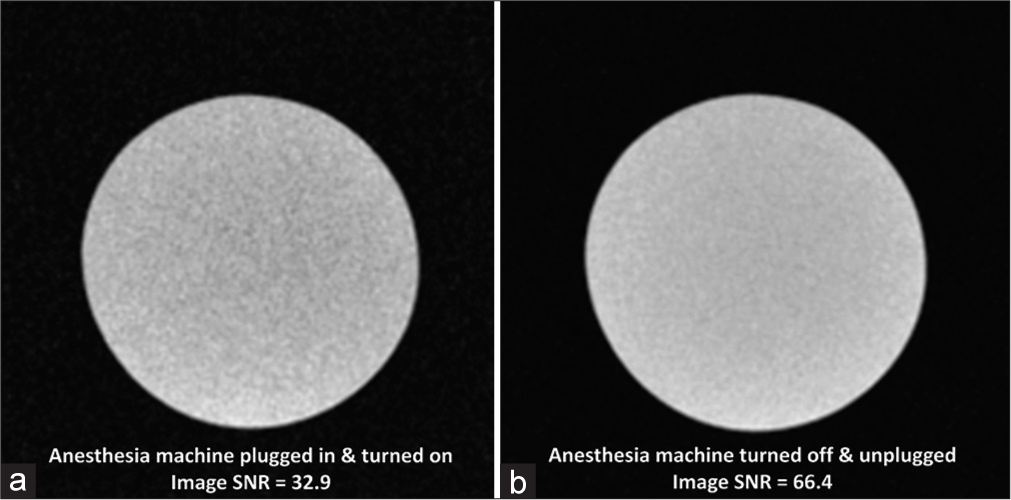
- Phantom testing showed (a) image signal-to-noise ratio was lower when an anesthesia machine positioned near the magnet was plugged into a wall outlet inside the magnetic resonance scanner room and turned on compared to (b) image signal-to-noise ratio after the anesthesia machine was turned off and unplugged.
UNRELATED MRI ARTIFACTS THAT RESEMBLE RF INTERFERENCE ARTIFACT
There are MRI artifacts that may resemble RF interference artifact but are actually not related to it, and there are traits that can separate them from RF interference. It is important for MRI personnel to have a good basic knowledge of these artifacts to be able to promptly distinguish them from RF interference artifact to effectively handle the artifact problem.
Spike artifact
Spike artifact, also called Herringbone artifact, crisscross artifact, or corduroy artifact, originates from electrical charge interference and appears as parallel bright and dark straight lines across the entire image [Figure 8].[1,3,4,7] It can be causedby a discharge of static electricity in the receiver, which may be a result of low humidity, or there may be defective or loose components in the RF circuitry. Besides, magnetic field gradients [Table 1] may also produce spikes, especially when applied at a very high-duty cycle. Unlike the RF artifact, the lines of the artifact can be in an orthogonal direction or at an oblique angle, with the orientation and separation of the lines depending on the position of the spike in the k-space data. Another difference from RF interference is that spike artifact is usually sporadic and limits to one or two images in a series while RF interference artifact tends to appear in all images within a series.[7]
- Head magnetic resonance image showing spike artifact (labeled by yellow arrows) across the whole image.
Motion artifact
Body tissue motion or fluid flow during an MR scan can produce image artifacts that manifest along the phase-encoding direction and extend over the entire image [Figure 9].[1-9,12] Motion artifacts may stem from blood vessel pulsations, respiratory/cardiac motion, swallowing, and physical movement of the patient. Random motion such as patient movement tends to produce artifact that has a smear appearance while periodic motion, such as cardiac/vascular pulsation or respiratory motion, typically produces artifacts that are more discrete. Motion artifact may be identified and distinguished from RF interference artifact by observing that the artifact is in line with the moving tissues or flowing vessels in the phase-encoding direction [Figure 9].
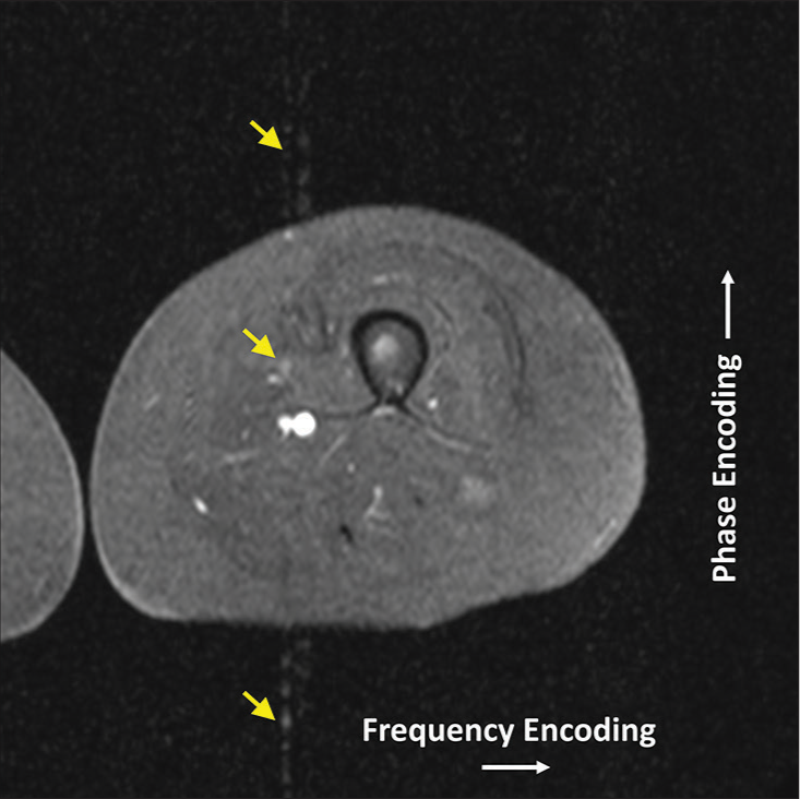
- Leg magnetic resonance image with pulsation blood flow artifact (labeled by yellow arrows). Note the artifacts are in line with the blood vessels in the phase encoding direction.
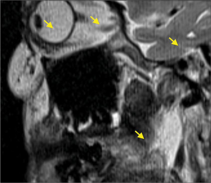
Free induction decay artifact
Free induction decay (FID) [Table 1] artifact appears as an intensity ripple, as seen in the subcutaneous fat at the top of the head in this 3D fast spin-echo (FSE) [Table 1] image [Figure 10]. This artifact is caused by the formation of FID signal from non-180° refocusing RF pulses [Table 1] that are typically used in 3D FSE sequences.[1,13] The FID signal creates an image containing only the edges of tissues with short longitudinal-relaxation time (T1) [Table 1] (such as fatty tissues), and this image super-imposes on the original image leading to the FID artifact. Unlike the RF interference artifact, spike artifact, and motion artifact, FID artifact is restricted to the affected tissues and does not extend into the surrounding image background.
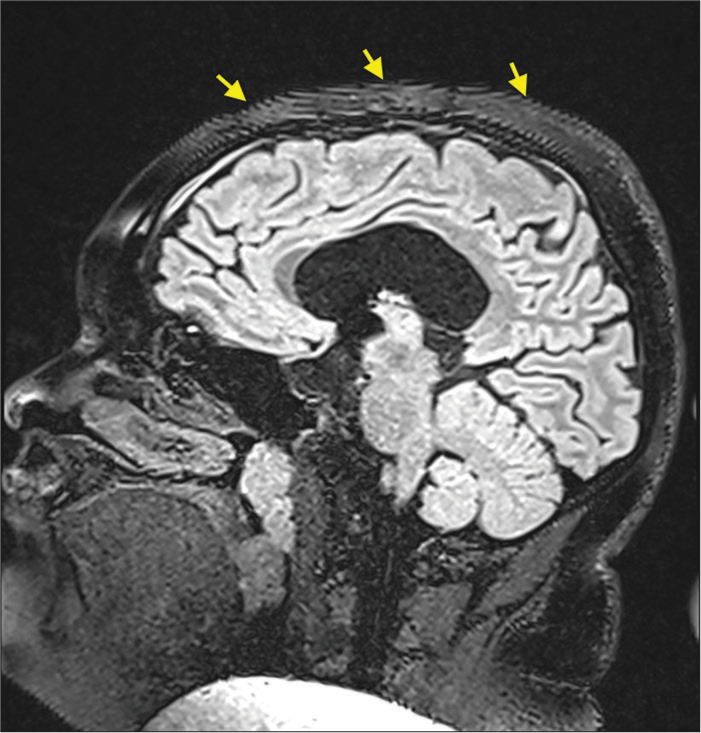
- Free induction decay artifact (labeled by yellow arrows) in the subcutaneous fat on top of the head fast-spin-echo magnetic resonance image.
IDENTIFICATION AND REMOVAL OF RF INTERFERENCE ARTIFACTS
To eliminate RF interference artifact, the RF field source must be correctly identified and removed. First, the artifact should be confirmed to be indeed caused by RF interference and not one that mimics RF interference artifact. Once that is done, we can look for the interference source. The first thing to check is whether the magnet room door is tightly closed as opened door is a common cause of RF leakage that is simple to fix. Another quick visual check is to see whether there is any electrically conductive cable or wire passing through the waveguide to outside the magnet room. If so, it should be removed or pulled back completely into the MR scanner room.
Next search for the interference source would be to check whether there is any equipment plugged into the power outlet and/or turned on. If so, turn off and unplug the equipment to see if the artifact problem is resolved, keeping in mind that turning off the equipment alone may not be sufficient to remove RF interference.
A common practice to deal with RF interference artifact is to contact the MR system manufacturer or RF shield company right away, but this causes delay in patient scanning and disruption in system usage. Besides, RF shielding test conducted by a shield company is generally expensive. With the exception of complicated cases such as RF shield damage, it is desirable to have the RF interference problem identified and rectified by the MRI facility personnel. This can alleviate the above issues and may also avoid unnecessary changes in the imaging procedure due to misidentification of the image artifact origin, e.g., switching from the extremity coil to the scanner body coil as mentioned in the power injector example that may lead to sub-optimal acquisition setting and inferior image quality.
Besides effective handling of RF interference problems when they occur, it is also important to periodically check and maintain the RF shielding to reduce the chance the problems occur. The American College of Radiology (ACR) weekly MRI test includes visual checking of the RF shielding integrity in the door contact and window screen.[14] Moreover, the door RF shield contact should be cleaned regularly, e.g. using denatured alcohol, to reduce rust and dust built-up and ensure good electrical contact. In addition, ACR weekly test includes checking the phantom images for ghosting that may reveal potential RF interference artifact. Uniform phantom testing is generally more sensitive in detecting RF interference artifact than in vivo imaging as the artifact in the latter may be obscured by biological structures and other artifacts such as blood pulsation artifact.
Recently, there have been researches to eliminate electromagnetic inference in MRI.[15-18] They generally utilize electromagnetic interference sensors to predict and cancel the RF interference detected by the MRI system. These developments are mainly targeted toward point-of-care very low-field MRI systems without effective RF shielding, though a newly published study includes the implementation on a 1.5 T system with incomplete RF shielding.[18] Since these developments are not available on common clinical MRI systems, they are beyond the scope of this article.
CONCLUSION
While there have been publications on MR image artifacts, this article aims to provide more focused and in-depth review of RF interference and its associated image artifacts in MRI. The information should facilitate prompt and correct identification of RF interference artifact and enable efficient handling of RF interference problems, avoiding unnecessary cancellation or delay in patient scanning and minimizing the associated adverse effect to image quality and interpretation. This article should serve as a useful educational reference to MR technologists and radiologists in dealing with MR image artifacts that may be caused by RF interference.
Ethical approval
The Institutional Review Board approval is not required.
Declaration of patient consent
Patient’s consent is not required as patients identity is not disclosed or compromised.
Conflicts of interest
There are no conflicts of interest.
Use of artificial intelligence (AI)-assisted technology for manuscript preparation
The author confirms that there was no use of artificial intelligence (AI)-assisted technology for assisting in the writing or editing of the manuscript and no images were manipulated using AI.
Financial support and sponsorship
Nil.
References
- Primer on commonly occurring MRI artifacts and how to overcome them. Radiographics. 2022;42:E102-3.
- [CrossRef] [PubMed] [Google Scholar]
- An image-based approach to understanding the physics of MR artifacts. Radiographics. 2011;31:849-66.
- [CrossRef] [PubMed] [Google Scholar]
- AAPM/RSNA physics tutorial for residents: MR artifacts, safety, and quality control. Radiographics. 2006;26:275-97.
- [CrossRef] [PubMed] [Google Scholar]
- From A as in aliasing to Z as in zipper: Artifacts in MRI. Clin Neuroradiol. 2008;18:25-36.
- [CrossRef] [Google Scholar]
- Application of basic physics principles to clinical neuroradiology: Differentiating artifacts from true pathology on MRI. AJR Am J Roentgenol. 2013;201:369-77.
- [CrossRef] [PubMed] [Google Scholar]
- Body MR imaging: Artifacts, k-space, and solutions. Radiographics. 2015;35:1439-60.
- [CrossRef] [Google Scholar]
- Optimizing cardiac MR imaging: Practical remedies for artifacts. Radiographics. 2008;28:1161-87.
- [CrossRef] [PubMed] [Google Scholar]
- Artifacts in musculoskeletal magnetic resonance imaging: Identification and correction. Skeletal Radiol. 2001;30:179-91.
- [CrossRef] [PubMed] [Google Scholar]
- The basics of MRI. 1996. Available from: http://www.cis.rit.edu/htbooks/mri/index.html [Last accessed on 2024 Jun 23]
- [Google Scholar]
- 2021. IEEE Letters on Electromagnetic Compatibility Practice and Applications. 3:43-6.
- [CrossRef] [Google Scholar]
- Basic principles of and practical guide to clinical MRI radiofrequency coils. Radiographics. 2022;42:898-918.
- [CrossRef] [PubMed] [Google Scholar]
- Optimized three-dimensional fast-spin-echo MRI. J Magn Reson Imaging. 2014;39:745-67.
- [CrossRef] [PubMed] [Google Scholar]
- Magnetic resonance imaging quality control manual Reston, VA: American College of Radiology; 2015. p. :51.
- [Google Scholar]
- A single-coil-based method for electromagnetic interference reduction in point-of-care low field MRI systems. J Magn Reson. 2023;346:107355.
- [CrossRef] [PubMed] [Google Scholar]
- External dynamic interference estimation and removal (EDITER) for low field MRI. Magn Reson Med. 2022;87:614-28.
- [CrossRef] [PubMed] [Google Scholar]
- Active EMI suppression system for a 50 mT unshielded portable MRI scanner. IEEE Trans Biomed Eng. 2022;69:3415-26.
- [CrossRef] [PubMed] [Google Scholar]
- Robust EMI elimination for RF shielding-free MRI through deep learning direct MR signal prediction. Magn Reson Med. 2024;92:112-27.
- [CrossRef] [PubMed] [Google Scholar]


