Translate this page into:
Primary Follicular Lymphoma of the Common Bile Duct Mimicking Cholangiocarcinoma
Address for correspondence: Dr. Khaled Youssef Elbanna, Department of Medical Imaging (Mail Code 1222), King Abdul-Aziz Medical City (KAMC), National Guard Health Affairs, National Guard Hospital, Riyadh - 11426, Saudi Arabia. E-mail: khaledbanna77@gmail.com
-
Received: ,
Accepted: ,
This is an open-access article distributed under the terms of the Creative Commons Attribution License, which permits unrestricted use, distribution, and reproduction in any medium, provided the original author and source are credited.
This article was originally published by Medknow Publications & Media Pvt Ltd and was migrated to Scientific Scholar after the change of Publisher.
Abstract
Primary non-Hodgkin's lymphoma of the common bile duct is extremely rare. We present a case with history of inflammatory bowel disease and clinical manifestations of obstructive jaundice. Abdominal magnetic resonance imaging with magnetic resonance cholangiopancreatography (MRCP) was done and demonstrated tight stricture at the middle part of common bile duct, and radiological findings were supportive of extra-hepatic cholangiocarcinoma. Whipple's procedure was performed and the case was histopathologically proven to be non-Hodgkin's lymphoma of follicular subtype involving the common bile duct. Lymphoma of the hepatobiliary system is usually present as secondary manifestation of systemic malignant lymphoma. However, primary malignant lymphomas arising from the hepatobiliary tree are extremely rare. The radiological appearance of common bile duct lymphoma is very similar to cholangiocarcinoma, making preoperative diagnosis very difficult, as in our present case. We also compare the imaging findings of our case to those seen in reported cases of follicular lymphoma of the common bile duct.
Keywords
Cholangiocarcinoma
common bile duct
lymphoma
magnetic resonance cholangiopancreatography
INTRODUCTION

The gastrointestinal tract is the most common site of extranodal non-Hodgkin's lymphoma (NHL) and the small bowel is considered the most commonly affected part of the gastrointestinal tract.[1] Lymphoma of the hepatobiliary system usually presents as secondary manifestation of systemic malignant lymphoma. However, primary malignant lymphomas arising from the hepatobiliary tree are extremely rare.[2]
CASE REPORT
A 71-year-old male patient with history of inflammatory bowel disease presented with progressive jaundice. On physical examination, no superficial lymphadenopathy was detected and his abdominal examination was normal.
Laboratory evaluation confirmed cholestasis with the following findings: Total bilirubin 86.6 μmol/l (normal: <26 μmol/l), direct bilirubin 66 μmol/l (normal <7 μmol/l), aspartate aminotransferase (AST) 95 U/l (normal: 0–35 U/l), alanine aminotransferase (ALT) 77 U/l (normal: 3–36 U/l), and alkaline phosphatase 438 U/l (normal: 35–100 U/l). In tumor marker studies, the cancer antigen 19-9 (CA 19-9) was elevated with a value of 215 U/ml (normal: <37 U/ml). Serologic markers of hepatitis B and C were negative. Magnetic resonance cholangiopancreatography (MRCP) was requested to rule out primary sclerosing cholangitis.
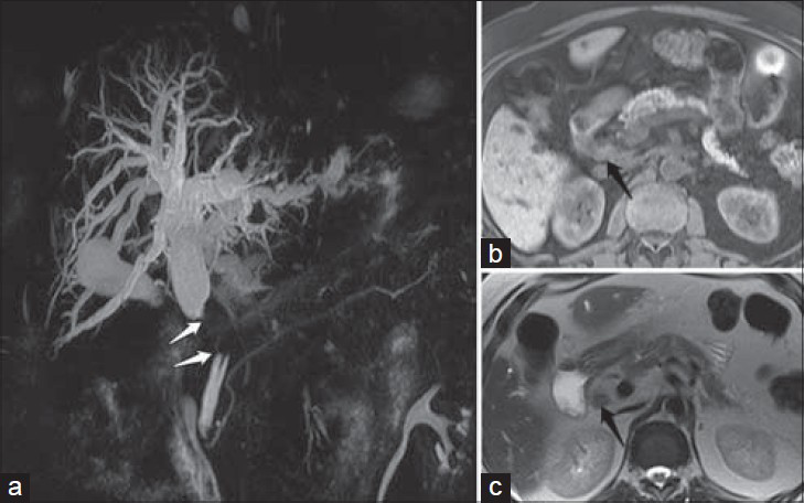
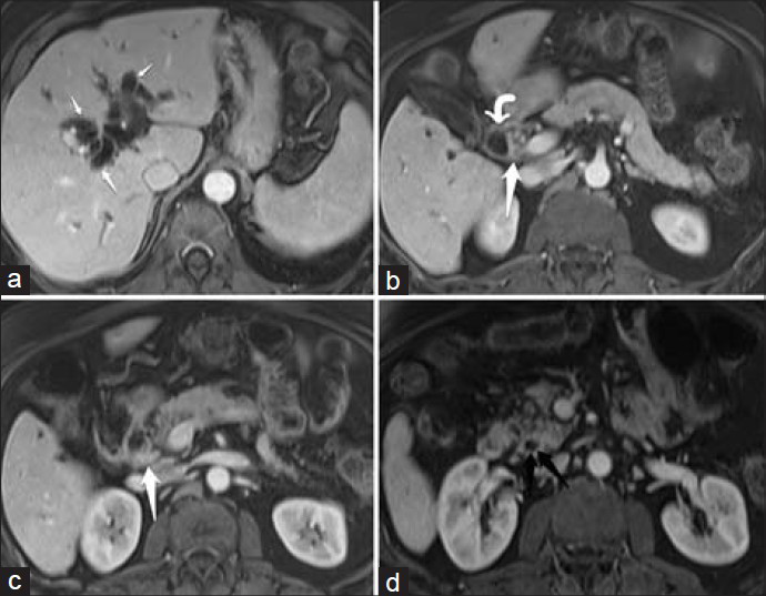
MRCP and contrast-enhanced magnetic resonance imaging (MRI) showed dilatation of the intrahepatic and extrahepatic bile ducts down to tight stenosis at the middle part of the common bile duct, measuring 2.3 cm and located almost 3.6 cm from the bifurcation of the common hepatic duct. At the area of stricture, there was circumferential mural thickening with intermediate signal intensity in T1- and T2-weighted images and homogeneous post-contrast enhancement [Figures 1 and 2]. Radiologic features favored the diagnosis of extrahepatic cholangiocarcinoma.

- 71-year-old male with obstructive jaundice secondary to CBD lymphoma. (a) Magnetic resonance cholangiopancreatography (MRCP) coronal projection demonstrate 2.3 cm tight stricture at the mid-CBD (white arrows) with severe upstream dilatation. (b and c) Non-contrast MRI upper abdomen axial T1- and T2-weighted images show circumferential mural thickening at the area of maximum stenosis with intermediate signal intensity (black arrows).

- 71-year-old male with obstructive jaundice secondary to CBD lymphoma. Contrast-enhanced MRI with axial post-contrast T1-weighted images at different levels: (a) at the level of the hepatic duct bifurcation, shows severe bilobar intrahepatic biliary dilatation (short white arrows); (b) at the level of the junction of the markedly dilated proximal CBD (curved white arrow) and the stenotic segment (straight white arrow); (c) at the level of mid-CBD stricture show that the lesion enhances homogeneously (white arrow); (d) at the level of distal CBD shows the normal caliber of the CBD within the pancreatic head (black arrow).
No further diagnostic studies were arranged as the radiological, clinical, and laboratory features were indicative of cholangiocarcinoma. The surgical plan was to perform Whipple's operation and the head of the pancreas, duodenum, common bile duct (CBD), and gallbladder were resected.
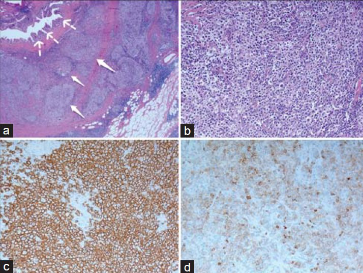
Gross examination showed a firm mass measuring 2.2 × 2 × 1.4 cm surrounding the duct and the cut surface showed grayish-white, lobulated tissue markedly compressing the distal CBD and showed a slit-like opening. Histopathologically, the mass was found to consist of lymphoid cell nodules surrounding and constricting the CBD. These nodules were composed of small cleaved lymphocytes (centrocytes) and larger lymphoid cells (centroblasts). The centroblasts counted more than 16 cells per high-power field [Figure 3a and b].

- Histological sections stained with hematoxylin and eosin (H and E) (a) low-power view, (×25) shows nodules of lymphoid cells (white arrows) surrounding the common bile duct (dotted white arrows). (b) medium-power view, (×200) shows the nodules with a mix of neoplastic centrocytes and centroblasts. Immunochemical tests (c) shows the neoplastic lymphoid cells are positive for CD20 marker and (d) the cells are positive for CD10 marker.
Pancreatic parenchyma and duodenum were free from tumor. Nine lymph nodes were identified and found negative for malignancy. Gallbladder showed chronic cholecystitis.
Immunohistochemical staining showed the neoplastic cells were positive for cluster of differentiation 20 (CD20), cluster of differentiation 10 (CD10), and B cell lymphoma 6 (BCL6) markers [Figure 3c and d]. Few cells were positive for cluster of differentiation 30 (CD30). They were negative for CD3, CD5, B cell lymphoma 2 (BCL2), and multiple myeloma oncogene 1 (MUM1). Ki-67 (named after the location where it was discovered; Kiel University) was expressed by 70% of cells within the follicles and 10% outside the follicles. CD23 highlighted follicular dendritic cell meshwork in the background. The diagnosis of follicular lymphoma, grade 3A was reached based on these findings.
DISCUSSION
Reviewing the English language medical literature on primary NHL originating from the bile duct, only 30 cases were found and most of them presented with jaundice and the most common subtype among these cases was diffuse large B-cell lymphoma (DLBCL).[2] There is increasing incidence of NHL, which may be primarily due to a variety of factors, particularly the rising incidence of human immunodeficiency virus (HIV) infection.[34] Follicular lymphoma of the gastrointestinal tract is extremely rare, accounting for only 1% of all gastrointestinal NHLs.[5]
Among those cases reviewed by Lee et al.,[2] three cases were follicular lymphoma, of which two were males aged 33 and 32 years and one was a female aged 53 years, with all of them having the lymphomatous infiltration affecting the bifurcation of the CBD with gross soft tissue masses [Table 1].[167] While in our case, the lymphoma involved the middle part of the of the CBD without sizable soft tissue mass and was mainly seen as stenotic area.

Follicular lymphoma is considered to be an indolent lymphoma, but the clinical behavior of this lymphoma depends on the histologic grade and the extent of disease at presentation. With time, approximately a third of follicular lymphomas transform into more aggressive lymphomas, particularly DLBCL.[8]
Accurate pre-surgical diagnosis may alter the treatment choice, and chemotherapy should be the first choice for primary biliary lymphoma. Surgery should play a role if an accurate preoperative diagnosis cannot be made, and the involved lesions produce complications, such as bile duct stricture, that are not amenable to non-surgical therapies. Also, failure of chemotherapy to eradicate localized disease is another situation where surgery may have a role.[9]
On the other hand, radiotherapy should be reserved for residual disease after primary chemotherapy, or to relieve symptoms of pain caused by the disease.[10]
Correct pre-surgical diagnosis is quite difficult, but smooth surface of the stricture segment of the biliary tree seen by endoscopic retrograde cholangiopancreatography (ERCP) or percutaneous transhepatic cholangiography (PTC) may indicate the extramural nature of the mass, and thus be suggestive of lymphoma.[69] Homogenous intensity in both pre- and post-contrast study at the stenotic site of bile duct is another suggestive feature of lymphoma.[9]
CONCLUSION
Primary NHL is one of the rare differential diagnoses of cholangiocarcinoma and shows similar imaging findings.
The clinical symptoms and signs, and the laboratory study are non-specific and sometimes misleading. Pre-surgical diagnosis is quite challenging due to non-specific clinical manifestations, laboratory profile, and imaging findings. If an accurate diagnosis is made before surgical intervention, chemotherapy is the first choice of treatment. Surgical resection should be reserved for those cases without a definite diagnosis, or for patients with complicating biliary obstruction, or who fail to respond to chemotherapy.
Available FREE in open access from: http://www.clinicalimagingscience.org/text.asp?2014/4/1/72/148267
Source of Support: Nil
Conflict of Interest: None declared.
REFERENCES
- Primary follicular lymphoma of the extrahepatic bile duct mimicking a hilar cholangiocarcinoma: Case report and review of the literature. Hum Pathol. 2009;40:1808-12.
- [Google Scholar]
- Primary non-Hodgkin's lymphoma originating from the common bile duct: A case report and literature review. J Gastroenterol. 2003;38:51-8.
- [Google Scholar]
- Changing incidence of non-Hodgkin lymphomas in the United States. Cancer. 2002;94:2015-23.
- [Google Scholar]
- Increasing incidence and descriptive epidemiology of extranodal non-Hodgkin lymphoma in parts of England and Wales. Hematol J. 2002;3:95-104.
- [Google Scholar]
- Low-grade follicular lymphoma of the small intestine. J Clin Gastroenterol. 2002;34:155-9.
- [Google Scholar]
- Follicular lymphoma of the extrahepatic bile duct mimicking cholangiocarcinoma. J Hepatobiliary Pancreat Surg. 2008;15:196-9.
- [Google Scholar]
- Primary non-Hodgkin's lymphoma of the extrahepatic bile duct. J Gastrointest Cancer 2011 [Epub ahead of print]
- [Google Scholar]
- Follicular lymphomas: Assessment of prognostic factors in 127 patients followed for 10 years. Ann Oncol. 1991;2(Suppl 2):123-9.
- [Google Scholar]
- CT and MRI findings of primary non-Hodgkin's lymphoma of the common bile duct mimicking cholangiocarcinoma. AJR Am J Roentgenol. 2004;182:1608-9.
- [Google Scholar]
- Primary non-Hodgkin's lymphoma of the common bile duct presenting as obstructive Jaundice. J Gastroenterol. 2004;39:692-6.
- [Google Scholar]


