More results...

Generic selectors
Exact matches only
Search in title
Search in content
Post Type Selectors
Filter by Categories
Abdominal Radiology, Original Research
Abdominal Radiology, Pictorial Essay
Anthropology and Dental Radiology, Original Research
Breast Imaging, Case Report
Breast Imaging, Original Research
Breast Imaging, Pictorial Essay
Breast Imaging, Review Article
Cardiopulmonary Imaging, Case Report
Cardiopulmonary Imaging, Case Series
Cardiopulmonary Imaging, Education
Cardiopulmonary Imaging, Original Research
Cardiopulmonary Imaging, Pictorial Essay
Cardiopulmonary Imaging, Review Article
Case Report
Case Report, Breast Imaging
Case Report, Cardiopulmonary Imaging
Case Report, Diagnostic Radiology
Case Report, Neuroradiology/Head and Neck Imaging
Case Report, Vascular and Interventional Radiology
Case Series
Case Series, Abdominal Radiology
Case Series, Diagnostic Radiology
Case Series, Vascular and Interventional Radiology
DENTAL ARTICLE
Dental Radiology, Case Report
Dental Radiology, Case Series
Dental Radiology, Original Research
Diagnostic Radiology, Case Report
Diagnostic Radiology, Case Series
Diagnostic Radiology, Original Research
Diagnostic Radiology, Pictorial Essay
Diagnostic Radiology, Review Article
Editorial
Education, Diagnostic Radiology
Education, Original Research
Emergency, Original Research
Erratum
Gastrointestinal Imaging, Case Report
Gastrointestinal Imaging, Case Series
Gastrointestinal Imaging, Original Research
Gastrointestinal Imaging, Pictorial Essay
General and Emergency Radiology, Original Research
General and Emergency Radiology, Review Article
Genitourinary and Gynecologic Imaging, Case Report
Genitourinary and Gynecologic Imaging, Original Research
Imaging Science, Original Research
Interventional Radiology, Original Research
Letter to Editor
Musculoskeletal Imaging, Case Report
Musculoskeletal Imaging, Original Article
Musculoskeletal Imaging, Original Research
Musculoskeletal Imaging, Pictorial Essay
Musculoskeletal Imaging, Review Article
Neuroradiology , Review Article
Neuroradiology Head and Neck Imaging, Pictorial Essay
Neuroradiology, Case Report
Neuroradiology, Pictorial Essay
Neuroradiology/Head and Neck Imaging, Case Report
Neuroradiology/Head and Neck Imaging, Case Series
Neuroradiology/Head and Neck Imaging, Original Research
Neuroradiology/Head and Neck Imaging, Review Article
Neuroradiology/Spine Imaging, Original Research
Nuclear Medicine, Case Report
Nuclear Medicine, Original Research
Nuclear Medicine, Pictorial Essay
Original Article
Original Article, Neuroradiology
Original Research
Original Research Article
Original Research, Dental Radiology
Original Research, Descriptive Study
Original Research, Diagnostic Radiology
Original Research, Gastrointestinal Imaging
Original Research, Genitourinary and Gynecologic Imaging
Original Research, Musculoskeletal Imaging
Original Research, Neuroradiology/Head and Neck Imaging
Original Research, Nuclear Medicine
Original Research, Vascular and Interventional Radiology
Pediatric Imaging, Case Report
Pediatric Imaging, Pictorial Essay
PICTORIAL ESSAY
PICTORIAL REVIEW
Radiologic-Pathologic Correlation
RADIOLOGICAL-PATHOLOGICAL CORRELATION
Radiology Business, Original Research
Research Article
Review Article
Review Article, Diagnostic Radiology
Review Article, Gastrointestinal imaging
Review Article, Musculoskeletal
Review Article, Vascular and Interventional Radiology
Technical Innovation
Technical Innovation, Gastrointestinal Imaging
Ultrasound, Case Report
Ultrasound, Original Research
Ultrasound, Review Article
Vascular and International Radiology, Case Report
Vascular and Interventional Radiology, Case Report
Vascular and Interventional Radiology, Case Series
Vascular and Interventional Radiology, Original Research
Vascular and Interventional Radiology, Pictorial Essay
Vascular and Interventional Radiology, Review Article
Vascular and Interventional Radiology, Short Communication
Vascular and Interventional, Original Research
View/Download PDF

Translate this page into:

RADIOLOGICAL-PATHOLOGICAL CORRELATION
2012
:2;
35
doi:
10.4103/2156-7514.97757

Primary Endobronchial Leiomyosarcoma of the Lung: Clinical, Gross and Microscopic Findings of Two Cases

Department of Pathology, Ibn Sina Hospital, Rabat, Morocco
Address for correspondence: Dr. Hafsa Elouazzani, Department of Pathology, Ibn Sina Hospital, rabat, Morocco. E-mail: hafoussa02@hotmail.fr
Licence

This is an open-access article distributed under the terms of the Creative Commons Attribution License, which permits unrestricted use, distribution, and reproduction in any medium, provided the original author and source are credited.

Disclaimer:
This article was originally published by Medknow Publications & Media Pvt Ltd and was migrated to Scientific Scholar after the change of Publisher.

Abstract

Primary leiomyosarcoma of the lung is an unusual malignant tumor. Among this entity, the endobronchial form is very rare and the preoperative diagnosis is extremely difficult. We present two different presentations and outcomes of primary endobronchial leiomyosarcoma of the lung. In both cases, the histological study and the immunohistochemical stain, of the surgical resection, provided the final diagnosis. Through those cases we present the diagnostic and therapeutic difficulties encountered.

Keywords

Endobronchial
leiomyosarcoma
pathology

INTRODUCTION

Primary lung leiomyosarcoma is a rare malignant tumor, arising from the smooth muscle of the bronchi, bronchioles or bronchial arterioles occurs in adults. It accounts for less than 0.5% of all primary malignant lung tumors. The endobronchial form is exceptional in that only 14 patients older than 20 years have been reported. The preoperative diagnosis is extremely difficult. We report two cases of primary endobronchial leiomyosarcoma diagnosed after thorachotomy through the histological and the immunohistochemical investigations. The first case is about a 45-year-old non-smoker woman who was admitted with cough, hemoptysis, and right paravertebral pain. There was no history of asbestosis, traffic exposure, or family history of lung cancer. The second case is about a 30-year-old non-smoker man who suffered from a productive cough, a massive hemoptysis, and painful sensations on the right side of the chest.

Radiological features

Case 1

Chest radiography showed a round opacity on the upper zone of the right lung [Figure 1a]. Computed tomography (CT) scan of thorax revealed a round tissue lesion in the dorsal segment of the right upper lobe (6 cm in diameter)obstructing the right lobar bronchus [Figure 1b]. Fiberoptic bronchoscopy showed a tumor partially obstructing the orifice of the right lobar bronchus. Bronchoscopic biopsy was not done to avoid further tumor bleeding.

Figure 1
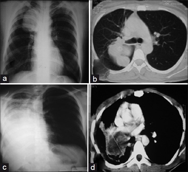
Case 1, a 45-year-old woman: (a) Anterior chest radiograph shows a round opacity on the upper zone of right lung. (b) Computed tomography of thorax reveals a round tissular lesion of dorsal segment of right upper lobe, obstructing the right lobar bronchus.Case 2, a 30-year-old man: (c) Anterior chest radiograph shows an almost total opacification of the right lung with a mediastinal shift to the same side. (d) Computed tomography of thorax reveals an endobronchial tumor in the right main bronchus, causing complete atelectasis in the right middle lobe.

Case 2

Chest radiography showed almost total opacification of the right lung, with a mediastinal shift in the same side [Figure 1c]. Computed tomography revealed an endobronchial tumor (7 cm of diameter) in the right main bronchus, causing complete atelectasis in the right middle lobe [Figure 1d]. Fiberoptic bronchoscopy showed a total obstruction in the orifice of the right main bronchus by a reddish tumor. Bronchoscopic biopsy was abandoned to avoid further tumor bleeding. Due to the persistent tumor bleeding, surgical intervention was arranged. Thoracothomy revealed an inextirpable and necrotic endobronchial tumor, arising from the right main bronchus. During intervention, a tumor fragment migrated into the endotracheal tube intubation. This fragment was submitted for pathological examination.

Pathological features

Case 1

Ultrasound-guided transthoracic biopsy found spindle-shaped cells without atypia or mitoses compatible with an endobronchial leiomyma of the lung. Uterine examination revealed normal findings. So a superior lobectomy was performed. In pathological examination, the surgical specimen showed a fleshy white lesion with necrosis, hemorrhage, and intraluminal polyp [Figure 2]. Microscopically, the tumor consisted of spindle cells with atypia and hyperchromatic nuclei. Occasional mitotic figures and foci of necrosis were also seen [Figure 3a]. The immunohistochemistry investigations showed a moderately differentiated tumor expressing smooth muscle actin [Figure 3c], h-caldesmone and vimentin with a negative reaction to cytokeratin and epithelial markers (AE1/AE3, KL1, CK7, CK20, CK5/6). The Ki-67 (Index of proliferation) was estimated at less than 5%. These pathological features allowed the diagnosis of primary endobronchial leiomyosarcoma Grade II of (French Fédération Nationale des Centres de Lutte Contre le Cancer) FNCLCC (intermediate grade). The postoperative course was uneventful for 2 years past treatment.

Figure 2
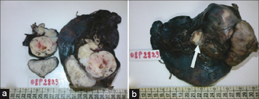
Case 1: (a) Grossly, the tumor shows a fleshy white lesion with necrosis, hemorrhage and (b) an intraluminal polyp.
Figure 3
Histopathology (Hematoxylin and Eosin, X40). Case1. (a) The tumor shows a fascicular proliferation of spindle cells with eosinophilic cytoplasm, mild to moderate nuclear atypia and brisk mitotic activity (10 mitoses per 10 hpf). Case 2. (b) The tumor shows a nuclear pleomorphism and atypia and high mitotic activity (17 mitoses per hpf). (c) Immuno-stain demonstrates positive smooth muscle actin in the tumor cells.

Case 2

The diagnosis of primary endobronchial leiomyosarcoma Grade III of FNCLCC (High-grade) [Figure 3b] was established through the histological study and immunohistochemistry investigations (immune-stains were positive for smooth muscle actin, h-caldesmon, vimentin and negative for cytokeratin AE1/AE3, KL1, CK7, CK20, CK5/6, or Epithelial Membrane Antigen (EMA) and for Cluster of Differenciation 45 CD45). In this case, the Ki-67 was estimated at 85%. Chemotherapy with Adriamycin was started. Three months later, recurrence of the atalactasis was observed. The patient died of acute respiratory distress and suffocation due to hemoptysis.

DISCUSSION

Primary leiomyosarcomas of the lung are a rare malignancy occurring in adults and represents less than 0.5% of all primary malignancy disease of the lung and 30% of primary sarcomas of the lung.[13] A few cases of endobronchial localization have been described. Since 1967, only 14 cases in patients older than 20 years have been reported.[4] In general, it occurs during middle age with a slight predominance of males. Due to the location of the endobronchial lesion, patients may present with symptom of pulmonary obstruction such as chest pain, cough, fever, wheezing, hemoptysis, dyspnea, and even expectoration of tumor fragments.[5] The radiological appearances include the findings of bronchial obstruction such as atelectasis, mediastinal shift and hyperlucency, or pneumothorax.[5]

A definitive preoperative diagnosis of endobronchial leiomyosarcoma is extremely difficult. In the literature reviewed, only five of the 14 (37%) cases had a definite preoperative diagnosis.[4] The morphologic features of these tumors recapitulate those seen in soft tissue, namely a fascicular proliferation of spindle cells with moderate amounts of eosinophilic cytoplasm, cigar-shaped nuclei, and inconspicuous nucleoli.[6] Pulmonary leiomyosarcoma can show a wide spectrum of differentiation. Low-grade tumors are characterized by a well-developed fascicular arrangement of tumor cells intersecting at right angles, with low mitotic rates (<3per 10 hpf) and absence of cellular atypia, necrosis and hemorrhage. Intermediate-grade tumors are characterized by increased cellularity with mild to moderate nuclear atypia and brisk mitotic activity (3-8 mitoses per 10 hpf). High-grade tumors show marked cellularity with nuclear pleomorphism and atypia, high mitotic activity (average 8-12 mitoses per 10 hpf), abundant necrosis, and hemorrhage.[6] In immunohistochemical studies, low-grade (well diff erentiated) tumors are more likely to show immunoreactivity for muscle markers, such as smooth muscleactin, h-caldesmone and desmin, while the less differentiated (high-grade tumors) can be negative for all muscle markers and require ultrastructural examination to confirm the diagnosis. It is important to note that some smooth muscle tumors may show a positive reaction for cytokeratin.

A panel of epithelial markers is therefore recommended when evaluating spindle-cell neoplasm of the lung to avoid misinterpretation as carcinosarcoma that contain a component of sarcomatoid differentiation.

Surgical resection of the tumor was a favorite modality of treatment for these tumors. However, to complete resection and prolong survival, sleeve lobectomy, pneumonectomy or carcinal resection may be necessary, depending on the anatomic location and size of the tumor.[7] Chemotherapy is suggested in case with metastases, but is very inefficient with a partial response and short duration. The protocols using adriamycin have shown a response rate of 16-27 %.[7]

The prognosis appears to correlate directly with tumor grade and degree of differentiation. The low-grade tumors generally follow a less aggressive course than those of intermediate or high–grade.[5] Other prognostic factors as the size and long-time stability of solitary lung nodules are not necessarily reliable criteria for assessing prognosis.[8]

In summary, primary endobronchial leiomyosarcoma is a very rare clinical and pathological entity. The surgical resection should be the favored method for definitive diagnosis and curative treatment.

Source of Support: Nil

Conflict of Interest: None declared.

REFERENCES

  1. , , , , . Primary sarcomas of the lung: A clinicopathologic study of 12 cases. Lung Cancer. 2002;38:283-9.
    [Google Scholar]
  2. , , , . Primary lung sarcomas: long survivors obtained with iterative complete surgery. Lung Cancer. 2001;31:241-5.
    [Google Scholar]
  3. , , , , . Le léomyosarcome primitif du poumon : à propos d’un cas. Revue des Maladies Respiratoires Janvier. 2007;24:82.
    [Google Scholar]
  4. , , , . Pulmonary leiomyosarcoma presenting as a pancoast tumor. Pathol Res Pract. 2007;203:745-8.
    [Google Scholar]
  5. , , , , . Primary endobronchial leiomyosarcoma: Diagnosis following expectoration of tumor fragment. Respiration. 2001;68:99-102.
    [Google Scholar]
  6. , , . Tumors of the lung and pleura.Diagnostic Histopathology of Tumors. Vol 1. (3rd ed). New York: Churchill Livingstone; . p. :193-4.
    [Google Scholar]
  7. , , , . Surgical treatment of endobronchial leiomyosarcoma with right main bronchus total obstruction: A case report. Ann Thorac Cardiovasc Surg. 2008;14:105-8.
    [Google Scholar]
  8. , , , , , . Long-term development of a primary lung sarcoma, probably lymphangiosarcoma-a case report. Thoracic Cardivasc Surg. 1984;32:178-81.
    [Google Scholar]
Show Sections