More results...

Generic selectors
Exact matches only
Search in title
Search in content
Post Type Selectors
Filter by Categories
Abdominal Radiology, Original Research
Abdominal Radiology, Pictorial Essay
Anthropology and Dental Radiology, Original Research
Breast Imaging, Case Report
Breast Imaging, Original Research
Breast Imaging, Pictorial Essay
Breast Imaging, Review Article
Cardiopulmonary Imaging, Case Report
Cardiopulmonary Imaging, Case Series
Cardiopulmonary Imaging, Education
Cardiopulmonary Imaging, Original Research
Cardiopulmonary Imaging, Pictorial Essay
Cardiopulmonary Imaging, Review Article
Case Report
Case Report, Breast Imaging
Case Report, Cardiopulmonary Imaging
Case Report, Diagnostic Radiology
Case Report, Gastrointestinal Imaging
Case Report, Genitourinary and Gynecologic Imaging
Case Report, Neuroradiology/Head and Neck Imaging
Case Report, Vascular and Interventional Radiology
Case Series
Case Series, Abdominal Radiology
Case Series, Diagnostic Radiology
Case Series, Musculoskeletal Imaging
Case Series, Vascular and Interventional Radiology
DENTAL ARTICLE
Dental Radiology, Case Report
Dental Radiology, Case Series
Dental Radiology, Original Research
Diagnostic Radiology, Case Report
Diagnostic Radiology, Case Series
Diagnostic Radiology, Original Research
Diagnostic Radiology, Pictorial Essay
Diagnostic Radiology, Review Article
Editorial
Education
Education, Diagnostic Radiology
Education, Imaging science
Education, Original Research
Emergency, Original Research
Erratum
Gastrointestinal Imaging, Case Report
Gastrointestinal Imaging, Case Series
Gastrointestinal Imaging, Original Research
Gastrointestinal Imaging, Pictorial Essay
General and Emergency Radiology, Original Research
General and Emergency Radiology, Review Article
Genitourinary and Gynecologic Imaging, Case Report
Genitourinary and Gynecologic Imaging, Original Research
Imaging Science, Original Research
Interventional Radiology, Original Research
Letter to Editor
Musculoskeletal Imaging, Case Report
Musculoskeletal Imaging, Original Article
Musculoskeletal Imaging, Original Research
Musculoskeletal Imaging, Pictorial Essay
Musculoskeletal Imaging, Review Article
Neuroradiology , Review Article
Neuroradiology Head and Neck Imaging, Pictorial Essay
Neuroradiology, Case Report
Neuroradiology, Pictorial Essay
Neuroradiology/Head and Neck Imaging, Case Report
Neuroradiology/Head and Neck Imaging, Case Series
Neuroradiology/Head and Neck Imaging, Original Research
Neuroradiology/Head and Neck Imaging, Review Article
Neuroradiology/Spine Imaging, Original Research
Notice of Retraction
Nuclear Medicine, Case Report
Nuclear Medicine, Original Research
Nuclear Medicine, Pictorial Essay
Original Article
Original Article, Neuroradiology
Original Research
Original Research Article
Original Research, Cardiopulmonary Imaging
Original Research, Dental Radiology
Original Research, Descriptive Study
Original Research, Diagnostic Radiology
Original Research, Gastrointestinal Imaging
Original Research, Genitourinary and Gynecologic Imaging
Original Research, Musculoskeletal Imaging
Original Research, Neuroradiology/Head and Neck Imaging
Original Research, Nuclear Medicine
Original Research, Vascular and Interventional Radiology
Pediatric Imaging, Case Report
Pediatric Imaging, Pictorial Essay
PICTORIAL ESSAY
Pictorial essay, Musculoskeletal Imaging
Pictorial essay, Neuroradiology/Head and Neck Imaging
PICTORIAL REVIEW
Radiologic-Pathologic Correlation
RADIOLOGICAL-PATHOLOGICAL CORRELATION
Radiology Business, Original Research
Research Article
Review Article
Review Article, Diagnostic Radiology
Review Article, Gastrointestinal imaging
Review Article, Musculoskeletal
Review Article, Musculoskeletal Imaging
Review Article, Vascular and Interventional Radiology
Technical Innovation
Technical Innovation, Cardiopulmonary Imaging
Technical Innovation, Gastrointestinal Imaging
Ultrasound, Case Report
Ultrasound, Original Research
Ultrasound, Review Article
Vascular and International Radiology, Case Report
Vascular and Interventional Radiology, Case Report
Vascular and Interventional Radiology, Case Series
Vascular and Interventional Radiology, Original Research
Vascular and Interventional Radiology, Pictorial Essay
Vascular and Interventional Radiology, Review Article
Vascular and Interventional Radiology, Short Communication
Vascular and Interventional, Original Research
View/Download PDF

Translate this page into:

CASE REPORT
2012
:2;
22
doi:
10.4103/2156-7514.95436

Prenatal Diagnosis of Amniotic Band Syndrome in the Third Trimester of Pregnancy using 3D Ultrasound

Department of Obstetrics, Federal University of Sāo Paulo (UNIFESP), Sāo Paulo, SP, Brazil

Address for correspondence: Prof. Edward Araujo Júnior, Department of Obstetrics, Federal University of Sāo Paulo (UNIFESP) Rua Carlos Weber, 956 apto. 113 Visage, Alto da Lapa, Sāo Paulo – SP, Brazil, CEP 05303-000. E-mail: araujojred@terra.com.br

Licence

This is an open-access article distributed under the terms of the Creative Commons Attribution License, which permits unrestricted use, distribution, and reproduction in any medium, provided the original author and source are credited.

Disclaimer:
This article was originally published by Medknow Publications & Media Pvt Ltd and was migrated to Scientific Scholar after the change of Publisher.

Abstract

Amniotic band syndrome is characterized by a build-up of bands and strings of fibrous tissue that adhere to the fetus and can compress parts of the fetus, thus causing malformations and even limb amputation while the fetus is still in the uterus. The clinical manifestations are extremely variable and their extent may range from a single abnormality, like a constriction ring, to multiple abnormalities. Such abnormalities are generally diagnosed at the end of the first or the beginning of the second trimester using two-dimensional ultrasonography (2DUS). Three-dimensional ultrasonography (3DUS) in rendering mode allows spatial analysis of the fetus and amniotic band, thus enabling better comprehension of this pathological condition and better counseling for the parents. There has not previously been any evidence to show that 3DUS would be useful in cases of late diagnosis (third trimester) of amniotic band syndrome. In the present case, a primigravid woman underwent her second obstetric ultrasound scan in the 34th week, from which we observed two bands in contact with the right forearm, but with normal movement of this limb and its fingers. 3DUS made it possible to see the spatial relationship of these bands to the fetal body, thereby confirming their adherence to the limb. After the birth, the prenatal diagnosis of amniotic band syndrome without limb constriction was confirmed. A surgical procedure was carried out on the third day after birth to excise the bands, and the newborn was then discharged in a good general condition.

Keywords

Amniotic band syndrome
prenatal diagnosis
three-dimensional ultrasonography

INTRODUCTION

Amniotic band syndrome has many synonyms such as, congenital constriction rings, amniotic bands, limb body wall complex, “adam complex” (amniotic deformity, adhesions, and mutilations) and amniotic band sequence.[1] This syndrome comprises a variety of anatomical abnormalities among newborns, and is associated with intrauterine struggling by the fetus in the uterus, thereby leading to deformations, malformations, or ruptures. Amniotic band syndrome occurs in around 1:1,200 to 1:1,500 live newborns,[2] but lack of correct diagnosis certainly underestimates the real occurrence rate.

The clinical manifestations of amniotic band syndrome are extremely variable, and their extent may range from a single abnormality, like a constriction ring, to multiple abnormalities. The syndrome occurs in both sexes equally, and there is no racial predisposition. Almost all cases are sporadic, even though some rare evidences of familial cases have been described.[3]

Limb constrictions occur most frequently, ranging in severity from deformity of the distal part of the limb to partial or total amputation. Craniofacial abnormalities are the most serious type, because of the importance of the organs involved. These are frequent and variable, including encephalosis and facial deformity of various degrees. Visceral abnormalities are a rare form of the syndrome, among which gastroschisis is the most frequent type.[4] Omphalocele, genital exstrophy, and ambiguous genitalia can occur.[5]

Generally the diagnosis is made by means of two-dimensional ultrasonography (2DUS) at the end of the first or the beginning of the second trimester of pregnancy.[6] Three-dimensional ultrasonography (3DUS) in rendering mode allows reconstruction of the surface of the fetus, thus, enabling spatial analysis. In cases of fetal malformation, 3DUS allows better understanding of the abnormalities and better family counseling. Only three case reports describing the use of 3DUS for diagnosing amniotic band syndrome exist in the literature. In two of them, 3DUS was used at the end of the first and the beginning of the second trimester,[78] and only one case was in the third trimester.[9] We present a case of amniotic band syndrome diagnosed in the 34th week, in which 3DUS enabled clear analysis of this malformation, as well as better understanding and better family counseling.

CASE REPORT

A 28-year-old primigravid woman underwent routine obstetric ultrasonography in the 34th week. Her prenatal care had begun late, and she had only undergone one ultrasound examination, in the 20th week, that had not shown any fetal abnormalities. All the prenatal laboratory tests were normal, and the patient said that she had not been making any personal use of medicines or using any illicit drugs, and that there were no cases of malformations or chronic diseases like arterial hypertension and diabetes mellitus among members of her family. 2DUS showed normal fetal growth, with a weight prediction of 2.242g, amniotic fluid index (AFI) of 140, and anterior placenta. There was an accessory lobe on the left lateral posterior wall, and a membrane (amniotic band) was present between this lobe and the main placental mass. Morphological analysis showed two bands adhering to the right forearm of the fetus, but with normal movements of this limb and its fingers [Figure 1]. No other fetal abnormalities were seen. 3DUS was performed using a multifrequency volumetric transducer (Accuvix V20; Samsung Medison, Seoul, Korea) and clearly showed the spatial relationship between the bands and the body of the fetus, thus proving that the bands had really adhered to the forearm, but without strangling it [Figure 2]. The 3D image allowed the parents to gain a better understanding of the condition and enabled better counseling toward a good prognosis for the fetus. The pregnant woman had her delivery scheduled as a caesarian section in the 38th week, and the postnatal phenotypic examination confirmed the diagnosis of amniotic band syndrome without injury to the forearm of the fetus. The newborn underwent surgery to remove the bands on the third day of life and was subsequently discharged in a good general condition.
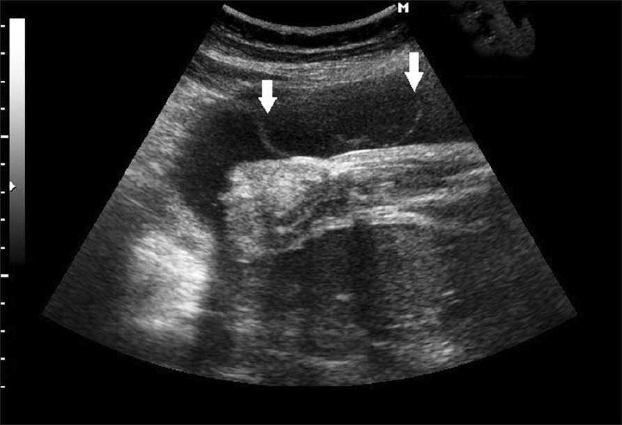
Two-dimensional ultrasonography shows adherence of two amniotic bands to the right forearm of the fetus (white arrows).
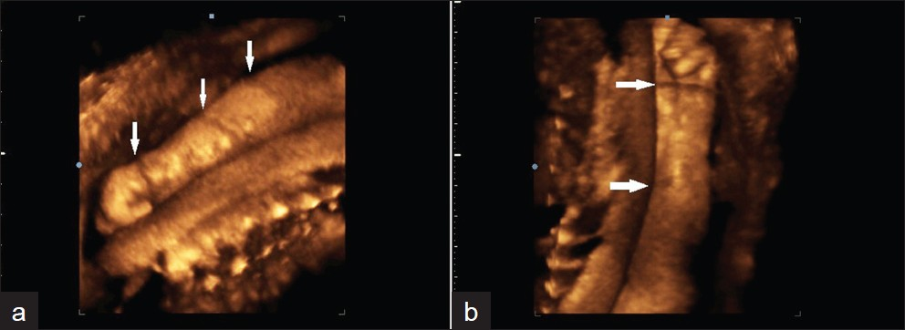
Figure 1
Two-dimensional ultrasonography shows adherence of two amniotic bands to the right forearm of the fetus (white arrows).
(a) and (b) Three-dimensional ultrasonography in rendering mode, shows amniotic bands adhering to the right forearm of the fetus (white arrows), without constriction rings.
Figure 2
(a) and (b) Three-dimensional ultrasonography in rendering mode, shows amniotic bands adhering to the right forearm of the fetus (white arrows), without constriction rings.

DISCUSSION

Two theories have been formulated in attempts to explain the pathogenesis of amniotic band syndrome. The Streeter theory,[1] which was accepted for a long time, takes the view that imperfect local embryonic development would indicate the abnormalities observed. The fibrotic bands would thus originate from imperfect histogenesis of the developing fetal tissue. However, this theory does not explain the wide diversity of abnormalities observed. The theory put forward by Torpin in 1965[10] is the one that is most accepted. This author took the view that the problem begins because of rupturing of the amniotic sac, thereby separating the chorion and amnion, such that some of the amniotic liquid goes out into the chorionic cavity. The fibrotic strings formed from the chorion would prevent normal development of some anatomical parts of the fetus, thus resulting in different kinds of abnormalities. According to Higginbottom et al.,[5] three mechanisms would explain the role of the bands in originating the malformations: a) interruption of the normal morphogenesis, for example, at the time of the facial fusion process; b) deformation due to compression of parts of the fetus (oligohidramnios) or interlacing of an anatomical part of the fetus by a band; or c) injury to a part of the fetus that had already formed, like in the cases with constriction rings around the limbs.

One of the possible causes of an amniotic lesion is maternal abdominal trauma; other, less likely causes might be drug use or ammonites. Some cases of amniotic band syndrome in patients that underwent amniocentesis on the beginning of the 2nd trimester[5] and chorionic villus sample have been described. Rare descriptions of intrafamilial repetitions of constriction rings or congenital limb amputation[3] give rise to the assumption that a genetic contribution could occur in some cases.

In our case, the patient did not have any antecedents, so we could assume that this case had a sporadic origin. The late diagnosis was a consequence of prenatal care that had begun late and of an ultrasound scan performed in the 20th week that did not show any fetal abnormalities. 3DUS enabled spatial analysis on the fetus proving that the bands had adhered to the surface of the right forearm of the fetus. Moreover, 3DUS provides proof that the prognosis for the fetus is good when it does not show any strangling of the limb, and it allows better understanding and better parent counseling. Inubashiri et al.[7] described a case of amniotic band syndrome diagnosed in the 14th week by 3DUS, which was associated with severe fetus malformations, and in which the parents decided to terminate the pregnancy. Likewise, Hata et al.[8] described two cases of this syndrome diagnosed in the 13th and 14th weeks, in which 3DUS made it possible to conduct analysis on the spatial relationship between the bands and the fetus, as well as enabling counseling for the parents. In both of these cases, the parents decided to terminate the pregnancy. The case of amniotic band syndrome with the latest diagnosis by 3DUS (and the only one in the third trimester until now) was described by Paladini et al.[9] These authors described a case in the 28th week, similar to ours, but in their case a constriction ring was observed on the forearm of the fetus and was confirmed after birth.

CONCLUSION

In summary, this is the second case description of late diagnosis of amniotic band syndrome by 3DUS. This method was shown to be important for proving that the bands had adhered to the limb of the fetus and showing that the prognosis is good when no constriction ring is demonstrated. In addition, 3DUS allowed better understanding of the pathology, better counseling for the parents, and a good prognosis for the newborn.

Source of Support: Nil

Conflict of Interest: None declared.

REFERENCES

  1. , , , , , . Amniotic band syndrome. Pediatria. 1990;17:26-9.
    [Google Scholar]
  2. , , . The amniotic band syndrome in monozygotic twins. Am J Obstet Gynecol. 1983;146:864-5.
    [Google Scholar]
  3. , , , , , . Familial amniotic bands. Am J Med Genet. 1983;14:81-7.
    [Google Scholar]
  4. , , , , , . Amniotic band malformations. Schweiz Rundsch Med Prax. 1985;74:358-62.
    [Google Scholar]
  5. , , , , . The amniotic band disruption complex - Timing of amniotic rupture and variable spectra of consequent defects. J Pediatr. 1979;95:544-9.
    [Google Scholar]
  6. , , , , . Typical body wall defect associated with craniofacial anomalies and amniotic bands diagnosed in early pregnancy. Taiwan J Obstet Gynecol. 2007;46:286-7.
    [Google Scholar]
  7. , , , , , , . 3D and 4D sonographic imaging of amniotic band syndrome in early pregnancy. J Clin Ultrasound. 2008;36:573-5.
    [Google Scholar]
  8. , , , . 3D/4D sonographic evaluation of amniotic band syndrome in early pregnancy: A supplement to 2D ultrasound. J Obstet Gynecol Res. 2011;37:656-60.
    [Google Scholar]
  9. , , , , . Congenital constriction band of the upper arm: The role of three-dimensional ultrasound in diagnosis, counseling and multidisciplinary consultation. Ultrasound Obstet Gynecol. 2004;23:520-2.
    [Google Scholar]
  10. , . Amniochorionic mesoblastic fibrous strings and amniotic bands: Associated constricting fetal malformations or fetal death. Am J Obstet Gynecol. 1965;91:65-75.
    [Google Scholar]

Fulltext Views
15,282

PDF downloads
3,396
View/Download PDF
Download Citations
BibTeX
RIS
Show Sections