
Translate this page into:
Percutaneous Coronary Intervention in Single Coronary Artery from Right Sinus: Radial Route is Right
Address for correspondence: Dr. Himanshu Mahla, Department of Cardiology, HRMC Institute of Cardiology, SP Medical College, Bikaner, Rajasthan, India. E-mail: himanshumahla@yahoo.in
-
Received: ,
Accepted: ,
This is an open access article distributed under the terms of the Creative Commons Attribution-NonCommercial-ShareAlike 3.0 License, which allows others to remix, tweak, and build upon the work non-commercially, as long as the author is credited and the new creations are licensed under the identical terms.
This article was originally published by Medknow Publications & Media Pvt Ltd and was migrated to Scientific Scholar after the change of Publisher.
Abstract
We present percutaneous coronary intervention (PCI) using radial approach in a rare case of single coronary artery originating from the right sinus. Although these anomalies and stenosis of anomalous vessels have been described previously, treatment of atherosclerotic lesions by PCI has rarely been reported. There is a definite procedural risk during PCI in patients with a single ostium because dissection with the guiding catheter would result in a catastrophic event. Additionally, technical difficulties may occur due to the ostial configuration and course of the branch to be stented. The patient suffered an acute coronary syndrome-inferior wall STEMI, and was thrombolysed elsewhere within a window period of 4 h. He had post myocardial infarction (MI) angina and was referred to our center after 3 days of thrombolysis. We present this technically challenging and rare case in which PCI of right coronary artery was performed through the radial route.
Keywords
Coronary artery disease
percutaneous coronary intervention
single coronary artery
STEMI


INTRODUCTION
Left main coronary artery arising from the right sinus of Valsalva as a single coronary ostium is an extremely rare anatomic anomaly occurring in approximately 0.06% of angiographic series.[12] Single coronary artery is encountered more frequently with other congenital cardiac malformations such as persistent truncus arteriosus, tetralogy of Fallot, transposition of the great arteries, or pulmonary atresia. Some single coronary ostium variants have been reported to carry a significant risk of severe cardiac events including myocardial infarction (MI) and sudden cardiac death, especially during exercise. We present a case of single coronary artery from right sinus with left main coronary artery originating from the same ostium, which underwent successful angioplasty with stenting to right coronary artery through the radial route.
CASE REPORT
A 60-year-old male was referred to our tertiary care institute for coronary angiography. Patient suffered acute coronary syndrome (ACS) - inferior wall MI 3 days prior to admission in our institute. He was thrombolysed with streptokinase at a peripheral center and referred to our institute on the 4th day post thrombolysis in view of post MI angina. Patient was a chronic smoker. Twelve-lead electrocardiogram revealed q wave with T inversion in the inferior leads. Transthoracic echocardiogram showed regional hypokinesia in the right coronary artery (RCA) territory with adequate left ventricular ejection fraction. Coronary angiography (CAG) by radial approach using Tiger 5F catheter (Terumo Corp., Kanagawa, Japan) was performed. CAG revealed blunt left sinus, with no artery originating from the left sinus [Figure 1 and Video 1]. Right coronary artery originated from the usual location and a long left main coronary artery arose from the same ostium [Figures 2, 3 and Video 2]. We exchanged the Tiger catheter with Judkin's right coronary catheter (Medtronic, Inc., Minneapolis, MN, USA) as it was not possible to selectively cannulate the ostium with the Tiger catheter. There was significant tubular stenosis in the mid-RCA region and non-critical plaque in the proximal left coronary artery (LCA) [Figures 2, 3 and Videos 2, 3]. As it is important to know the course of anomalous coronaries before any intervention, CT angiogram was done which revealed retroaortic course of the left coronary [Figures 4 and 5]. It was decided to do percutaneous coronary intervention (PCI) with stenting to the mid-RCA in view of post MI angina and was performed on the 5th day post thrombolysis. RCA was selectively cannulated with Judkin's right coronary catheter (Medtronic, Inc.). The lesion was crossed with 0.014″ × 190 cm BMW wire (Abbott Vascular, Santa Clara, CA, USA) and predilated with 2.5 × 12 mm Maverick balloon (Boston Scientific, Florida, USA) [Figure 6 and Video 4]. During predilatation, guide sucked in the vessel and pressure damping was noted due to obstruction of supply to the left coronary and this was managed by meticulous decannulation and avoiding deep intubation. A 3.25 × 33 mm Xience V stent (Abbott Vascular, Florida, USA) was deployed at 14 atm [Figure 7 and Video 5]. There were no complications [Figure 8 and Video 6]. Patient is doing well on follow-up.
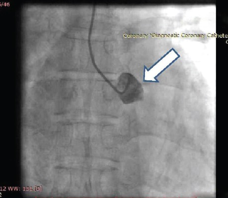
- 60-year-old male with ACS-inferior wall MI,
post thrombolysis and post MI angina. Coronary angiography-AP view fluoroscopy shows blunt left sinus (arrow)
with no coronary origin.

- 60-year-old male with
ACS-inferior wall MI, post thrombolysis and post MI angina. Coronary angiography-AP view fluoroscopy shows blunt
left sinus with no coronary origin.
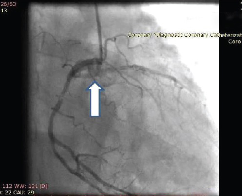
- 60-year-old male
with ACS-inferior wall MI, post thrombolysis and post MI angina. Coronary angiography-right anterior oblique
angiographic view shows both right and left coronaries originating from the right sinus (arrow).
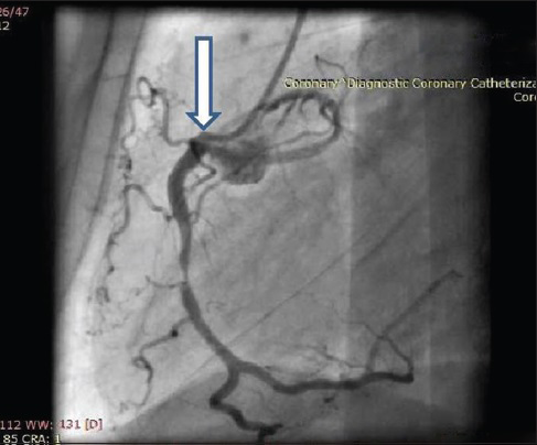
- 60-year-old male with ACS-inferior wall MI, post thrombolysis and post MI
angina. Coronary angiography-lateral angiographic view shows both RCA and LCA originating from same ostium
(arrow).

- 60-year-old male with ACS-inferior wall MI, post
thrombolysis and post MI angina. Coronary angiography-right anterior oblique angiographic view shows both right
and left coronaries originating from the right sinus.

- 60-year-old male with ACS-inferior wall MI, post thrombolysis and post MI angina.
Coronary angiography-lateral angiographic view shows both RCA and LCA originating from same ostium.
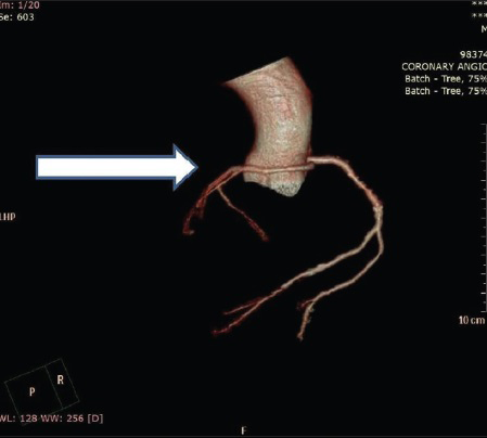
- 60-year-old male with ACS-inferior wall MI, post thrombolysis and
post MI angina. CT coronary angiography-CT image with 3D reconstruction shows retroaortic course of LCA
(arrow).
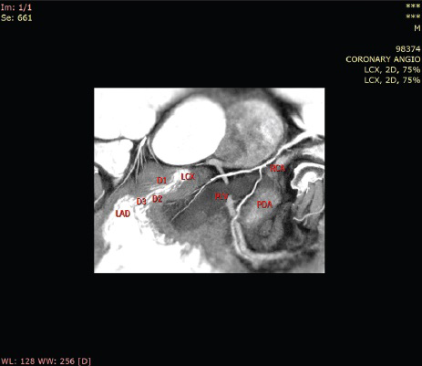
- 60-year-old male with ACS-inferior wall MI, post
thrombolysis and post MI angina. CT coronary angiography-CT axial image shows all three coronary arteries.
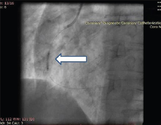
- 60-year-old male with ACS-inferior wall MI, post thrombolysis and
post MI angina. Coronary angiography shows left anterior oblique fluoroscopic view of RCA during predilatation
(arrow).

- 60-year-old male with ACS-inferior wall MI, post
thrombolysis and post MI angina. Coronary angiography shows left anterior oblique fluoroscopic view of RCA
during predilatation.
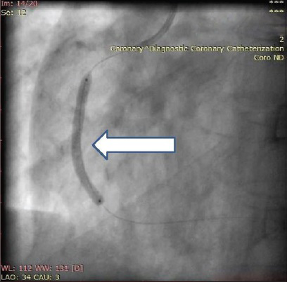
- 60-year-old male
with ACS-inferior wall MI, post thrombolysis and post MI angina. Coronary angiograph shows left anterior oblique
fluoroscopic view of RCA during stent deployment (arrow).

- 60-year-old male with ACS-inferior wall MI, post thrombolysis and post MI angina.
Coronary angiography shows left anterior oblique fluoroscopic view of RCA during stent deployment.
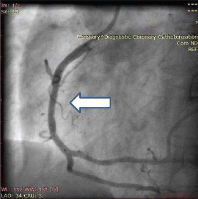
- 60-year-old male with ACS-inferior wall MI post thrombolysis and
post MI angina. Coronary angiography-left anterior oblique angiographic view of RCA shows end result post
stenting (arrow).

- 60-year-old male
with ACS-inferior wall MI, post thrombolysis and post MI angina. Coronary angiography - left anterior oblique
angiographic view of RCA shows end result post stenting.
DISCUSSION
Several classification systems for coronary artery abnormalities exist. Lipton et al., classified coronary variations based on origin and anatomical course relating to the ascending aorta and pulmonary trunk.[2] Type L represents an RCA originating from the left main stem and type R indicates that the coronary artery originates from the RCA. These types are then classified as I-III. Class I follows the anatomical course of either an RCA or LCA. Class II indicates one coronary artery arising from the proximal part of the normally located opposite coronary artery. In class III, the left anterior descending (LAD) and left circumflex (LCx) arise separately from the proximal part of a normal RCA. Classes II and III are then designated as anterior (type A) to pulmonary artery or posterior (type P) to aorta, or interarterial (type B) if it courses between the ascending aorta and the pulmonary trunk. Type B morphology has been associated with a high risk of clinical consequences when associated with an intramural course.[2] Angelini et al., proposed a slightly different classification according to the anatomical course within the interventricular sulcus and atrioventricular groove, as well as the location of penetrating side branches.[3] According to Lipton's classification, our patient had R II P subgroup (single coronary artery from the right sinus with LCA arising from the proximal part of RCA and a posterior course to aorta). Lipton's classification has been modified by others, adding to this classification the “S” septal (through the interventricular septum) and “C” combined types.[1] R II P subgroup is rare.[4] Though there are several case reports of PCI in single coronary artery, most are through the femoral route.[56] To the best of our knowledge, this case report is one of a few similar cases described in the literature. We did not have much difficulty during PCI as we were using right radial artery approach and Judkin's right catheter. For right coronary cannulation, we always start with a JR curve. Other catheters like internal mammary artery (IMA), Multipurpose, and Amplatz left (AL) can also be used according to the situation.[7]
CONCLUSION
The present case merits mention because of several points: 1) Intervention in a single coronary artery through radial approach has been rarely reported and in type R II, it is all the more rare. 2) CT 3D reconstruction is a useful tool to understand the ostial configuration and course of anomalous coronary. 3) Radial PCI is as good as femoral PCI for anomalous coronaries, provided good hardware is selected and operator has experience in radial interventions. 4) Meticulous attention should be given to pressure damping while doing PCI in cases with both coronaries originating from single ostium. 5) We should take non-selective shoot if there is no coronary originating from either sinus (left/right).
Financial support and sponsorship
Nil.
Conflicts of interest
There are no conflicts of interest.
Acknowledgment
We are thankful to Dr. Neha Godara for her kind support.
Available FREE in open access from: http://www.clinicalimagingscience.org/text.asp?2015/5/1/65/170735
REFERENCES
- Coronary artery anomalies in 126,595 patients undergoing coronary arteriography. Cathet Cardiovasc Diagn. 1990;21:28-40.
- [Google Scholar]
- Isolated single coronary artery: Diagnosis, angiographic classification, and clinical significance. Radiology. 1979;130:39-47.
- [Google Scholar]
- Coronary anomalies: Incidence, pathophysiology, and clinical relevance. Circulation. 2002;105:2449-54.
- [Google Scholar]
- Isolated single coronary artery: A review of 50,000 consecutive coronary angiographies. Eur Heart J. 1992;13:1637-40.
- [Google Scholar]
- Angioplasty of the right coronary artery with origin of all three coronary arteries from a single ostium in the right sinus of Valsalva. Am Heart J. 1993;126:985-7.
- [Google Scholar]
- Primary coronary angioplasty with stenting for acute coronary syndrome in patients with isolated single coronary artery: A report of 2 cases. Jpn Heart J. 2003;44:759-65.
- [Google Scholar]
- Patel's Atlas of Transradial Intervention: The Basics.


